Whatโ€™s In YOUR Protein? Optimizing Protease Digestions to Get the Inside Scoop

Itโ€™s time to analyze your protein and you are trying to decide where to begin. You are asking questions like: Which protease do I choose? How much enzyme should I use in my digest? How long should I perform my digest?

Unfortunately, there is no one-size fits all answer to this type of question other thanโ€ฆ โ€œwell it depends.โ€ All protease digests will be a balance between denaturing the protein sample to allow access to cleavage sites, optimizing conditions for the protease to function, and compatibility with your workflow and downstream applications. We provide general guidelines that work for most samples, but frequently you will need to optimize the conditions need for your specific sample and application.

Here, I use the example of a trypsin digest for downstream mass spectrometry to highlight key questions to ask and factors that can be optimized for any digest.

Continue reading “Whatโ€™s In YOUR Protein? Optimizing Protease Digestions to Get the Inside Scoop”

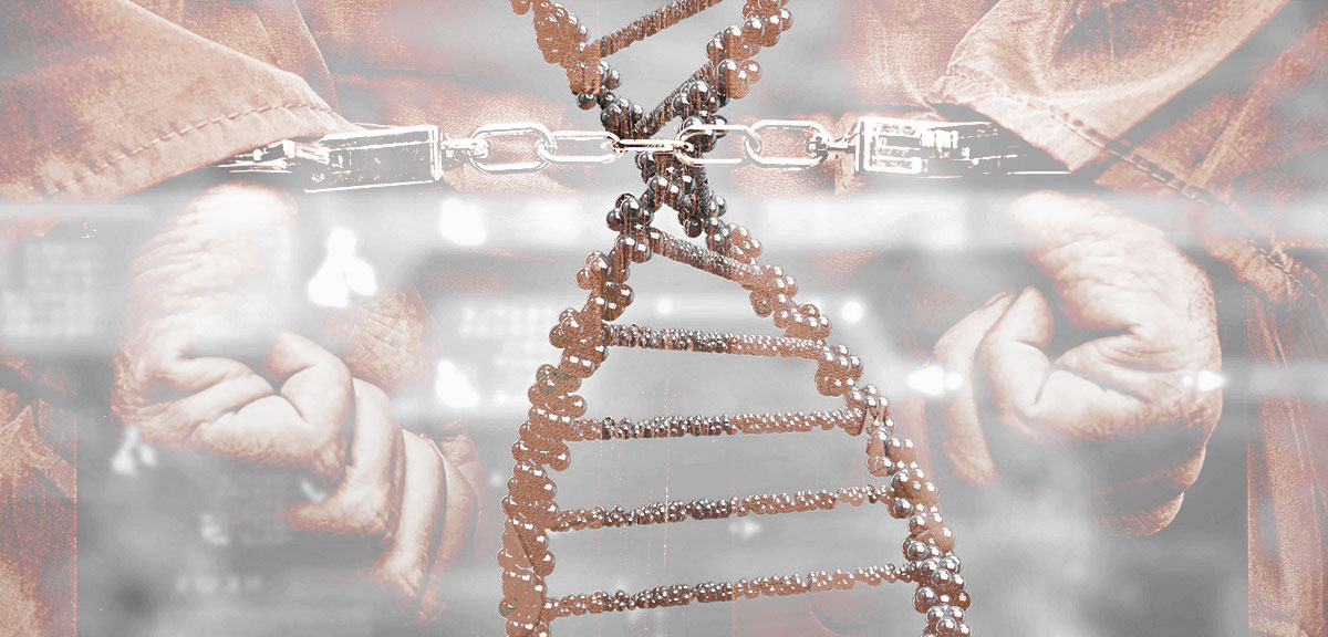
Conflict, CRISPR and the Scientific Method

Scientific inquiry is a process that is revered as much as it is misunderstood. As I listened to a TED talk about the subject, I was reminded that for the general public the foundation of science is the scientific methodโ€”the linear process of making an observation, asking a question, forming an hypothesis, making a prediction and testing the hypothesis.

While this process is integral to doing science, what gives scientific findings credibility and value is consensus from the scientific community. Building consensus is the time-consuming process that includes peer review, publication and replication of results. It is also the part of scientific inquiry that so often leads the public to misunderstand and mistrust scientific findings.

Continue reading “Conflict, CRISPR and the Scientific Method”

A Crash Course in Fighting Lab Contamination

When I first started in my undergraduate lab, one of the first things I learned was how to prepare agar plates for growing yeast. My supervisor, a grad student, looked over my shoulder as I added the yeast extract, bacto peptone, and other ingredients. I sealed the pitcher tightly with aluminum foil and autoclaved it until sterile. When I was ready to pour the plates, I carried the pitcher to the โ€œplate-pouringโ€ room, ripped the foil off, and started to pour an even layer of agar into each of the plastic dishes, leaving the lids off so they could cool. After Iโ€™d poured a dozen or so, my grad student supervisor burst into the room.

โ€œWhat are you doing?โ€ she demanded.

โ€œIโ€™m pouring plates,โ€ I stammered back.

She took a deep breath and explained. By fully uncovering the pitcher and leaving my plates uncovered, I had left my precious media at high risk for contamination. The open containers were far too inviting for potential contaminants floating through the air. In the end, we ended up throwing away several of the plates that had been exposed the longest.

Now, I donโ€™t share this story to demonstrate how clueless when I first started in the research lab as an undergrad. We all have those โ€œuh-ohโ€ moments when we realize for the first time that something that seemed so obvious was, in fact, more complicated than weโ€™d expected. However, that day I learned how easily I could sabotage my own work by unwittingly inviting contaminants into my experiments.

Whether you work with yeast, bacteria, mammalian cells or anything else in a molecular biology lab, preventing contamination is crucial to getting desired results. Fortunately, minimizing your risk can be incredibly easy.

Letโ€™s start with your lab bench. Everyone has their own organization system, but if yours is โ€œout-of-control chaos,โ€ you might want to reevaluate. Benchtop clutter makes it difficult to thoroughly clean the bench as often as needed. All those bottles of solutions, empty tip boxes, and wrinkled protocol sheets harbor dust and other unwelcome particles that you want to keep away from your cultures and reactions.

Once your benchtop is tidy (or at least somewhat tidy), make sure you keep the surface as clean as possible. Immediately clean up any spills or drips that happen while youโ€™re working. Wiping your workspace with a 10% bleach solution will sterilize it, and following that up with 70% ethanol will dry it quickly. This wash should be performed at least once a day. Ideally you should also regularly remove everything from your workspace and perform a deeper cleaning of your benchtop, as well as any shelves and containers in your area.

Now that your bench is in good shape, itโ€™s time to gear up . You should always follow standard safety procedures (lab coat and goggles, closed-toe shoes, hair tied back), but above all, make sure you never forget your gloves. Gloves protect you from harmful chemicals, but they also protect your experiments from anything that could be on your hands. Skin can carry reagents, bacteria, and enzymes that are good for your body but bad for your experiments. Change your gloves regularly to prevent potential carryover of reagents or samples between containers. A good rule is, โ€œWhen in doubt, change your gloves.โ€

Finally, to guard against airborne contaminants, do your best to keep everything covered when you arenโ€™t immdiately using it. I learned this rule the hard way when several of my yeast plates developed fuzzy patches of mold several days after I poured them. Bacteria and other undesirables floating through the air can affect stock solutions, cultures, plates, tubes, and basically anything else you rely on. Keep your lids on and cover open containers to minimize air exposure to reduce the chances of nefarious particles finding their way in.

Thereโ€™s no way to guarantee youโ€™ll never experience some form of contamination in your lab, but smart practices can help reduce your risk. Develop an anti-contamination routine that meets your needs and make sure you stick to it every day in the lab.
From Drug Screening to Agriculture to Cardiac Development, A Dual-Luciferase Reporter Brings You the Story

Today’s blog was written by guest blogger Katarzyna Dubiel, marketing intern in Cellular Analysis and Proteomics. Last updated 02/12/2021

Reporter gene assays have been critical for the study of a wide-range of biological questions, from regulation of gene expression to cellular signaling. While reporter gene assays constitute a large group of technologies, here we highlight the diversity of new discoveries enabled by highly quantitative and easily measured bioluminescent luciferase-based reporter assays. Below are our top picks of exciting research discoveries involving the Dual-Luciferase Reporter Assay format using firefly and Renilla luciferases.

Continue reading “From Drug Screening to Agriculture to Cardiac Development, A Dual-Luciferase Reporter Brings You the Story”

Fished to the Edge: How DNA Identification Can Help Fight the Illegal Trade of Threatened Shark Species

Whether your first encounter was peering through the thick glass of an aquarium tank or peeking through your fingers in a darkened theater, there is something about sharks that captures our imagination. These fierce, and sometimes fearsome, creatures have existed in our oceans for over 400 million years, andย  survived multiple mass extinction events, including the one that killed the dinosaurs. They are not, however, the vicious, vengeful villain that some movies would have us believe. Sharks are apex predators, who play an important role in the world’s ocean ecosystem by regulating the population of prey species below them. ย Unfortunately, they are also part of one of the most threatened group of marine fish in the world. Of the more than 400 species of sharks that exist in our oceans today, approximately 15% are considered vulnerable, endangered or critically endangered. Continue reading “Fished to the Edge: How DNA Identification Can Help Fight the Illegal Trade of Threatened Shark Species”
Of Mice and Microbes: The Science Behind Food Analysis

In general, people like to know that their food is what the label says it is. It’s a real bummer to find out that beef lasagna you just ate was actually horsemeat. Plus, there are many religious, ethical and medical reasons to be cognizant of what you eat. Someone who’s gluten intolerant and Halal probably doesn’t want a bite of that BLT.

Labels donโ€™t always accurately reflect what is in food. So how do we confirm that we are in fact buying crab, and not whitefish with a side of Vibrio contamination?

For the most part, it comes down to separation science. Scientists and technicians use various chromatographic methods, such as gas chromatography, liquid chromatography, and mass spectrometry, to separate the complex mixture of molecules in food into individual components. By first mapping out the molecular profile of reference samples, they can then take an unknown sample and compare its profile to what it should look like. If the two don’t match up, an analyst would assume that the unknown is not what it claims to be. Continue reading “Of Mice and Microbes: The Science Behind Food Analysis”

How Autophagy Feeds Cancer’s Need for Metabolites

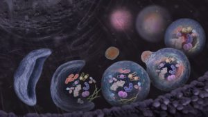
Illustration of energy metablism in cell.

Metabolism underpins numerous cellular processes. Without it, cells would not grow, divide, synthesize or secrete. Another pathway, autophagy, degrades unwanted cellular materials, helping to maintain cell health. With these opposing roles, is there a connection between autophagy and metabolism? As it turns out, the answer is yes. Because molecules degraded by autophagy are recycled and fed into metabolism pathways as precursor compounds. There are interesting implications as a result of this connection, ones that affect cancer cells as described in a recent Cell Metabolism review article.

Autophagic flux, the process by which molecules and organelles are directed to the autophagosome, fuse with the lysosome and are degraded, involves a selective process that determines the cargo carried within the autophagosome. Autophagy-related genes (ATGs) direct the process and particular receptor proteins bind the cargo. What is interesting about the connection among cancer, autophagy and metabolism is the complexity of the role that autophagy plays in cancer. While autophagy was thought to act in a more tumor suppressive manner as shown when one copy of an ATG6 analogous gene in mice was deleted and the other left unaltered, and malignant tumors developed, but in mice mosaic for ATG5 deletions, the inhibition of autophagy resulted in benign tumors in the liver. This latter experiment suggested autophagy was needed for cancer progression, a hypothesis reinforced by the lack of ATG mutations in human cancers.

Continue reading “How Autophagy Feeds Cancer’s Need for Metabolites”

Autophagy: The Poem

Roberta A. Gottlieb, MD, is the Director of Molecular Cardiobiology at Cedar-Sinai, a nonprofit academic healthcare organization. She is interested in the role of autophagy in myocardial ischemia, a kind of heart diseaseย in which blood flow to the heart is blocked. (Studies have shown that autophagy is upregulated during myocardial ischemia, but why this happens is not entirely clear.) Her ultimate goal is to understand and mitigate ischemic injury, with the hope of developing therapeutics for humans.

Andโ€”sheโ€™s a poet.

Continue reading “Autophagy: The Poem”

“Aha! Moments” in Science Education

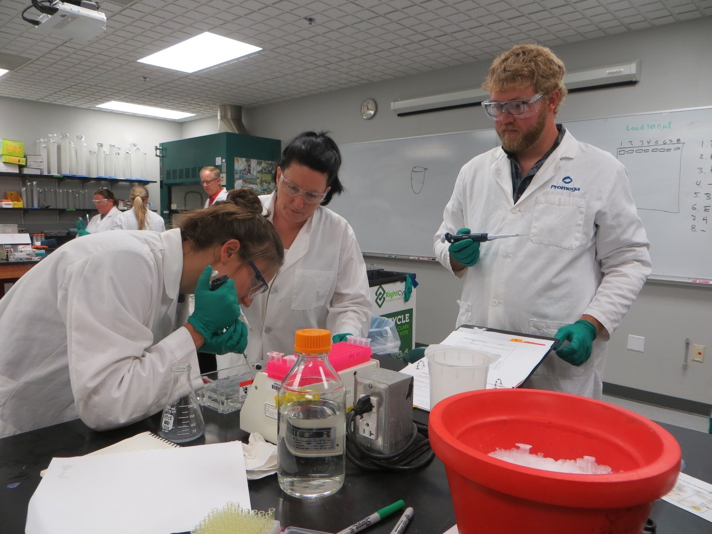
Significant resources are required to deliver high-quality science experiences for students and their teachers. In addition to generous amounts of staff time, for both preparation and program delivery, often there are costly lab supplies. Access to a well-equipped laboratory designed to facilitate educational experiences is also important.

Of course, hands-on experiences are related to learning: for example, becoming scientifically literate, meeting science standards, preparing for AP tests. That said, many of us involved in science outreach activities will tell you that perhaps the most significant justification for these investments is that you never know when one of the students will experience that ‘Aha!’ moment which proves to be life-changing for them.

Over the years, we have heard many testimonials from students, teachers, school-to-career coordinators and other school district personnel, mentors and parents that speak to this experience. There just seems to be something about getting into the lab and engaging directly in โ€œdoing scienceโ€ that stays with some participants as they head back to school, continue with their studies and on to their careers. Continue reading ““Aha! Moments” in Science Education”

Forensic Scientists Improve Sexual Assault Kit Turnaround Time with Y-Screening

The backlog of sexual assault kit samples in crime laboratories across the nation is a topic that hit the spotlight when a group of journalists uncovered the issue in an open records search of crime lab records in 2015. Reasons for the backlog include lack of staff, lack of funding, and simply, lack of time or a decision not to prosecute the case. Processing samples can be a labor-intensive process.

We recently interviewed Lynndsey R. Simon, Forensic Scientist II and Alternate CODIS Administrator from the Columbus Police Forensic Services Center to discuss some recent changes in sample processing in their laboratory that are helping to alleviate some of the backlog. She will be presenting a talk at the upcoming International Symposium on Human Identification (ISHI) in September.

The Columbus Police Forensic Services Center is a smaller forensic laboratory and according to Simon, one of the biggest challenges they face is strained resources. The DNA extraction and processing kits that forensic laboratories use are very expensive, and the number of DNA samples that laboratories are getting for DNA analysis are increasing. With limited resources and funding, maximizing efficiency and finding the best solutions for the laboratory becomes critical.

Continue reading “Forensic Scientists Improve Sexual Assault Kit Turnaround Time with Y-Screening”