A Historic Milestone for PROTAC Research: What Vepdegestrant’s FDA Decision Means for Drug Discovery

Protein degrader research has yielded its first approved therapeutic for specific breast cancer patients: Vepdegestrant received FDA approval on May 1, 2026 (1). Vepdegestrant is an oral PROteolysis TArgeting Chimera (PROTAC) that targets the estrogen receptor for degradation in breast cancer patients with ESR1-mutated ER+/HER2– advanced breast cancer (2) produced by Arvinas, Inc. in collaboration with Pfizer Inc.

A Different Kind of Drug Development

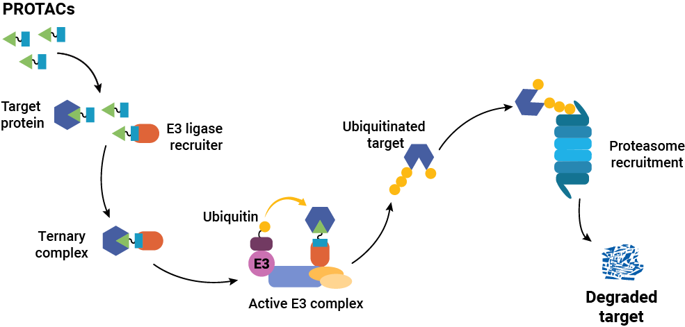
Targeted protein degraders (or PROTACs) have opened new possibilities in drug discovery research. Instead of inhibiting protein function or interaction, degraders cause the removal of the target protein itself. Traditional small molecule drugs work by binding a protein to inhibit it or block function, and they must remain bound to work. That means that the target protein should be well-characterized in terms of binding and activity sites, and the drug must bind specifically only to the target protein. In contrast, degraders only need to bind long enough to recruit cellular protein degradation machinery to the target protein, and the method does not rely on an accessible and specific binding site on the target protein. Once degradation occurs, the degrader is released and can engage with the next target.

The approval of vepdegestrant is a landmark moment for the entire TPD and induced proximity field, demonstrating that it is possible to rationally design molecules whose pharmacology is categorically distinct from traditional drugs, relying on a catalytic rather than occupancy-driven mechanism of action.  More importantly, this translates to meaningful clinical outcomes in patients. —Dr. Kristin Riching, Promega R&D Scientist

The first peptide-based PROTAC was described in 2001 in the laboratories of Craig Crews and Ray Deshaies (3), but translating the concept into orally bioavailable, clinically viable molecules took nearly two decades, using tools that did not exist when the field began. More than 40 PROTAC degraders have now entered clinical trials (4), with vepdegestrant the most advanced, supported by Phase 3 data from the VERITAC-2 trial demonstrating statistically significant improvement in progression-free survival in ESR1-mutant patients. That progress required solving a measurement problem as much as a chemistry one: how do you quantify target protein degradation at endogenous levels, with enough sensitivity and throughput to drive a screening campaign? CRISPR-engineered protein tagging combined with the small bioluminescent reporter tag, HiBiT solved that problem, providing a sensitive, HTS-compatible readout of endogenous target levels without relying on laborious, artifact-prone western blots. Critically, HiBiT also enabled researchers to watch target protein degradation unfold in real time in living cells.

“Seeing it happen in real time, frankly, may have been what convinced many people that the modality had genuine merit.”
—Dr. Kristin Riching

Developing a PROTAC is not like developing a traditional inhibitor. Success requires successful completion of a complex cascade of cellular events: the molecule must enter the cell, engage the target protein and the E3 ligase simultaneously, form a productive ternary complex in the right geometry, trigger ubiquitination, and drive proteasomal degradation, all while competing with cellular noise that can blunt each step. “PROTACs are large molecules, so they are often not very permeable,” Riching explains. “They also need to simultaneously engage both the target and the E3 ligase machinery, but they need to do so in a productive geometry that leads to ubiquitination, which is not easily predicted. In cells, many compounding factors can limit activity, making it difficult to identify which parameters most need improvement. Event-driven modalities like PROTACs rely on robust tools to tease apart each mechanistic step to aid SAR optimization.”

Getting that data means adopting a screening framework built around mechanistic understanding of the full degradation cascade from the earliest stages of optimization, while preserving the native biology and the stoichiometric relationships that govern degradation efficiency. It also means going beyond endpoint measurements. Knowing whether a target is degraded is a starting point; knowing how fast, how completely, and how durably it degrades is what distinguishes a development candidate from a dead end. Riching’s research has shown that different proteins in the same family can respond to the same PROTAC with dramatically different kinetic profiles (5,6), which is a distinction that endpoint assays cannot capture, and one that can determine which compounds are worth advancing.

What’s Next after Vepdegestrant?

The approval of Vepdegestrant validates more than just a single drug, it validates the PROTAC drug category and the tools and methods that enabled it. For researchers working on next-generation degraders, the signal is clear: the modality works. Now the question is how far we can push it.

Riching points to E3 ligase diversity as the field’s most pressing unresolved problem. “The greatest challenge will be expanding beyond the two E3 ligases — CRBN and VHL — that have driven most PROTAC progress to date,” she says. “We don’t yet fully understand the scope of targets accessible through these ligases, but it stands to reason that additional ligases will be necessary to unlock a larger portion of the degradable proteome. Their broad distribution also limits opportunity for tissue-selective targeting. Developing the tools and chemistry to recruit a wider repertoire of E3 ligases remains one of the most important unsolved problems the field faces.”

Beyond ligase diversity, the field is expanding its conception of what a degrader can be. Molecular glues, LYTACs, and other induced proximity strategies are broadening the range of accessible targets — including extracellular and membrane-bound proteins that sit outside the reach of classical PROTACs. Each new modality brings its own characterization challenges, and the same principle holds: understanding mechanism at the cellular level, early and rigorously, is what separates the compounds worth advancing from those that look promising in a tube.

The approval of vepdegestrant is a landmark. But researchers working in this space know it is a beginning as much as it is a culmination — proof that the approach is sound, and a starting line for everything that follows.


Read more about Innovative Imaging Solutions for Targeted Protein Degradation on our website.


Literature Cited

  1. Arvinas, Inc. Arvinas Announces FDA Approval of VEPPANU (vepdegestrant) for the Treatment of ESR1m, ER+/HER2– Advanced Breast Cancer Accessed: May 5, 2026 
  1.  Arvinas, Inc. (2025) Arvinas Announces FDA Acceptance of the New Drug Application for Vepdegestrant for the Treatment of ESR1m, ER+/HER2– Advanced Breast Cancer. August 8. Accessed: April 27, 2026.  
  1. Sakamoto, K.M. et al. (2001) Protacs: Chimeric Molecules that Target Proteins to the Skp1-Cullin-F Box Complex for Ubiquitination and DegradationProc. Natl. Acad. Science USA 98, 8554-–9. Accessed: May 4, 2026. 
  1. Chen, S. (2026) Protein Degraders (PROTACS & Molecular Glues) in 2026: The Emergining Challenge to Traditional Drug Development Accessed: May 5, 2026 
  1. Riching, K.M et al. (2018) Quantitative Live-Cell Kinetic Degradation and Mechanistic Profiling of PROTAC Mode of ActionACS Chem. Biol13, 2758–70. Accessed: May 4, 2026 
  1. Riching, K.M. et al.  (2022) The Importance of Cellular Degradation Kinetics for Understanding Mechanisms in Targeted Protein DegradationChem. Soc. Rev. 51, 6210–6221. 

This article was written with AI assistance.

Why BRETSA™ Target Engagement Matters for Drug Discovery

Drug discovery researchers face a fundamental constraint in their work to develop safe, effective therapeutics: the vast majority of the human proteome remains inaccessible to conventional small molecule approaches. Proteins without defined binding pockets, those lacking known chemical probes, and protein targets that fail to translate from biochemical assays into cellular models have long been considered out of reach of standard drug discovery screening tools. As Dixit et al. describe, developing biochemical or cellular assays for all genome-encoded targets “is not scalable and likely impossible as most proteins have ill-defined or unknown activity” — these are what the authors call “the dark undruggable expanses” of the proteome [1].

That gap is now narrowing. Promega Corporation recently launched the TarSeer™ BRETSA™ Target Engagement System, a live-cell target engagement platform designed to bring previously challenging targets within reach of early-stage drug discovery.

The Problem: A Translation Gap in Early Discovery

Drug discovery teams regularly encounter a frustrating disconnect. A compound may show strong binding activity in a biochemical assay, only to fail when tested in a cellular environment. Without target-specific cellular assays, which generally aren’t available for poorly characterized proteins, researchers face difficult choices when deciding which compounds to advance through the drug development pipeline.

Continue reading “Why BRETSA™ Target Engagement Matters for Drug Discovery”

What Shelter Dogs Can Tell Us About Emerging Zoonotic Diseases

Why Are Zoonotic Diseases Becoming a Bigger Risk?

As of September 9, 2025, the Worldometer listed the human global population as 8.3 billion people (1). This population growth means that humans will be living and working in previously uninhabited or minimally disturbed environments, increasing interactions between humans, domestic animals, wildlife, and their pathogens. This intensifying human-animal interface heightens the risk of zoonotic disease transmission, where pathogens cross species barriers (from wildlife to domestic livestock or from wildlife to humans), potentially leading to outbreaks and even pandemics.

How Do Urbanization and Climate Change Amplify Zoonotic Threats?

Urbanization, habitat disruption, and climate change further exacerbate these risks by altering ecosystems and facilitating the spread and emergence of vector-borne and zoonotic diseases. Understanding and addressing these threats requires robust surveillance, effective diagnostics, and proactive strategies to prevent and mitigate disease emergence and spread.

In urban areas, public health officials are already using wastewater to monitor known pathogens and identify “hot spots” of activity to predict increases in illness within local populations (2). Animal shelters are another place where there is an opportunity to monitor for emerging infectious diseases that could affect domestic pet animals.

Continue reading “What Shelter Dogs Can Tell Us About Emerging Zoonotic Diseases”

Developing an Experimental Model System to Understand the Tumor Microenvironment of Melanoma Brain Metastases

Cancer’s greatest threat is its ability to spread to other tissues—a process known as metastasis. Melanoma, a form of skin cancer, exemplifies this devastating progression. Although treatable when caught early—with surgical removal resulting in over 99% survival at five years—once melanoma metastasizes, five-year survival rates plummet dramatically to around 27%. Even more concerning, melanoma exhibits a particularly high tendency to invade the central nervous system, causing melanoma brain metastases (MBMs) that are incurable and reduce median survival to just 13 months.

To understand metastasis, we need reliable and realistic experimental models. Traditional cell cultures on plastic dishes are limited, failing to replicate the intricate spatial organization and biochemical interactions within living tissues. Animal models are informative but expensive, ethically complex, and not always accurate for human diseases. Addressing this critical gap, Reed-McBain and colleagues (2025) introduced an innovative microphysiological system (MPS) designed to simulate the tumor microenvironment in the brain affected by metastatic melanoma.

Continue reading “Developing an Experimental Model System to Understand the Tumor Microenvironment of Melanoma Brain Metastases”

IL-6/STAT3-Regulated Long Non-Coding RNA Is Involved in Colorectal Cancer Progression

Researchers from Wenzhou Medical University in China have identified a mechanism involving long non-coding RNAs (lncRNA) that contributes to colorectal cancer (CRC) progression. CRC is the third most common cancer worldwide and is one of the most lethal cancers across the globe. Understanding the molecular mechanisms that underlie the development and progression of CRC is critical to developing biomarkers to detect it and new therapeutics to treat it. 

Continue reading “IL-6/STAT3-Regulated Long Non-Coding RNA Is Involved in Colorectal Cancer Progression”

Using Dual-Luciferase Assays to Identify the Role of Non-Coding RNAs in Disease

In recent years, non-coding RNAs—especially microRNAs (miRNAs) and long non-coding RNAs (lncRNAs)—have emerged as powerful regulators of cellular behavior. These molecules modulate gene expression, often by targeting mRNAs for translational suppression or degradation. Two recent studies—one focused on osteoarthritis and the other on 5-Fu-resistant colorectal cancer—illustrate how these non-coding, regulatory RNAs operate within disease-relevant signaling networks, providing new points for therapeutic intervention.

lncRNA, long noncoding RNA

Both studies use the pmirGLO Dual-Luciferase miRNA Target Expression Vector to evaluation predicted miRNA activity.  This dual-luciferase system offers a clean and quantifiable way to validate miRNA–mRNA interactions using a simple bioluminescent readout. By cloning the 3´ untranslated regions (UTRs) of suspected targets downstream of a firefly luciferase reporter and normalizing against Renilla luciferase, researchers can rapidly confirm whether a miRNA directly regulates its target.

Continue reading “Using Dual-Luciferase Assays to Identify the Role of Non-Coding RNAs in Disease”

An Unexpected Role for RNA Methylation in Mitosis Leads to New Understanding of Neurodevelopmental Disorders

Traditionally, RNA methylation has been studied in the context of gene expression regulation, RNA stability and translation efficiency, with its primary role thought to be in modulating cellular homeostasis and protein synthesis. However, a 2025 study by Dharmadkikari and colleagues uncovers an unexpected and critical function for RNA methylation in mitotic spindle integrity.

False color transmission electron microscope (TEM) micrograph of a mitotic cell in metaphase stage showing chromosomes (purple) in the equatorial plane and one of the mitotic spindle poles (blue). Mutations in SPOUT1/CENP-32 affect RNA methylation which is necessary for proper cell division.
False color transmission electron microscope (TEM) micrograph of a mitotic cell in metaphase stage showing chromosomes (purple) in the equatorial plane and one of the mitotic spindle poles (blue).

The study identifies a critical role for SPOUT1/CENP-32-dependent methylation in mitotic spindle formation and accurate chromosome segregation. Originally identified in a large-scale analysis of proteins associated with mitotic chromosomes, SPOUT1/CENP-32 encodes a putative RNA methyltransferase. The protein localizes to mitotic spindles, and when it is absent centrosome detachment from the spindle poles, delayed anaphase, and chromosome segregation errors are observed. Further, CRISPR experiments in human cells show that the protein is essential for cell viability.

Continue reading “An Unexpected Role for RNA Methylation in Mitosis Leads to New Understanding of Neurodevelopmental Disorders”

Unlocking the Secrets of ADP-Ribosylation with Arg-C Ultra Protease, a Key Enzyme for Studying Ester-Linked Protein Modifications 

Post-translational modifications of proteins are critical for proper protein function. Modifications such as phosphorylation/dephosphorylation can act as switches that activate or inactivate proteins in signaling cascades. The addition of specific sugars to membrane proteins on cells are critical for recognition, interaction with the extracellular matrix and other activities. While we know volumes about some types of protein modifications, ADP-ribosylation on aspartate and glutamate residues has been more difficult to study because of the chemical instability of these ester-linked modifications. 

Matić Lab (Eduardo José Longarini and Ivan Matić) recently published a study that explored mono-ADP-ribosylation (ADPr) on aspartate and glutamate residues by the protein PARP1 and its potential reversal by PARG. PARP1 and PARG signaling are central to DNA repair and apoptosis pathways, making them potentially powerful therapeutic targets in cancer or neurodegenerative diseases in which DNA repair processes are often disrupted. 

Continue reading “Unlocking the Secrets of ADP-Ribosylation with Arg-C Ultra Protease, a Key Enzyme for Studying Ester-Linked Protein Modifications “

Exploring the Respiratory Virus Landscape: Pre-Pandemic Data and Pandemic Preparedness

influenza viruses are part of the worldwide respiratory virus landscape

Since the COVID-19 pandemic, public health researchers and research scientists have sought more urgently to understand the worldwide respiratory virus landscape. The COVID-19 pandemic has forced us to re-evaluate our global public health priorities and activities. Additionally, acute respiratory tract infections are one of the leading causes of illness and death worldwide, particularly in developing countries. To really understand what changed with the pandemic and how we can best respond going forward, we need to understand what the baseline landscape was before the pandemic. Studies using samples that were collected prior to the pandemic are essential to this effort.

Continue reading “Exploring the Respiratory Virus Landscape: Pre-Pandemic Data and Pandemic Preparedness”

From Fins to Genes: DNA Barcoding Unlocks Marine Diversity Along Mozambique’s Coast

DNA Barcding unlocks marine diversity along Mozambique's coast

The Mozambique Channel, which is located between the Madagascar and Mozambique on the African coast, is an important hot spot for biodiversity because its many coastal ecosystems provide a range of habitats that support diverse plant and animal species. Understanding the biodiversity of an ecosystem, particularly biodiversity hot spots, is important for many reasons. For marine systems, accurate classification and reporting of fish species supports fisheries research, natural resource surveys, forensic studies, conservation studies, and enables discovery of new or under-reported species. Studies have been limited along the west coast of Africa and are only now in their early stages.

A 2024 research study by Muhala and colleagues applied DNA barcoding to evaluate the composition of marine and coastal fish diversity from the Mozambican coast. In the study, the Wizard® Genomic DNA Purification Kit was used to extract DNA from both teleost (ray-finned) and elasmobranch (sharks, rays and skates) fish classes, with a total of 143 species sampled from local artisanal fisheries along the Mozambican coast. The samples were primarily composed of muscle or fin tissues, which are ideal for genetic analysis due to their higher DNA yield. These tissue samples were collected from various fish species captured along the coast of Mozambique, stored in ethanol (96%) to preserve DNA integrity, and then processed using the Wizard kit. Total genomic DNA was extracted from the muscle or fin tissues, as per the manufacturer’s protocol. This method ensures the isolation of high-quality genomic DNA, which is crucial for subsequent polymerase chain reaction (PCR) amplification and sequencing. The COI gene (cytochrome c oxidase subunit I) was targeted for DNA barcoding, enabling species identification and assessment of genetic diversity.

Continue reading “From Fins to Genes: DNA Barcoding Unlocks Marine Diversity Along Mozambique’s Coast”