Small RNA Transfection: How Small Players Can Make a Big Impact

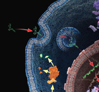
When looking at small aspects of living things, especially cells, it can often be difficult to fully grasp the magnitude of regulation employed within them. We first learn the central dogma in high school biology. This is the core concept that DNA makes RNA and RNA makes protein. Despite this early education, it can be lost on many the biological methods that are employed to regulate this process. This regulation is very important when one considers the disastrous things that can occur when this process goes askew, such as cancer, or dysregulated cell death. Therefor it is very important to understand how these regulatory mechanisms work and employ tools to better understand them.

Continue reading “Small RNA Transfection: How Small Players Can Make a Big Impact”

Insects and Science: Optimizing Work with Sf9 Insect Cells

Insects are a keystone species in the animal kingdom, often providing invaluable benefits to terrestrial ecosystems and useful services to mankind. While many of them are seen as pests (think mosquitos), others are important for pollination, waste management, and even scientific research.

Insect biotechnology, or the use of insect-derived molecules and cells to develop products, is applied in a diverse set of scientific fields including agricultural, industrial, and medical biotechnology. Insect cells have been central to many scientific advances, being utilized in recombinant protein, baculovirus, and vaccine and viral pesticide production, among other applications (5).

Therefore, as the use of insect cells becomes more widespread, understanding how they are produced, their research applications, and the scientific products that can be used with them is crucial to fostering further scientific advancements.

Primary Cell Cultures and Cell Lines

Cell culture - Cell lines - Insect Cells

In general, experimentation with individual cells, rather than full animal models, is advantageous due to improved reproducibility, decreased space requirements, less ethical concerns, and a reduction in expense. This makes primary cell cultures and cell lines essential contributors to basic scientific research.

Continue reading “Insects and Science: Optimizing Work with Sf9 Insect Cells”

I Have My Luciferase Vector, Now What?

Choosing and Optimizing Transfection Methods

Here in Technical Services we often talk with researchers at the beginning of their project about how to carefully design and get started with their experiments. It is exciting when you have selected the Luciferase Reporter Vector(s) that will best suit your needs; you are going to make luminescent cells! But, how do you pick the best way to get the vector into your cells to express the reporter? What transfection reagent/method will work best for your cell type and experiment? Do you want to do transient (short-term) transfections, or are you going to establish a stable cell line?

Continue reading “I Have My Luciferase Vector, Now What?”

How to Reduce Cell Culture Variability

Scenario 1: Jake needs a flask of MCF-7 cells for an assay, so he sends an email to the graduate student listserv asking for cells. Melissa replies that she has an extra flask of cells that she could share. Jake happily accepts the cells and begins his experiment.

Scenario 2: Michael passaged his cells yesterday and, according to the protocol, was supposed to plate cells today for treatment. However, his previous experiments were delayed, so he decides to plate them tomorrow instead. The cells look healthy, so it should be ok.

What is wrong with the above scenarios? These actions may seem harmless, but they could be the cause of variability, leading to irreproducible results. Continue reading “How to Reduce Cell Culture Variability”

The Cell Line Identity Crisis: Old problems, new concerns

If you work with cell lines you may have paid attention to the dramatic headline published last month in the online journal STAT, Thousands of studies used the wrong cells, and journals are doing 29981164-Whistlenothing.” In their column The Watchdogs (“Keeping an eye on misconduct, fraud, and scientific integrity”), Ivan Oransky and Adam Marcus call out the fact that scientists continue to publish research using cell lines that are contaminated or misidentified. Recent estimates have found that the percentage of misidentified cell lines used by scientists is as high as 20 to 36. The blame here is being placed on the peer reviewed journals for not blowing the whistle. The authors call for journals to put some “kind of disclaimer on the thousands of studies affected.”

This is not a new claim. The continuing problem of cell line misidentification, of lack of authentication, has been covered before in various channels. It’s easy to find news publicizing yet another retracted publication. Promega Connections has published a number of blog posts addressing this, one as recently as last year: Do You #Authenticate? This post describes the bold move by the journal Nature to adopt a new policy around cell line authentication. Beginning in May 2015 the journal required authors of all submitted manuscripts to confirm the identity of cell lines used in their studies and provide details about the source and testing of their cell lines. Continue reading “The Cell Line Identity Crisis: Old problems, new concerns”

Improving the Success of Your Transfection

12150558-plasmid_with_cell_membrane3

Not every lab has a tried and true transfection protocol that can be used by all lab members. Few researchers will use the same cell type and same construct to generate data. Many times, a scientist may need to transfect different constructs or even different molecules (e.g., short-interfering RNA [siRNA]) into the same cell line, or test a single construct in different cultured cell lines. One construct could be easily transfected into several different cell lines or a transfection protocol may work for several different constructs. However, some cells like primary cells can be difficult to transfect and some nucleic acids will need to be optimized for successful transfection. Here are some tips that may help you improve your transfection success.

Transfect healthy, actively dividing cells at a consistent cell density. Cells should be at a low passage number and 50–80% confluent when transfected. Using the same cell density reduces variability for replicates. Keep cells Mycoplasma-free to ensure optimal growth.

Transfect using high-quality DNA. Transfection-quality DNA is free from protein, RNA and chemical contamination with an A260/A280 ratio of 1.7–1.9. Prepare purified DNA in sterile water or TE buffer at a final concentration of 0.2–1mg/ml.

Continue reading “Improving the Success of Your Transfection”

General Considerations for Transfection

Many studies, from reporter assays to protein localization to BRET and FRET, require successful transfection first. Yet, transfection can be tricky and difficult. There are many considerations when planning transfection of your cells including reagent selection, stable or transient experiment, type of molecule and endpoint assay used. Here we discuss these considerations to help you plan a successful transfection scheme for your experimental system. Continue reading “General Considerations for Transfection”

Get More Out of Your Lentiviral Production

fugene6_lvv_blogThis review is a guest blog by Amy Landreman, Product Specialist in Cellular Analysis at Promega Corporation.
Lentiviral vectors (LVV) have become a valuable research tool for delivering genetic content into a wide range of cell types. Commonly derived from the HIV-1 genome, LVV have the advantage of being able to infect both dividing and non-dividing cells. They can be particularly valuable for introducing genetic material into cell lines that are difficult to transfect using other methods and are also being used in gene therapy applications.

Unlike other gene delivery tools, transducing mammalian cells with LVV requires significant upfront effort since the LVV particles carrying the desired genetic content first need to be created. In general this involves co-transfecting a packaging cell line, such as HEK293T, with a set of three to four separate plasmids that encode the protein content required to generate the LVV particles: the transfer plasmid, which contains the transgene of interest, a packaging plasmid, and an envelope plasmid. After co-transfection, the packaging cell line is allowed to incubate for a couple of days during which time the LVV particles are produced and accumulating in the culture supernatant. The supernatant containing the recombinant LVV is then harvested and, following several concentration steps, the LVV particles are ready to be used for introducing the desired genetic content into the mammalian target cells. Continue reading “Get More Out of Your Lentiviral Production”

Practical Tips for HEK293 Cell Culture When Using cAMP-Glo™ Assay

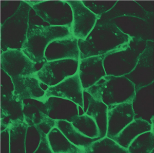
HEK293 cells stably expressing HaloTag®-ECS (ExtraCellular Surface; comprised of a signal sequence and single transmembrane domain of β1-integrin) fusion protein labeled with HaloTag® Alexa Fluor® 488 Ligand and then imaged.
HEK293 cells stably expressing HaloTag®-ECS (ExtraCellular Surface; comprised of a
signal sequence and single transmembrane domain of β1-integrin) fusion protein labeled
with HaloTag® Alexa Fluor® 488 Ligand and then imaged.
G Protein Coupled Receptors represent one of the largest classes of cell surface receptors and one of the most important classes for drug targets. Fifty of the top 200 drugs target GPCRs. GPCRs respond to various stimuli like light, odors, hormones, neurotransmitters and others. They cover virtually all therapeutic areas. When a particular GPCR is implicated in a disease, researchers screen the GPCR and its signaling pathways, the hope being that promising therapeutic targets might be identified. Major G-protein families signal via secondary messengers like cAMP, which in turn activate a range of effector systems to change cell behavior and/or gene transcription. There are various approaches and methods to study GPCRs and measure the increase or decrease of intracellular cAMP. However, the fastest and the most sensitive among all methods is a plate based cAMP-Glo™ Assay. Continue reading “Practical Tips for HEK293 Cell Culture When Using cAMP-Glo™ Assay”

Cell Line Misidentification Rears Its Ugly Head

Cancer cell illustrationBack in 2009, we reported on the problem of cell line contamination (1). In that article we reported the statistics that an estimated 15–20% of the time, the cell lines used by researchers are misidentified or cross-contaminated with another cell line (1). This presents a huge problem for the interpretation of data and the reproducibility of experiments, a key pillar in the process of science. We have revisited this topic several times, highlighting the issues cell and tissue repositories have discovered with cell lines submitted to them (2) and discussing the new guidelines issued by ANSI (3,4) for researchers regarding when during experimental processes cell lines should be authenticated and what methods are acceptable for identifying cell lines.

Just recently two papers were voluntarily retracted by their authors because of cross contamination among cell lines used in the laboratories. The first that came to my attention represented the first retraction from Nature Methods in its nine years of publication. In this paper, cross contamination of a primary gliomasphere cell lines with HEK cells expressing GFP resulted in “unexplained autofluorescence” associated with tumorigenicity (5). The second paper, retracted from Cancer Research by the original authors, was also another cross contamination story involving HEK cells (6). In this story a gene was incorrectly described as a tumor suppressor, that when silenced led to the formation of tumors in nude mice. It turns out that the contaminating HEK cells also failed to express this same gene.

So because of cross contamination of cell lines, two groups have voluntarily retracted papers. Being open and honest about what had happened with the cell lines and reaching the decision to retract the papers could not have been an easy thing, but these decisions benefit the scientific community in many ways. Obviously they benefit the researchers doing work on the specific research questions addressed by the papers by preventing researchers from pursuing paths that lead to dead ends. But in the bigger picture these retractions reinforce the argument that cell line authentication needs to become a routine and accepted part of any experimental process that depends on cell culture if we are to have confidence in the experimental results.

References

  1. Dunham, J.H. and Guthmiller, P.  (2009) Doing good science: Authenticating cell line identity. Promega Notes 101, 15–18.
  2. Duham, J.H. and Guthmiller, P. (2012) Doing good science: Authenticating cell line identity. Promega PubHub. [Internet: Accessed September 2013]
  3. Gopal, A. (2013) Fingerprinting  your cell lines. Promega Connections blog [Internet: Accessed September 2013]
  4. Sundquist, T. (2013) Preventing the heartache of cell line contamination. Promega Connections blog [Internet: Accessed September 2013]
  5. Evanko, D. (2013) A retraction resulting from cell line contaminationMethagora blog. [Internet Accessed September 2013]
  6. Negorev, D. (2013) Retraction: Sp100 as a potent tumor suppressor: Accelerated senescence and rapid malignant transformation of human fibroblasts through modulation of an embryonic stem cell program. Can. Res. 73, 4960.