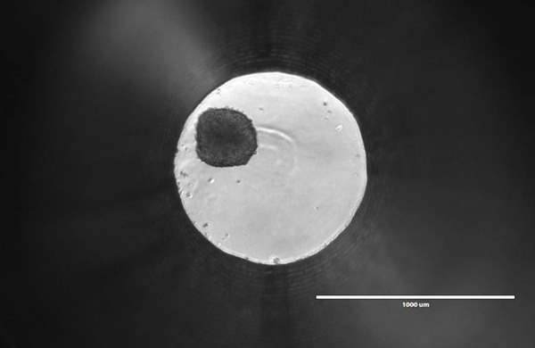
Traditionally, scientists have relied on flat,
two-dimensional cell cultures grown on substrates such as tissue culture
polystyrene (TCPS) to study cellular physiology. These models are simple and
cost-effective to culture and process. Within the last decade, however, three-dimensional
(3D) cell cultures have become increasingly popular because they are more
physiologically relevant and better represent in vivo conditions.
This past weekend was the 9th Annual Wisconsin Science Festival, and we at Promega were excited to join in the celebration of science throughout the state. We participated in the Discovery Expo on Thursday and Friday, where dozens of demonstrations and exhibits were scattered throughout the Wisconsin Institute for Discovery building. Thousands of children on field trips filled the halls, eager to poke and prod at strange and exciting new things.
At our table, we talked about the science of bioluminescence. With 3D-printed firefly luciferase models in hand, we showed the glow of recombinant luciferase to the incoming children and explained to them how scientists could use bioluminescence like a tiny “flashlight” to look inside of cells and watch what’s happening. Our learners received a nice little reward for their attentiveness in the form of glow-in-the-dark firefly stickers.
With average sea surface temperatures increasing around the world, coral bleaching events are growing in extent and severity. More than two thirds of the corals in the Great Barrier Reef, the world’s largest coral reef, have already bleached. While the physiological consequences of coral bleaching are well-studied, we still don’t fully understand how bleaching happens on a cellular level.
Corals living on shallow patch reefs in Palau. The Palau International Coral Reef Center is the staging ground for the research on mechanisms allowing corals to thrive in warming waters.
Steve Palumbi at Stanford University is delving deeper into the mechanisms by which coral bleaching occurs. In 2018, Promega pledged $3 million over three years to the nonprofit Revive & Restore Catalyst Science Fund, to identify and develop advanced techniques for conservation, enhancing biodiversity, and genetic rescue. Palumbi was awarded the first grant from this fund to study the genomic stress trigger that causes corals to bleach in warming oceans.
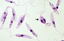
The Medicinal Chemistry Center (CQMED), headquartered at Campinas State University in Brazil, recently started a project in partnership with Promega to develop drugs that can be used against Leishmania. This genus of protozoans is the etiological agent of leishmaniasis, transmitted to humans by sandflies.
Microscopic image of Leishmania tropica. Credit: Brian E. Keas at Michigan State University.
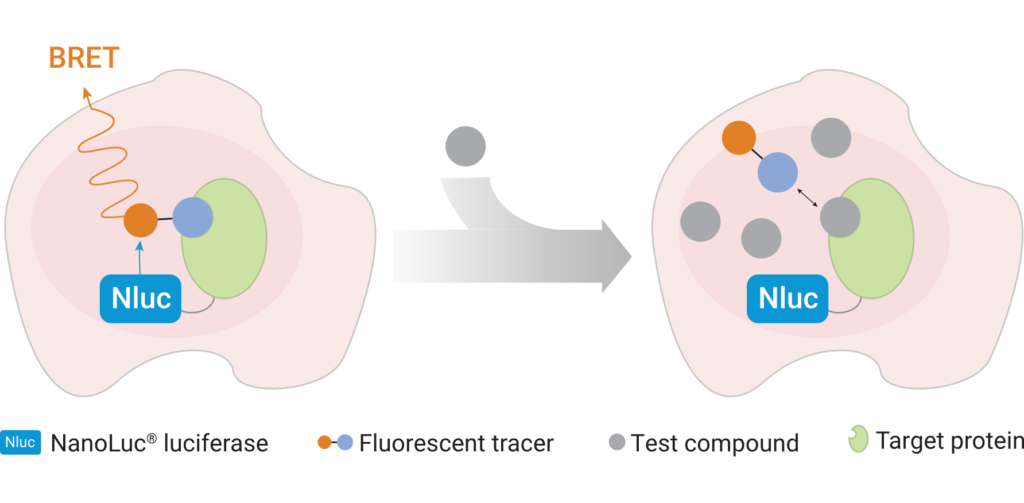
Leishmaniasis is classified as a neglected tropical disease that mainly affects poor communities. Symptoms include large skin sores and an enlarged spleen. The challenge in developing drugs to treat Leishmania is finding appropriate therapeutic targets. These targets are normally proteins whose inhibition leads to death of the parasite. In addition to pharmaceutical company Eurofarma, whose goal is to develop drugs for Leishmania, Promega was chosen to help solve this problem because of our NanoBRET™ Target Engagement (TE) assay*, a well-established technique for measuring protein interactions. In this assay, NanoLuc® luciferase is attached to the protein of interest, and a fluorescent NanoBRET™ tracer molecule is added to the cells. This produces a BRET signal. When a competing ligand is added, it will displace the tracer molecule, enabling quantification of the strength of the interaction compared to the tracer molecule..
A challenge that researchers will face will be ensuring that the NanoBRET™ tracer reaches the inside of the parasite cells; because Leishmania is an intracellular parasite, molecules need to cross the host cell membrane, the membrane of the vacuole containing the parasites, and the membrane of the parasite itself. Another challenge the slow reproduction of Leishmania within macrophages. On top of that is the fact that the parasite’s metabolism varies depending on its biological cycle, meaning that there could be long periods of time during which a drug’s therapeutic target is not expressed in the cell, during which time the drug would have no effect. The ideal target would be expressed at high levels throughout the cell cycle.
The project is being led by Rafael Couñago, a researcher at CQMED, and Promega scientists Matt Robers and Jean-Luc Vaillaud.
*An earlier version of this blog incorrectly said that these experiments are based on the NanoBRET™ assay using HaloTag® protein.
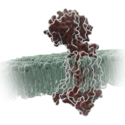
G Protein-Coupled Receptors (GPCRs) are a very large, diverse family of transmembrane receptors in eukaryotes. These receptors detect molecules outside the cell and activate internal signaling pathways by coupling with G proteins. Once a GPCR is activated, β-arrestins translocate to the cell membrane and bind to the occupied receptor, uncoupling it from G proteins and promoting its internalization.
Reporter tags are useful for studying the dynamics of GPCRs and associated proteins, but large tags can disrupt the receptors’ native functioning, and often overexpression of the tagged protein is required to obtain sufficient signal. Here is one example of how researchers have used the small, bright NanoLuc® luciferase to overcome these common challenges and answer questions about GPCRs.
The stage is set. You’ve spent days setting up this experiment. Your bench is spotless. All the materials you need to finally collect data are laid neatly before you. You fetch your cells from the incubator, add your detection reagents, and carefully slide the assay plate into the luminometer. It whirs and buzzes, and data begin to appear on the computer screen. But wait!
These data are garbage!
Don’t let this dramatic person be you. Here are 8 tips from us on things to watch out for before you start your next luminescent assay. Make sure you’ll be getting good data before wasting precious sample!
We have published 130 blogs here at Promega this year (not including this one). I diligently reviewed every single one and compiled a list of the best 8.5%, then asked my coworkers to vote on the top 5 out of that subset. Here are their picks:
This past weekend, I had the opportunity to be a part of “Once Upon a Christmas Cheery in the Lab of Shakhashiri”. Bassam Z. Shakhashiri is a professor of chemistry at the University of Wisconsin–Madison who is well-known for his fun science demonstrations and a fervent dedication to public science communication. Once Upon a Christmas Cheery started in 1970 as an end-of-semester treat for Dr. Shakhashiri’s freshman chemistry class; by 1973, the Christmas lecture had become so popular that Wisconsin Public Television offered to broadcast it during Christmas week, and this collaboration has continued uninterrupted ever since.
That’s 49 years of Christmas lectures, commemorated by making indium, the 49th element, the Sesame Street-esque “sponsor” of the show. It helps that indium burns bright violet, the name of Dr. Shakhashiri’s granddaughter and hence his favorite color. The color purple made a firm foundation for many aspects of the show: The chrysanthemums frozen in liquid nitrogen were purple, as was the balloon I inflated during my spiel on air movement. Most of the set was various shades of purple, too.
The set was whimsical and very purple. Photo by Eric Baillies.
In my second or third year as a graduate student, I had to ship some microfluidic masters to a collaborator in Kenya. The masters were extremely fragile and took me several days in a cleanroom gown to make. I was horrified at having to send them on a perilous journey overseas, and somewhat flabbergasted that they made it to Nairobi whole and well. And yet, every day thousands of delicate items zoom around the world and arrive at their destinations in one piece. How?
Have you ever thought about plant viruses? Unless you’re a farmer or avid gardener, probably not. And yet, for many people the battle against agricultural viruses never ends. Plant viruses cause billions of dollars in damage every year and leave millions of people food insecure (1–2), making viruses a major barrier to meeting the United Nations’ global sustainable development goal of Zero Hunger by 2030.
At the University of Western Australia, Senior Research Fellow Dr. Laura Boykin is using genomics and supercomputing to tackle the problem of viral plant diseases. In a recent study, Dr. Boykin and her colleagues used genome sequencing to inform disease management in cassava crops. For this work, they used the MinION, a miniature, portable sequencer made by Oxford Nanopore Technologies, to fully sequence the genomes of viruses infecting cassava plants.
Cassava (Manihot esculenta) is one of the 5 most important calorie sources worldwide (3). Over 800 million people rely on cassava for food and/or income (4). Cassava is susceptible to a group of viruses called begomoviruses, which are transmitted by whiteflies. Resistant cassava varieties are available. However, these resistant plants are usually only protected against a small number of begomoviruses, so proper deployment of these plants means farmers must know both whether their plants are infected and, if so, the strain of virus that’s causing the infection.
XWe use cookies and similar technologies to make our website work, run analytics, improve our website, and show you personalized content and advertising. Some of these cookies are essential for our website to work. For others, we won’t set them unless you accept them. To learn more about our approach to Privacy we invite you to Read More
By clicking “Accept All”, you consent to the use of ALL the cookies. However you may visit Cookie Settings to provide a controlled consent.
We use cookies and similar technologies to make our website work, run analytics, improve our website, and show you personalized content and advertising. Some of these cookies are essential for our website to work. For others, we won’t set them unless you accept them. To find out more about cookies and how to manage cookies, read our Cookie Policy.
If you are located in the EEA, the United Kingdom, or Switzerland, you can change your settings at any time by clicking Manage Cookie Consent in the footer of our website.
Necessary cookies are absolutely essential for the website to function properly. These cookies ensure basic functionalities and security features of the website, anonymously.
Cookie
Duration
Description
cookielawinfo-checbox-analytics
11 months
This cookie is set by GDPR Cookie Consent plugin. The cookie is used to store the user consent for the cookies in the category "Analytics".
cookielawinfo-checbox-functional
11 months
The cookie is set by GDPR cookie consent to record the user consent for the cookies in the category "Functional".
cookielawinfo-checbox-others
11 months
This cookie is set by GDPR Cookie Consent plugin. The cookie is used to store the user consent for the cookies in the category "Other.
cookielawinfo-checkbox-advertisement
1 year
The cookie is set by GDPR cookie consent to record the user consent for the cookies in the category "Advertisement".
cookielawinfo-checkbox-necessary
11 months
This cookie is set by GDPR Cookie Consent plugin. The cookies is used to store the user consent for the cookies in the category "Necessary".
cookielawinfo-checkbox-performance
11 months
This cookie is set by GDPR Cookie Consent plugin. The cookie is used to store the user consent for the cookies in the category "Performance".
gdpr_status
6 months 2 days
This cookie is set by the provider Media.net. This cookie is used to check the status whether the user has accepted the cookie consent box. It also helps in not showing the cookie consent box upon re-entry to the website.
lang
This cookie is used to store the language preferences of a user to serve up content in that stored language the next time user visit the website.
viewed_cookie_policy
11 months
The cookie is set by the GDPR Cookie Consent plugin and is used to store whether or not user has consented to the use of cookies. It does not store any personal data.
Analytical cookies are used to understand how visitors interact with the website. These cookies help provide information on metrics the number of visitors, bounce rate, traffic source, etc.
Cookie
Duration
Description
SC_ANALYTICS_GLOBAL_COOKIE
10 years
This cookie is associated with Sitecore content and personalization. This cookie is used to identify the repeat visit from a single user. Sitecore will send a persistent session cookie to the web client.
vuid
2 years
This domain of this cookie is owned by Vimeo. This cookie is used by vimeo to collect tracking information. It sets a unique ID to embed videos to the website.
WMF-Last-Access
1 month 18 hours 24 minutes
This cookie is used to calculate unique devices accessing the website.
_ga
2 years
This cookie is installed by Google Analytics. The cookie is used to calculate visitor, session, campaign data and keep track of site usage for the site's analytics report. The cookies store information anonymously and assign a randomly generated number to identify unique visitors.
_gid
1 day
This cookie is installed by Google Analytics. The cookie is used to store information of how visitors use a website and helps in creating an analytics report of how the website is doing. The data collected including the number visitors, the source where they have come from, and the pages visted in an anonymous form.
Advertisement cookies are used to provide visitors with relevant ads and marketing campaigns. These cookies track visitors across websites and collect information to provide customized ads.
Cookie
Duration
Description
IDE
1 year 24 days
Used by Google DoubleClick and stores information about how the user uses the website and any other advertisement before visiting the website. This is used to present users with ads that are relevant to them according to the user profile.
test_cookie
15 minutes
This cookie is set by doubleclick.net. The purpose of the cookie is to determine if the user's browser supports cookies.
VISITOR_INFO1_LIVE
5 months 27 days
This cookie is set by Youtube. Used to track the information of the embedded YouTube videos on a website.
Performance cookies are used to understand and analyze the key performance indexes of the website which helps in delivering a better user experience for the visitors.
Cookie
Duration
Description
YSC
session
This cookies is set by Youtube and is used to track the views of embedded videos.
_gat_UA-62336821-1
1 minute
This is a pattern type cookie set by Google Analytics, where the pattern element on the name contains the unique identity number of the account or website it relates to. It appears to be a variation of the _gat cookie which is used to limit the amount of data recorded by Google on high traffic volume websites.