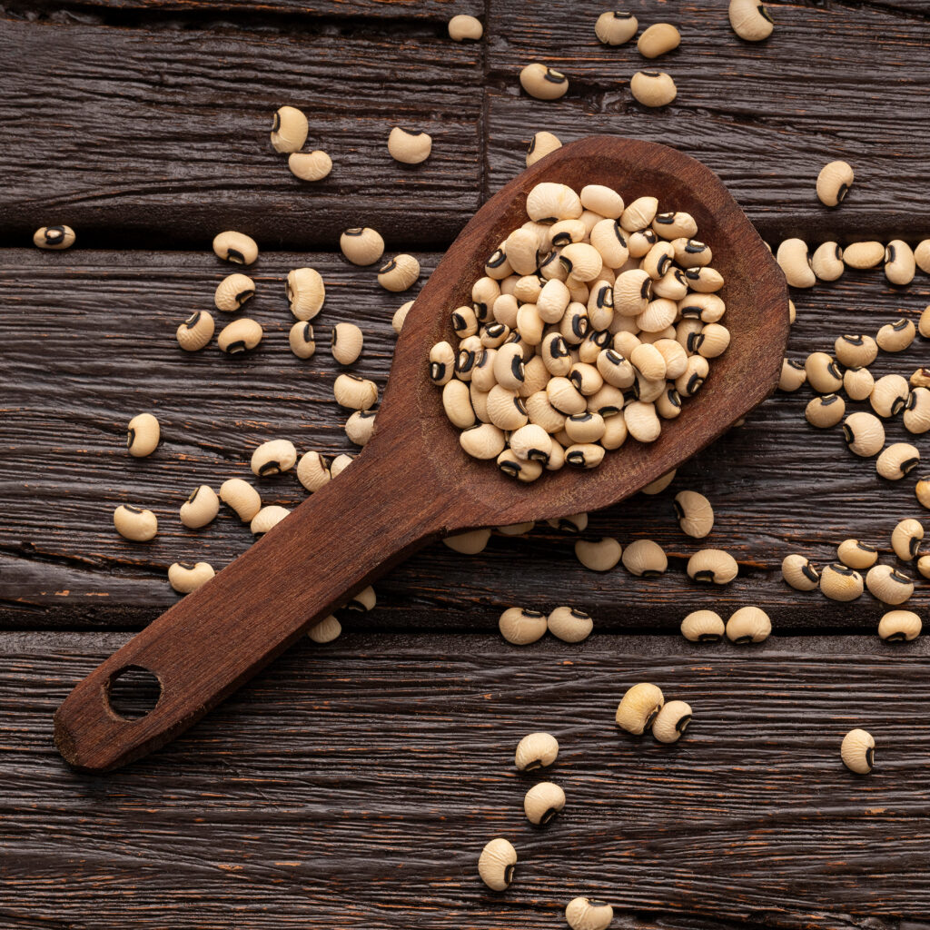
Cowpea (Vigna unguiculata), a humble tan and black legume, is one of the most important food crops in the world. Grown across sub-Saharan Africa, Asia, and parts of the Americas, Cowpea provides protein-rich nutrition for hundreds of millions of people, making it a cornerstone of smallholder agriculture. But cowpea production faces a persistent threat: the cowpea aphid-borne mosaic virus (CABMV), a common virus that can devastate yields across entire growing regions.
What makes CABMV particularly difficult to combat is how the virus infects its host. Instead of relying on viral translational machinery, the virus hijacks the plant’s systems to replicate. CABMV targets a protein called eIF4E, a translation initiation factor that the plant needs to read its own genetic instructions and produce proteins. The virus produces a protein, VPg, that binds directly to eIF4E and redirects the plant’s translational machinery to produce viral proteins instead. The plant can’t simply get rid of eIF4E. Without it, protein synthesis stalls. So how can cowpea defend itself against a virus that exploits one of its most essential proteins?
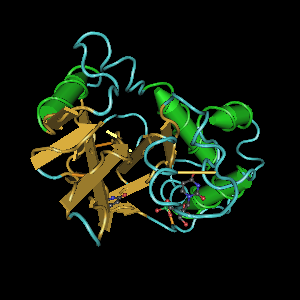
A new study published in Agronomy by researchers at the Federal University of Pernambuco, the Federal University of Minas Gerais, and Embrapa Recursos Genéticos e Biotecnologia takes a comprehensive look at this problem from the inside out1. The team characterized all three members of the eIF4E gene family in cowpea (eIF4E, eIF(iso)4E, and nCBP) across six cultivated varieties (cultivars) with known contrasting responses to CABMV infection. Two of those cultivars (Bajão and IT85F-2687) are resistant to the virus; the other four (Boca Negra, BR14 Mulato, Pingo de Ouro, and Santo Inácio) are susceptible to the virus.
Using a multi-omics approach that combined genomic, evolutionary and structural analyses, the researchers set out to answer a fundamental question: what makes some versions of eIF4E exploitable by the virus, and others not?
Continue reading “The Molecular Blueprint for Virus-Resistant Cowpea”