What Makes a “Good” Buffer?

Use of buffers, pour one solution into another.
Use of buffer aims to make pH remain nearly constant in solution.

Buffers are often overlooked and taken for granted by laboratory scientists, until the day comes when a bizarre artifact is observed and its origin is traced to a bad buffer.

The simplest definition of a buffer is a solution that resists changes in hydrogen ion concentration as a result of internal and environmental factors. Buffers essentially maintain pH for a system. The effective buffering range of a buffer is a factor of its pKa, the dissociation constant of the weak acid in the buffering system. Many things, such as changes in temperature or concentration, can affect the pKa of a buffer.

In 1966, Norman Good and colleagues set out to define the best buffers for biochemical systems (1). By 1980, Good and his colleagues identified twenty buffers that set the standard for biological and biochemical research use (2,3).  Good set forth several criteria for the selection of these buffers:

Continue reading “What Makes a “Good” Buffer?”

Converting RPM to g Force (RCF) and Vice Versa

Rotational Radius
Rotational radius of centrifuge for converting RPM to g force (RCF).

g Force or Relative Centrifugal Force (RCF) is the amount of acceleration to be applied to the sample. It depends on the revolutions per minute (RPM) and radius of the rotor, and is relative to the force of Earth’s gravity.

A good, precise protocol for centrifugation instructs you to use the g force rather than RPMs because the rotor size might differ, and g force will be different while the revolutions per minute stay the same. Unfortunately, many protocols are written in hurry and instructions are given in RPMs. Therefore, you have to convert g force (RCF) into revolutions per minute (rpms) and vice versa.

Modern centrifuges have an automatic converter but older ones do not. There is a simple formula to calculate this, but it takes some time to do the calculation. Meanwhile, your cells might die or the biochemical reaction goes on for three times longer than it should.

There are several ways to make conversion:

Continue reading “Converting RPM to g Force (RCF) and Vice Versa”

Control Samples: 3 Terrifying Tales for Scientists

Lab science cartoon
Carl may not scare her…but did she remember the controls?

Warning: This blog contains stories about phantom serial killers, frankenfoods, mysteriously phosphorylated bands and unrequited ligations that may be disturbing to some people. Children or scientists prone to anxiety over irreproducible results should read this with their eyes shut.

I

Clouds hung low in the sky, and the late October wind howled between the buildings, rattling the window panes of the basement laboratory. The grackles cawed in desperate warning, their flocks changing the evening color palette from gray to black. I was as unsettled as the weather, watching my blot slosh back and forth.

Continue reading “Control Samples: 3 Terrifying Tales for Scientists”

A Crash Course in Fighting Lab Contamination

When I first started in my undergraduate lab, one of the first things I learned was how to prepare agar plates for growing yeast. My supervisor, a grad student, looked over my shoulder as I added the yeast extract, bacto peptone, and other ingredients. I sealed the pitcher tightly with aluminum foil and autoclaved it until sterile. When I was ready to pour the plates, I carried the pitcher to the “plate-pouring” room, ripped the foil off, and started to pour an even layer of agar into each of the plastic dishes, leaving the lids off so they could cool. After I’d poured a dozen or so, my grad student supervisor burst into the room.

“What are you doing?” she demanded.

“I’m pouring plates,” I stammered back.

She took a deep breath and explained. By fully uncovering the pitcher and leaving my plates uncovered, I had left my precious media at high risk for contamination. The open containers were far too inviting for potential contaminants floating through the air. In the end, we ended up throwing away several of the plates that had been exposed the longest.

Now, I don’t share this story to demonstrate how clueless when I first started in the research lab as an undergrad. We all have those “uh-oh” moments when we realize for the first time that something that seemed so obvious was, in fact, more complicated than we’d expected. However, that day I learned how easily I could sabotage my own work by unwittingly inviting contaminants into my experiments.

Whether you work with yeast, bacteria, mammalian cells or anything else in a molecular biology lab, preventing contamination is crucial to getting desired results. Fortunately, minimizing your risk can be incredibly easy.

Let’s start with your lab bench. Everyone has their own organization system, but if yours is “out-of-control chaos,” you might want to reevaluate. Benchtop clutter makes it difficult to thoroughly clean the bench as often as needed. All those bottles of solutions, empty tip boxes, and wrinkled protocol sheets harbor dust and other unwelcome particles that you want to keep away from your cultures and reactions.

Once your benchtop is tidy (or at least somewhat tidy), make sure you keep the surface as clean as possible. Immediately clean up any spills or drips that happen while you’re working. Wiping your workspace with a 10% bleach solution will sterilize it, and following that up with 70% ethanol will dry it quickly. This wash should be performed at least once a day. Ideally you should also regularly remove everything from your workspace and perform a deeper cleaning of your benchtop, as well as any shelves and containers in your area.

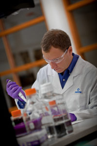
Now that your bench is in good shape, it’s time to gear up . You should always follow standard safety procedures (lab coat and goggles, closed-toe shoes, hair tied back), but above all, make sure you never forget your gloves. Gloves protect you from harmful chemicals, but they also protect your experiments from anything that could be on your hands. Skin can carry reagents, bacteria, and enzymes that are good for your body but bad for your experiments. Change your gloves regularly to prevent potential carryover of reagents or samples between containers. A good rule is, “When in doubt, change your gloves.”

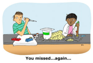
Finally, to guard against airborne contaminants, do your best to keep everything covered when you aren’t immdiately using it. I learned this rule the hard way when several of my yeast plates developed fuzzy patches of mold several days after I poured them. Bacteria and other undesirables floating through the air can affect stock solutions, cultures, plates, tubes, and basically anything else you rely on. Keep your lids on and cover open containers to minimize air exposure to reduce the chances of nefarious particles finding their way in.

There’s no way to guarantee you’ll never experience some form of contamination in your lab, but smart practices can help reduce your risk. Develop an anti-contamination routine that meets your needs and make sure you stick to it every day in the lab.

How to Take Care of Your Pipettes

Last updated: 1/22/21

what not to do with your pipettes

Pipettes are such a routine part of everyday life in the lab that it can be easy to take them for granted.  Their accuracy is vital, and there are many things we can adopt as best practices for success. Here are a few tips (no pun intended) gathered from around the Web by Kim Steinhauser of the Promega Metrology Department–the group charged with keeping our pipettes and other lab equipment functional and accurate.

Continue reading “How to Take Care of Your Pipettes”

Weird samples? Contact Tech Serv to find the right DNA purification kit for you.

“Dear Tech Serv,
We would like to detect DNA collected from swabs rubbed on the inside thighs of frogs. What would be the best DNA extraction kit to use for this?”

“Hi Tech Serv,
I need to find out a suitable kit for extracting DNA from bird fecal samples. Can I use ReliaPrep™ gDNA Tissue Miniprep System for that?”

These are just some examples of unconventional sample type inquiries that the Promega Technical Services Team receives regularly from scientists around the world. Many of these inquiries land in the hands of Technical Services Scientist, Paraj Mandrekar (a.k.a. “sample type guru”). Continue reading “Weird samples? Contact Tech Serv to find the right DNA purification kit for you.”

Six (and a Half) Reasons to Quantitate Your DNA

Knowing how much DNA you have is fundamental to successful experiments. Without a firm number in which you are confident, the DNA input for subsequent experiments can lead you astray. Below are six reasons why you should quantitate your DNA.

6. Saving time by knowing what you have rather than repeating experiments. If you don’t quantitate your DNA, how certain can you be that the same amount of DNA is consistently added? Always using the same volume for every experiment does not guarantee the same DNA amount goes into the assay. Each time there is a new purified DNA sample, the chances that you have the same quantity as before are lessened. Consequently, without knowing the DNA concentration of the sample you are using, the amount of input DNA cannot be guaranteed and experiments may have to be repeated.

Continue reading “Six (and a Half) Reasons to Quantitate Your DNA”

Methods for Quantitating Your Nucleic Acid Sample

Nucleic acid quanitation webinarFor most molecular biology applications, knowing the amount of nucleic acid present in your purified sample is important. However, one quantitation method might serve better than another, depending on your situation, or you may need to weigh the benefits of a second method to assess the information from the first. Our webinar “To NanoDrop® or Not to NanoDrop®: Choosing the Most Appropriate Method for Nucleic Acid Quantitation” given by Doug Wieczorek, one of our Applications Scientists, discussed three methods for quantitating nucleic acid and outlined their strengths and weaknesses. Continue reading “Methods for Quantitating Your Nucleic Acid Sample”

Cell Line Misidentification Rears Its Ugly Head

Cancer cell illustrationBack in 2009, we reported on the problem of cell line contamination (1). In that article we reported the statistics that an estimated 15–20% of the time, the cell lines used by researchers are misidentified or cross-contaminated with another cell line (1). This presents a huge problem for the interpretation of data and the reproducibility of experiments, a key pillar in the process of science. We have revisited this topic several times, highlighting the issues cell and tissue repositories have discovered with cell lines submitted to them (2) and discussing the new guidelines issued by ANSI (3,4) for researchers regarding when during experimental processes cell lines should be authenticated and what methods are acceptable for identifying cell lines.

Just recently two papers were voluntarily retracted by their authors because of cross contamination among cell lines used in the laboratories. The first that came to my attention represented the first retraction from Nature Methods in its nine years of publication. In this paper, cross contamination of a primary gliomasphere cell lines with HEK cells expressing GFP resulted in “unexplained autofluorescence” associated with tumorigenicity (5). The second paper, retracted from Cancer Research by the original authors, was also another cross contamination story involving HEK cells (6). In this story a gene was incorrectly described as a tumor suppressor, that when silenced led to the formation of tumors in nude mice. It turns out that the contaminating HEK cells also failed to express this same gene.

So because of cross contamination of cell lines, two groups have voluntarily retracted papers. Being open and honest about what had happened with the cell lines and reaching the decision to retract the papers could not have been an easy thing, but these decisions benefit the scientific community in many ways. Obviously they benefit the researchers doing work on the specific research questions addressed by the papers by preventing researchers from pursuing paths that lead to dead ends. But in the bigger picture these retractions reinforce the argument that cell line authentication needs to become a routine and accepted part of any experimental process that depends on cell culture if we are to have confidence in the experimental results.

References

  1. Dunham, J.H. and Guthmiller, P.  (2009) Doing good science: Authenticating cell line identity. Promega Notes 101, 15–18.
  2. Duham, J.H. and Guthmiller, P. (2012) Doing good science: Authenticating cell line identity. Promega PubHub. [Internet: Accessed September 2013]
  3. Gopal, A. (2013) Fingerprinting  your cell lines. Promega Connections blog [Internet: Accessed September 2013]
  4. Sundquist, T. (2013) Preventing the heartache of cell line contamination. Promega Connections blog [Internet: Accessed September 2013]
  5. Evanko, D. (2013) A retraction resulting from cell line contaminationMethagora blog. [Internet Accessed September 2013]
  6. Negorev, D. (2013) Retraction: Sp100 as a potent tumor suppressor: Accelerated senescence and rapid malignant transformation of human fibroblasts through modulation of an embryonic stem cell program. Can. Res. 73, 4960.

Preventing the Heartache of Cell Line Misidentification

Golden maskIt’s a scientist’s nightmare: Spending time and resources to investigate a biological phenomenon only to learn later that your cells are not what you think they are—their true identities hidden. As a result, all of the data that you’ve generated with those cells, published and unpublished, are cast into doubt. You thought that you knew your cells, that you could trust them, but your trust was misplaced. At some point, perhaps even before the traitorous cell line entered your laboratory, the cells were mislabeled, misidentified or contaminated with another cell line. It didn’t have to be this way. There are easy steps you can take to prevent the headache and heartache of cell line misidentification and contamination.

Continue reading “Preventing the Heartache of Cell Line Misidentification”