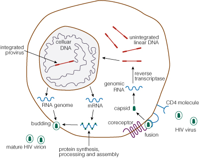
Scientists investigating the human immunodeficiency virus (HIV) have learned much about the retrovirus’s lifecycle, but their ultimate goals were to discover a cure and prevent infection. In the decades since HIV was discovered, basic research and pharmaceutical drug development have expanded the antiviral toolbox, but these HIV treatments do not provide a functional cure, only manage the infection. However, two techniques may offer a potential cure for HIV infection using CRISPR and a possible vaccine using mRNA.
Alternatives to animal testing have long been explored when it comes to studying the safety of various chemical compounds for use in food, medicine and cosmetics. With the advent of three-dimensional (3D) cell culture to create organoids, more relevant human organoid models are being explored as one way to provide safe and effective compound testing while minimizing the need for testing in animals. The international project Physiologically Anchored Tools for Realistic nanOmateriaL hazard aSsessment (PATROLS) led by the Swansea University Medical School aims to establish a battery of innovative, next-generation safety testing tools that can more accurately predict the effects of engineered nanomaterial (ENM) exposure in humans and the environment.
One project being researched by Samantha Llewellyn, a research assistant at Swansea University, is developing predictive physiologically relevant 3D liver models for ENM safety assessment. By having a model to evaluate realistic ENM exposures, a researcher can study liver function, hepatic metabolism and microtissue cell viability after acute (24 hours) or prolonged (several days) exposure. A microtissue model for assessing ENM hepatotoxicity needs to mimic primary hepatocytes and be amenable to assays used to test cell viability and metabolism.
The right tools for testing this 3D liver model include the bioluminescent-based CellTiter-Glo® 3D Viability and P450-Glo® Assays. When creating organoids, having reagents that can penetrate to the center of the dense and complex 3D liver spheroids is important so that the cell viability readout encompasses the entire microtissue. The CellTiter-Glo® 3D Viability Assay accomplishes this task, providing accurate assessment of 3D tissue cell health. Measuring cytochrome P450 (CYP450) activity is necessary for studying liver function. The P450-Glo® Assays have the flexibility to assess CYP450 activity while preserving the liver spheroids; thus, researchers can gather more data from a single experiment.
The importance of Samantha Llewellyn’s research as part of PATROLs is establishing a 3D liver model that could evaluate realistic ENM exposures and reduce the need for animal testing. Bioluminescent assays for assessing cell health and liver enzyme function are necessary to reach this goal.
To learn more about the last 30 years of bioluminescent innovations and the discoveries they’ve enabled, please visit our 30th anniversary celebration page.
When it comes to blocking the spread of viral pathogens that cause human disease, epidemiologists—people who study disease outbreaks—like to talk about herd immunity. But what do they mean when discussing the herd and their immunity? Today, I will tackle this subject but with a side jaunt: I am going to co-opt the word “herd” and replace it with “flock” thus making chickens the center of attention rather than cattle for this analogy about immunity in a population. (Disclaimer: I am utterly biased toward chickens and enjoy talking about my flock of 24 hens and pullets).
Who is the Herd Flock They Keep Talking About?
By using a collective term for a number of individuals such as “herd” or “flock”, epidemiologists and public health experts are referring to a population or community. Doing some investigation, I learned herd immunity was a term first used in 1917 and referred to…cows. That makes sense, right? When we talk about groups of cattle, the term used is “herd”. Turns out there was an infection that caused spontaneous miscarriages in cattle and became epidemic in American herds. Farmers managed this threat by destroying or selling the infected cows. However, a livestock veterinarian had a different view, describing this pathogen as “…a fire, which, if new fuel is not constantly added, soon dies down. Herd immunity is developed, therefore, by retaining the immune cows, raising the calves, and avoiding the introduction of foreign cattle” (1). Essentially, this veterinarian was noting that keeping the infected cows who had immunity against the contagion meant the herd were less likely to be reinfected and, thus, put an end to the epidemic.
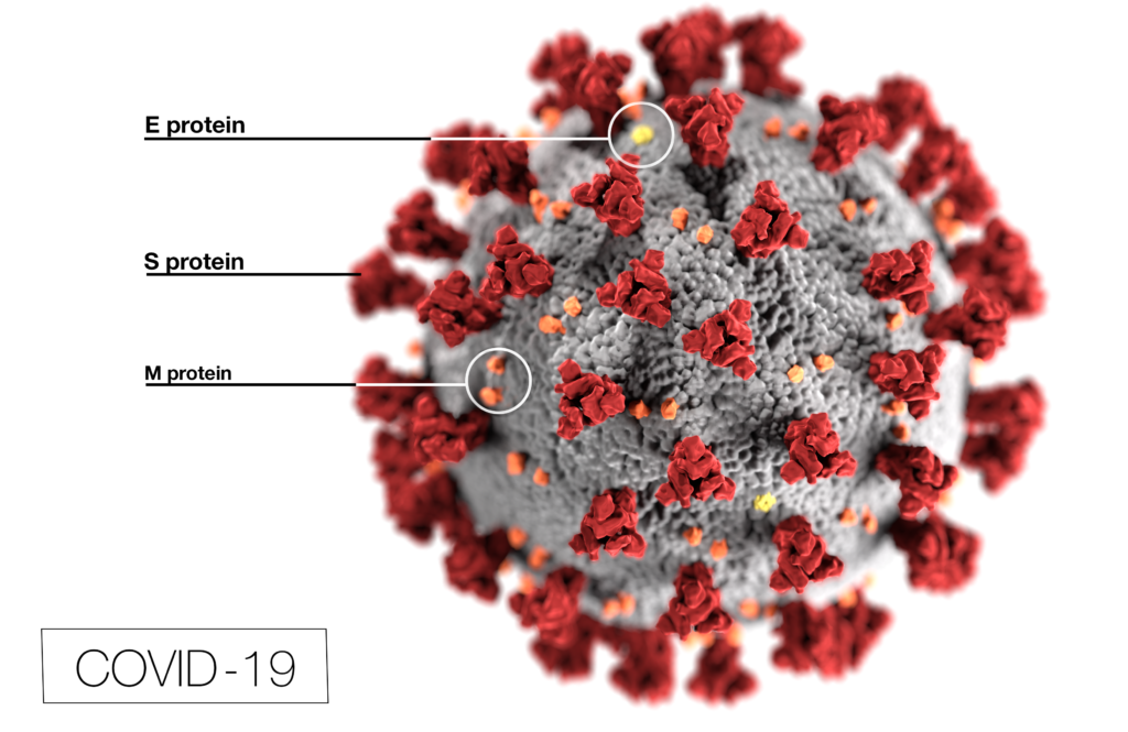
SARS-CoV-2 illustration from CDC; Photo Credit: Alissa Eckert, MS; Dan Higgins, MAM E = envelope; M = membrane
A worldwide pandemic requires scientific research to understand the viral pathogen. The focused efforts of global scientists are even more necessary in the face of a novel coronavirus like SARS-CoV-2, the causative agent of COVID-19. However, because SARS-CoV-2 causes human disease, research efforts are restricted by the need for physical laboratories that are equipped to handle the required level of containment and personnel trained to handle pathogens in these facilities. But what if we could bypass the restrictive facility requirements by engineering a synthetic, replication-defective version of SARS-CoV-2 that more researchers could use to study the pandemic coronavirus, expanding the capacity to test and develop methods to attenuate its devastating effect on humans?
The challenge is to develop a derivative of SARS-CoV-2 that reflects how it behaves in the cell but is compromised such that it is unable to infect cells more than a single time. That is, the virus can get into a cell or be introduced into cells and replicate but is unable to produce infectious virus would offer a pathway to expand research capacity without the use of special laboratory facilities. This replication-defective SARS-CoV-2 could be created to encode as much or as little of the genome needed to examine its lifecycle without becoming a fully infectious virus. In fact, this replication-defective version of SARS-CoV-2 could include additional genetic elements that could be used to control its expression, track the virus in cells and measure the level of its replication. This task has been undertaken by Dr. Bill Sudgen’s group at the University of Wisconsin–Madison McArdle Laboratory for Cancer Research, explained by graduate student Rebecca Hutcheson during her presentation “Making the Virus Causing COVID-19 Safe for Research”.
Centrosaurus is a herbivorous Ceratopsian dinosaur that lived in Canada in the Cretaceous Period.
Did dinosaurs get cancer? That isn’t an easy question to answer. Finding and diagnosing cancer in dinosaur fossils has proven difficult. Any soft tissue, the typical location of tumors, has degraded over the millennia. Fossilized bones millions of years old are subject to wear and tear, making it hard to distinguish bone damage from possible pathology. By using the knowledge and expertise gained from diagnosing cancer in humans, a team reported in The Lancet Oncology that they found the first known case of osteosarcoma in a lower leg bone from a horned dinosaur found in southern Alberta, Canada.
This case of bone cancer discovered in a specimen of Centrosaurus apertus found in the Canadian Dinosaur Park Formation was confirmed by examining the bone surface along with radiographic and histological analysis. The 77–75.5-million-year-old case was compared to both a normal C. apertus fibula from the Oldman formation also in southern Alberta, Canada, as well as a human fibula with an osteosarcoma.
A recent article published in Cancers demonstrates a new method for targeting glial cells using a lentiviral packaging system that incorporated Zika virus envelope proteins. By using the reporter gene firefly luciferase, researchers demonstrated that a pseudotyped virus could infect cultured glioblastoma cells.
Introduction
Viruses enjoy a fearsome reputation. SARS-CoV-2 is only the latest infectious agent that has garnered attention by becoming a worldwide pandemic. Even the viral name suggests that SARS-CoV-2 was not the first of its type [SARS-CoV is the virus behind the severe acute respiratory syndrome (SARS) that spread worldwide in the early 2000s]. There are many different families of viruses (e.g., coronavirus for SARS-CoV-2 or lentiviruses for HIV-1) and each show a preference to the cell types they want to infect. By investigating the life cycle of viruses to better understand their mechanisms, researchers can discover new opportunities that may be exploited.
In 2015 and 2016, the virus that concerned health authorities was Zika virus (ZIKV). While this virus generally caused mild disease, the babies of women who were infected during pregnancy were at increased risk for microcephaly and other brain defects. These defects were traced back to Zika virus infecting nerve tissue, specifically, glial cells. This discovery provided an opportunity to explore how Zika virus might affect the brain tumor, glioblastoma multiforme (GMB), especially the glioblastoma stem cells (GSCs) that resist conventional treatment and contribute to the poor prognosis for GMB. Studies suggested that Zika virus infection prolonged survival in animal glioma models and selectively killed GSC with minimal effects on normal cells. In fact, the molecules used by ZIKV to enter cells were predominantly found on tumors, not normal cells. Knowing that the ZIKV envelope proteins prM and E provide the target specificity for glial cells, Kretchmer et al. wanted to explore if ZIKV envelope proteins substituted in lentivirus packaging systems would be able to enter glioblastoma cells.
What animal can be found around the globe that outnumbers humans three to one? Gallus gallus domesticus, the humble chicken. The human appetite for eggs and lean meat drive demand for this feathered bird, resulting in a poultry population of over 20 billion.
Controversy over the origin of the domestic chicken (when, where and which species) have lead some researchers to look for that information in the genomes of contemporary chicken breeds and wild jungle fowl, the candidates from which chickens were derived. By sequencing over 600 genomes from Asian domestic poultry as well as 160 genomes from all four wild jungle fowl species and the five red jungle fowl subspecies, Wang et al. wanted to understand and identify the relationships and relatedness among these species and derive where domesticated chickens first arose.
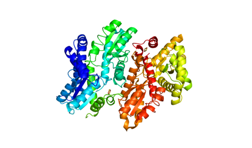
Human Ferrochelatase 2 angstrom crystal structure. Generated from 1HRK (RCSB PDB) using Pymol. Copyright: Sarah Wilson / CC BY-SA
Understanding how disease states arise from genetic variants is important for understanding disease resistance and progression. What can complicate our understanding of disease development is when two people have the same genetic variant, but only one has the disease. To investigate what might be happening with ferrochelatase (FECH) variant alleles that result in erythropoietic protoporphyria (EPP), scientists used next-generation sequencing (NGS) along with RNA analysis and DNA methylation testing to assess the FECH locus in 72 individuals from 24 unrelated families with EPP.
What is FECH and its relationship to EPP?
FECH is the gene for ferrochelatase, the last enzyme in the pathway that synthesizes heme. The inherited metabolic disorder, EPP, is caused when the activity of FECH is reduced to less than a third of normal levels thus, increasing the levels of protoporphyrin (PPIX) without metal in erythrocytes. The consequences of the low-metal PPIX include severe phototoxic skin reactions and hepatic injury due to PPIX accumulation in the liver.
How does FECH expression affect EPP?
The EPP disease state is not simply the lack of two functional FECH genes. Disease occurs with a hypomorphic allele, mutations in FECH that reduce its function, in trans to a null FECH allele. Researchers focused on three common variants called the GTC haplotype that are associated with expression quantitative trait loci (eQTL) that reduce FECH activity. Interestingly, these three variants have been found in trans, but researchers wanted to learn if there were individuals who were homozygous for the GTC allele and how EPP manifested for them.
When I encounter my cat fixated on specific locations in my kitchen, her behavior shows me that she has heard some mice in those areas. In fact, mice have been attributed as a reason that cats became companions to humans. Mice start gathering and reproducing so cats followed the food source and hunted the rodents, thus endearing themselves to humans, who were storing food for their own use. However, new evidence described in Scientific Reports has shown that mice have been associated with humans even before grain storage was widespread. In fact, by making our dwellings comfortable, we also created an inviting place for mice to live.
Heading into 2020, we realized that our Cartoon Lab was reaching a milestone: the 100th cartoon! We asked the “official” Promega Cartoonist Ed Himelblau to list his Top Five Cartoons and what inspired them. See what he has chosen in his own words:
This was the first of my cartoons that Promega published and it’s still one of my favorites. The file on my computer is dated February, 1999. I have been an undergraduate in a lab. I’ve mentored undergraduates in lab. Today I have lots of undergraduates working in my plant genetics lab at Cal Poly in San Luis Obispo. For the record, I enjoy having undergraduates in the lab and I never make them dress like robots. In this cartoon, I particularly like the centrifuge and stir plate on the right. I’ve always tried to put something in each cartoon (a tube rack, an enzyme shipping box, a desiccator) that make molecular biologists say, “I know that!”
XWe use cookies and similar technologies to make our website work, run analytics, improve our website, and show you personalized content and advertising. Some of these cookies are essential for our website to work. For others, we won’t set them unless you accept them. To learn more about our approach to Privacy we invite you to Read More
By clicking “Accept All”, you consent to the use of ALL the cookies. However you may visit Cookie Settings to provide a controlled consent.
We use cookies and similar technologies to make our website work, run analytics, improve our website, and show you personalized content and advertising. Some of these cookies are essential for our website to work. For others, we won’t set them unless you accept them. To find out more about cookies and how to manage cookies, read our Cookie Policy.
If you are located in the EEA, the United Kingdom, or Switzerland, you can change your settings at any time by clicking Manage Cookie Consent in the footer of our website.
Necessary cookies are absolutely essential for the website to function properly. These cookies ensure basic functionalities and security features of the website, anonymously.
Cookie
Duration
Description
cookielawinfo-checbox-analytics
11 months
This cookie is set by GDPR Cookie Consent plugin. The cookie is used to store the user consent for the cookies in the category "Analytics".
cookielawinfo-checbox-functional
11 months
The cookie is set by GDPR cookie consent to record the user consent for the cookies in the category "Functional".
cookielawinfo-checbox-others
11 months
This cookie is set by GDPR Cookie Consent plugin. The cookie is used to store the user consent for the cookies in the category "Other.
cookielawinfo-checkbox-advertisement
1 year
The cookie is set by GDPR cookie consent to record the user consent for the cookies in the category "Advertisement".
cookielawinfo-checkbox-necessary
11 months
This cookie is set by GDPR Cookie Consent plugin. The cookies is used to store the user consent for the cookies in the category "Necessary".
cookielawinfo-checkbox-performance
11 months
This cookie is set by GDPR Cookie Consent plugin. The cookie is used to store the user consent for the cookies in the category "Performance".
gdpr_status
6 months 2 days
This cookie is set by the provider Media.net. This cookie is used to check the status whether the user has accepted the cookie consent box. It also helps in not showing the cookie consent box upon re-entry to the website.
lang
This cookie is used to store the language preferences of a user to serve up content in that stored language the next time user visit the website.
viewed_cookie_policy
11 months
The cookie is set by the GDPR Cookie Consent plugin and is used to store whether or not user has consented to the use of cookies. It does not store any personal data.
Analytical cookies are used to understand how visitors interact with the website. These cookies help provide information on metrics the number of visitors, bounce rate, traffic source, etc.
Cookie
Duration
Description
SC_ANALYTICS_GLOBAL_COOKIE
10 years
This cookie is associated with Sitecore content and personalization. This cookie is used to identify the repeat visit from a single user. Sitecore will send a persistent session cookie to the web client.
vuid
2 years
This domain of this cookie is owned by Vimeo. This cookie is used by vimeo to collect tracking information. It sets a unique ID to embed videos to the website.
WMF-Last-Access
1 month 18 hours 24 minutes
This cookie is used to calculate unique devices accessing the website.
_ga
2 years
This cookie is installed by Google Analytics. The cookie is used to calculate visitor, session, campaign data and keep track of site usage for the site's analytics report. The cookies store information anonymously and assign a randomly generated number to identify unique visitors.
_gid
1 day
This cookie is installed by Google Analytics. The cookie is used to store information of how visitors use a website and helps in creating an analytics report of how the website is doing. The data collected including the number visitors, the source where they have come from, and the pages visted in an anonymous form.
Advertisement cookies are used to provide visitors with relevant ads and marketing campaigns. These cookies track visitors across websites and collect information to provide customized ads.
Cookie
Duration
Description
IDE
1 year 24 days
Used by Google DoubleClick and stores information about how the user uses the website and any other advertisement before visiting the website. This is used to present users with ads that are relevant to them according to the user profile.
test_cookie
15 minutes
This cookie is set by doubleclick.net. The purpose of the cookie is to determine if the user's browser supports cookies.
VISITOR_INFO1_LIVE
5 months 27 days
This cookie is set by Youtube. Used to track the information of the embedded YouTube videos on a website.
Performance cookies are used to understand and analyze the key performance indexes of the website which helps in delivering a better user experience for the visitors.
Cookie
Duration
Description
YSC
session
This cookies is set by Youtube and is used to track the views of embedded videos.
_gat_UA-62336821-1
1 minute
This is a pattern type cookie set by Google Analytics, where the pattern element on the name contains the unique identity number of the account or website it relates to. It appears to be a variation of the _gat cookie which is used to limit the amount of data recorded by Google on high traffic volume websites.