Drug Target Confirmed? Tivantinib’s Lesson on the Importance of Cellular Target Engagement

This guest blog post is written by Tian Yang, Associate Product Manager at Promega.

There are often challenges with translating results from a test tube into a living system, demanding more physiologically relevant assays. In drug discovery, demonstrating a compound’s ability to modulate its target protein in live cells is a critical step in the hit-to-lead workflow. A variety of cell-based assays can be used to assess a compound’s activity in live cells. Take kinase inhibitors as an example, these assays can range from substrate phosphorylation assays that more directly report on the activity of target kinases, to genetic reporter assays or cell viability assays that assess the downstream effects of target modulation.
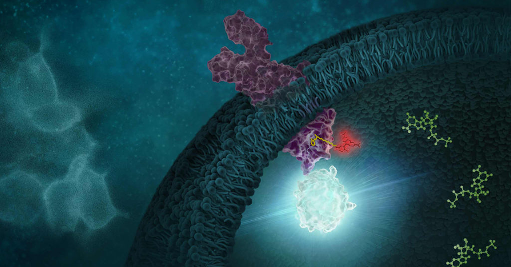
In the case of Tivantinib, several pieces of data from its development were used to establish its role as an inhibitor of MET kinase. MET Kinase is a prominent target for anti-cancer therapeutics due to frequent MET dysregulation in a wide range of tumors. For example, over-activation of MET drives cancer proliferation and metastasis. In the initial report on Tivantinib, in addition to biochemical activity assays performed with purified MET, the activity of Tivantinib in cells was verified by several methods, including: 1) inhibition of phosphorylation of MET and downstream signaling pathways, 2) cytotoxicity in cancer cell lines expressing MET, and 3) antitumor activity in xenograft mouse models (1). Additionally, a co-crystal structure of the MET-Tivantinib complex was solved, seemingly confirming that Tivantinib is a bona fide MET inhibitor capable of engaging MET in live cells (2). Based on these observations and other pre-clinical data, Tivantinib appeared to be a promising drug candidate and was taken through phase 3 clinical trials targeting cancers with MET overexpression. However, Tivantinib ultimately was not approved as a new therapeutic, failing to show efficacy in these phase 3 clinical trials (3,4).  

Within three years of the initial publication on Tivantinib, two separate articles challenged the mechanism of action in Tivantinib-induced cytotoxicity of tumor cells (5,6). Authors for both articles showed that Tivantinib can kill both MET-addicted and nonaddicted cells with similar potency. Both articles also concluded that perturbation of microtubule dynamics, instead of MET inhibition, is likely responsible for the cytotoxicity observed with Tivantinib. Considering the failed clinical trials and uncertainties regarding the mechanism of action, one may wonder if the original pre-clinical work adequately determined if Tivantinib effectively binds and inhibits MET in cells? If Tivantinib’s cellular engagement to MET was assessed directly rather than by MET phosphorylation analysis, would a different pre-clinical recommendation have been made?

Continue reading “Drug Target Confirmed? Tivantinib’s Lesson on the Importance of Cellular Target Engagement”
Base Editing Brilliance: David Liu’s Breakthrough Prize and Its Impact

On April 5, 2025, Dr. David R. Liu stood in the spotlight at the Barker Hangar in Santa Monica, California, to receive the Breakthrough Prize in Life Sciences—one of the most prestigious honors in science.
Dubbed the “Oscars of Science,” the Breakthrough Prizes were launched in 2012 by tech philanthropists including Sergey Brin, Mark Zuckerberg and Priscilla Chan, Yuri and Julia Milner and Anne Wojcicki. These prizes recognize groundbreaking achievements in life sciences, physics, and mathematics, with each laureate receiving a $3 million award—more than twice the amount of a Nobel Prize.

The winners are selected by panels of previous Breakthrough Prize recipients, ensuring peer-driven recognition. The annual ceremony brings together not only the best minds in science but also celebrities, filmmakers, and tech industry leaders, creating an uncommon crossover between pop culture and research, in an effort to bring more public attention as well as funding to scientific achievement.

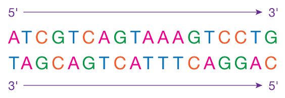
Dr. Liu was honored for inventing base editing and prime editing, technologies that allow precise, programmable rewriting of DNA to correct mutations linked to genetic disease—without introducing double-stranded breaks. These tools have rapidly transitioned from the bench to the clinic, with at least 15 clinical trials currently underway worldwide targeting diseases like sickle cell anemia, T-cell leukemia, and others.

Continue reading “Base Editing Brilliance: David Liu’s Breakthrough Prize and Its Impact”

Non-Pharmacological Approaches to ADHD: Exploring Inflammation and Omega-3s

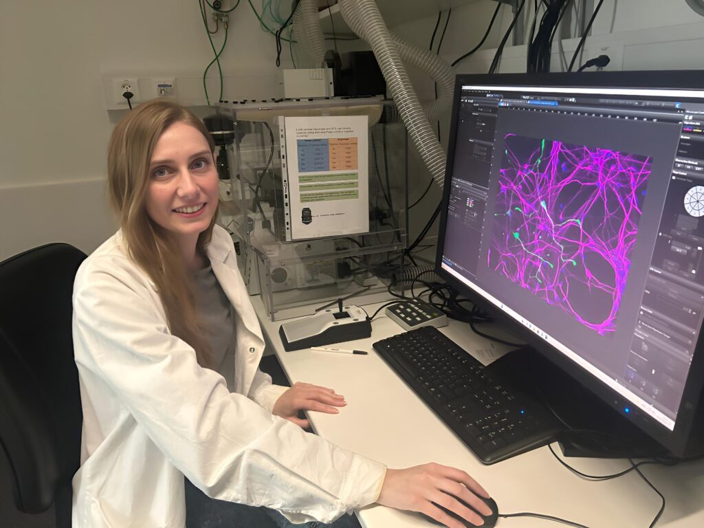
Attention-Deficit/Hyperactivity Disorder (ADHD) is a complex neurodevelopmental disorder that affects millions worldwide. Current therapeutic treatment relies on pharmaceutical approaches, but emerging research suggests that dietary supplements, such as omega-3 fatty acids, may offer complementary therapeutic options. A recent study published in the Journal of Psychiatric Research explores the relationship between inflammation and dietary supplements to determine how they might influence ADHD pathology. This work was conducted in Dr. Edna Grünblatt’s lab at the University of Zurich and was supported through Promega’s Academic Access Program. I had the chance to interview Dr. Natalie Walter, the lead author, to learn more about how her work offers potential opportunities for non-pharmacological interventions.

Continue reading “Non-Pharmacological Approaches to ADHD: Exploring Inflammation and Omega-3s”

Modified Nucleotides in IVT: Small Changes, Big Impact 

In our final blog post on double-stranded RNA (dsRNA), we turn our attention to the chemical building blocks of mRNA therapeutics—modified nucleotides. These seemingly minor changes to the RNA sequence play a crucial role in the success of mRNA-based vaccines and treatments. However, they also introduce complexities in accurately detecting and quantifying unwanted dsRNA byproducts— key steps in ensuring the therapeutic efficacy of your mRNA product. 

What Are Modified Nucleotides? 

Modified nucleotides are ribonucleotides containing chemically altered nucleosides — like specialty ingredients swapped into a classic recipe to improve taste and nutrition. Just as a chef might use a lactose-free milk or gluten-free flour to make a dish easier to digest without changing its core structure, scientists use chemically altered nucleosides during in vitro transcription (IVT) to improve how mRNA therapies perform. These modifications replace their natural counterparts (e.g., uridine or cytidine) in the final RNA product. Their incorporation improves the performance and safety of mRNA therapeutics in several ways: 

Continue reading “Modified Nucleotides in IVT: Small Changes, Big Impact “

dsRNA QC Considerations: How I Learned to Stop Worrying and Love my IVT Reactions

As mRNA therapeutics continue to expand across clinical pipelines, one persistent challenge remains for developers: reducing double-stranded RNA (dsRNA) contaminants that can compromise safety and efficacy. These unintended byproducts of in vitro transcription (IVT) can trigger unwanted immune responses and reduce the potency of the final product. Developers must prioritize dsRNA detection and control as essential steps in the process. In our previous blog post we offered a high-level discussion of what is double-stranded RNA (dsRNA), its biological function, and importance of detection in a therapeutic context.  Here, we’ll take a closer look at origins of dsRNA contamination, quality control measures, and improvement strategies.

Large-scale production of single-stranded RNA (ssRNA) for mRNA-based therapeutics is primarily done through in vitro transcription (IVT), an enzymatic process designed to generate high-yield, functional mRNA transcripts from a DNA template. This process uses purified RNA polymerase enzymes, such as T7, that recognize specific promoter sequences in the DNA template, generating the RNA transcripts of interest. However, IVT reactions also generate unwanted dsRNA byproduct. Below, we delve into some of the major quality control (QC) considerations and strategies to reduce dsRNA byproducts.

Continue reading “dsRNA QC Considerations: How I Learned to Stop Worrying and Love my IVT Reactions”

Bioluminescence vs. Fluorescence: Choosing the Right Assay for Your Experiment 

From enzyme activity to gene expression, light-based assays have become foundational tools in life science research. Among these, fluorescence and bioluminescence are two of the most widely-used approaches for detecting and quantifying biological events. Both rely on the emission of light, but the mechanisms generating that light—and the practical implications for experimental design—are quite different. 

Choosing between a fluorescence or bioluminescence assay isn’t as simple as picking between two reagents off the shelf. Each has strengths and limitations depending on the application, instrumentation, and biological system. In this blog, we’ll walk through how each method works, where they shine (and where they don’t), and what to consider when deciding which approach is right for your experiment. 

Continue reading “Bioluminescence vs. Fluorescence: Choosing the Right Assay for Your Experiment “

What 32,000 3D Spheroids Revealed About Culture Conditions

3D Spheroid Cell Culture

Three-dimensional (3D) cell culture systems have become essential tools in cancer research, drug screening and tissue engineering—offering a more physiologically relevant alternative to traditional 2D cultures, which often fail to replicate key in vivo microenvironment features. But as the field has evolved, variability in experimental outcomes has become a key challenge, limiting their reproducibility and translation into clinical settings. While spheroids offer layered architecture, nutrient gradients and multicellular interactions, inconsistent culture methods have made it difficult to draw reliable conclusions across labs.

Continue reading “What 32,000 3D Spheroids Revealed About Culture Conditions”

No More Dead Ends: Improving Legionella Testing with Viability qPCR

Image of cooling towers.

Legionella is the causative agent of Legionnaires’ disease, a severe form of pneumonia with a mortality rate of around 10%​. Contaminated water systems, including cooling towers and hot water systems, serve as primary reservoirs for this opportunistic pathogen. Traditional plate culture methods remain the regulatory standard for monitoring Legionella, but these methods are slow—often requiring 7–10 days for results—and suffer from overgrowth by non-Legionella bacteria​. Additionally, traditional methods fail to detect viable but non-culturable (VBNC) bacteria—cells that remain infectious but do not grow on standard culture media. 

Molecular methods like PCR-based detection provide faster and more sensitive Legionella identification. However, a key limitation persists: PCR detects DNA from both live and dead bacteria, leading to false positives and unnecessary or even wasteful remediation efforts​. To address this challenge, Promega has developed a viability qPCR method that retains the speed of molecular testing while distinguishing viable bacteria from non-viable remnants. In this third blog in our Legionella blog series, we cover how molecular detection methods can be refined to provide actionable results for Legionella monitoring. 

Continue reading “No More Dead Ends: Improving Legionella Testing with Viability qPCR”

Overcoming qPCR Inhibitors: Strategies for Reliable Quantification 

Today’s blog is written by guest blogger, Gabriela Saldanha, Senior Product Marketing Manager at Promega.

Quantitative PCR (qPCR) is an indispensable tool for nucleic acid analysis, widely used in research, clinical diagnostics and applied sciences. Its sensitivity and specificity make it a powerful method for detecting and quantifying DNA and RNA targets. However, qPCR reactions are highly susceptible to inhibitors—substances that interfere with enzyme activity, primer binding, or fluorescent signal detection. These inhibitors can originate from biological samples, environmental contaminants, or laboratory reagents, potentially leading to inaccurate quantification, poor amplification efficiency, or complete reaction failure.

Continue reading “Overcoming qPCR Inhibitors: Strategies for Reliable Quantification “

IC50, EC50 and Kd: What is the Difference and Why Do They matter?

A modern computer monitor displays a data analytics graph with an upward-trending line in orange and red. The screen has a dark theme with a grid overlay and numerical values. The monitor is set on a desk with a keyboard and mouse, illuminated by warm ambient lighting in the background, creating a professional, high-tech atmosphere.

Three of the most common metrics in drug discover are Kd, IC50 and EC50. At first glance it can seem that they measure the same thing, but they don’t. Kd measures how tightly a molecule or compound binds to its target. IC50 measures inhibition of a function and conversely, EC50 measures activation or induction of a response. Confusing these values can lead to misinterpretation of assay results and costly rework. Let’s take a closer look at each one.

Continue reading “IC50, EC50 and Kd: What is the Difference and Why Do They matter?”