Monitoring Mass Spec Instrument Performance and Sample Preparation

Proteomics, the analysis of the entire protein content of a living system, has become a vital part of life science research, and mass spectrometry (MS) is the method for analyzing proteins.  MS analysis of protein content allows researchers to identify proteins, sequence them and determine the nature of post translational modifications.

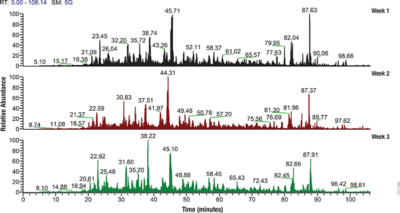
LC/MS performance monitoring. Each run used 1μg of human predigested protein extract injected into the instrument (Waters NanoAquity HPLC System interfaced to a Thermo Fisher Q Exactive™ Hybrid Quadrupole-Orbitrap Mass Spectrometer). Peptides were resolved with a 2-hour gradient. Weekly monitoring with the human extract ensured consistent analytical performance of the instrument.

Mass spectrometry allows characterization of molecules by converting them to ions so that they can be manipulated in electrical and magnetic fields. Basically a small sample (analyte) is ionized, usually to cations by loss of an electron. After ionization, the charged particles (ions) are separated by mass and charge;  the separated particles are measured and data displayed as a mass spectrum. The mass spectrum is typically presented as a bar graph where each peak represents a single charged particle having a specific mass-to-charge (m/z) ratio. The height of the peak represents the relative abundance of the particle. The number and relative abundance of the ions reveal how different parts of the molecule relate to each other.

For the study of large, organic macromolecules, matrix associated laser desorption/ionization (MALDI) or tandem mass spec/collision induced dissociation (MS/MS) techniques are often used to generate the charged particles from the analyte. MS analysis brings sensitivity and specificity to proteome analysis. The technique has excellent resolution and is able to distinguish one ion from another, even when their m/z ratios are similar. Macromolecules are present in extremely different concentrations in the cells, and MS analysis can detect biomolecules across five logs of concentration.

Continue reading “Monitoring Mass Spec Instrument Performance and Sample Preparation”

Optimize Your Western Blot

0447MA09_0A
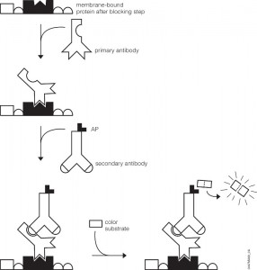
Western Blot Detection.

You’ve probably been there. You’ve got a new antibody or you’re testing out one you’ve made yourself. After weeks or months of work, your antibody is going to help move your research project forward. As you excitedly head to the dark room to develop your film, your mood is crushed when you see…bands, more bands, and smears. Alas, science has played one more cruel joke on you as you experience what so many of your fellow scientists have before. Despite such a dismal beginning, you often can still get good western blots by changing steps in your protocol.

Several steps in the western blot protocol can be optimized.

Continue reading “Optimize Your Western Blot”

Optimization of Western Blots Detecting Proteins Synthesized Using Cell-Free Expression #2

Detection of protein expressed using cell-free systems is required for most applications such as protein:protein interaction and protein:nucleic acid interaction studies. Traditionally, one adds radioactive [35S]methionine to cell-free expression reactions, and the methionine is incorporated into the expressed protein, allowing detection by autoradiography. Many researchers are moving away from radioactivity. Traditional Western blot analysis provides the researcher a nonradioactive method for detection but, if performed improperly, can result in high background, which can mask expressed proteins and affect downstream applications.

One critical step in producing low-background, high-signal Western blots is choosing the correct dilution of the primary antibody. Typically the manufacturer recommends antibody dilution from 1:1,000 to 1:2,500 for standard western blotting experiments. However when using crude lysates as a source of the target protein, these recommendations exhibit significant background. When the antibody was diluted 1:50,000, background was decreased significantly, and the positive signal was a large percentage of the total signal.

As a general recommendation when performing Western blot analysis of proteins expressed in cell-free systems, one must experimentally determine the optimal dilution of the primary antibody. In the Western blots performed in this study, primary antibodies were diluted ~50-fold more than the provider’s recommended dilution.

For additional technical details refer to this recent article published in Promega’s PubHub:

Hook, B and Schagat, T. (2011) Non-Radioactive Detection of Proteins Expressed in Cell-Free Expression Systems Promega Corporation Web site. Accessed August 17, 2011.

6X His Protein Pulldowns: An Alternative to GST

ResearchBlogging.orgPull-down assays probe interactions between a protein of interest that is expressed as fusion protein (e.g.,
(e.g., bait) and the potential interacting partners (prey).

In a pull-down assay one protein partner is expressed as a fusion protein (e.g., bait protein) in E. coli and then immobilized using an affinity ligand specific for the fusion tag. The immobilized
bait protein can then be incubated with the prey protein. The source of the prey protein depends on whether the experiment is designed to confirm an interaction or to identify new interactions. After a series of wash steps, the entire complex can be eluted from the affinity support using SDS-PAGE loading buffer or by competitive analyte elution, then evaluated by SDS-PAGE.

Successful interactions can be detected by Western blotting with specific antibodies to both the prey and bait proteins, or measurement of radioactivity from a [35S] prey protein. bait) and potential interacting partners (prey).

The most commonly used method to generate a bait protein is expression as a fusion protein contain a GST (glutathione-S transferase) tag in E. coli. This is followed by immobilization on particles that contain reduced glutathione, which binds to the GST tag of the fusion protein. The primary advantage of a GST tag is that it can increase the solubility of insoluble or semi-soluble proteins expressed in E. coli.

Among fusion tags, His-tag is the most widely used and has several advantages including: 1) It’s small in size, which renders it less immunogenically active, and often it does not need to be removed from the purified protein for downstream applications; 2) There are a large number of commercial vectors available for expressing His-tagged proteins; 3) The tag may be placed at either the N or C terminus; 4) The interaction of the His-tag does not depend on the tag structure, making it possible to purify otherwise insoluble proteins using denaturing conditions. Continue reading “6X His Protein Pulldowns: An Alternative to GST”

Use of Multiple Proteases for Improved Protein Digestion

One of the approaches to identify proteins by mass spectrometry includes the separation of proteins by gel electrophoresis or liquid chromatography. Subsequently the proteins are cleaved with sequence-specific endoproteases. Following digestion the generated peptides are investigated by determination of molecular masses or specific sequence. For protein identification the experimentally obtained masses/sequences are compared with theoretical masses/sequences compiled in various databases.

Trypsin is the favored enzyme for this application, for the following reasons: A) the peptides contain a basic residue (Arg or Lys) on the C terminus and thus are good candidates for collision induced activation (CAD) in tandem experiments (low charge states and high mass-to-charge ratios); B) it is relatively Inexpensive; and C) optimal digestion conditions have been well characterized.

An inherent limitation of trypsin is the size of the peptides that it generates. For most organisms > 50% of tryptic peptides are less than 6 amino acids, too small for mass spectrometry based sequencing.

Are you looking for proteases to use in your research?
Explore our portfolio of proteases today.

One recent publication examined the use of multiple proteases (trypsin, LysC, ArgC , AspN and GluC) in combination with either CAD or electron-based fragmentation (ETD) to improve protein identification (1). Their results indicated a significant improvement from a single protease digestion (trypsin), which yielded 27,822 unique peptides corresponding to 3313 proteins. In contrast using a combination of proteases with either CAD or ETD fragmentation methods yielded 92,095 unique peptides mapping to 3908 proteins.

Swaney DL, Wenger CD, & Coon JJ (2010). Value of using multiple proteases for large-scale mass spectrometry-based proteomics. Journal of proteome research, 9 (3), 1323-9 PMID: 20113005

Trypsin: Innovative Applications

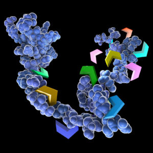
3D model of protein and protease cleavage

Tryptic digestion of samples and subsequent analysis by mass spectrometry is a popular technique for the identification of proteins typically those related to interaction partners or biomarkers characterization. This powerful tool can also be used for less traditional experimental designs. Three examples are:

Continue reading “Trypsin: Innovative Applications”