Optimizing Pressure Cycling Sample Preparation for Bottom-Up Proteomics

Large-scale analyses of the proteome have revealed proteomic changes in response to disease, and these changes hold great promise for diagnostics and treatment of complex diseases if proteomic analysis can be brought into the clinical laboratory. Successful and reliable large-scale proteomics requires sample preparation workflows that are reproducible, reliable and show little variability. To bring proteomics into the clinical laboratory, standardized procedures and workflows for sample prep and analysis are required to generate valid, actionable results on a time scale useful for the clinic.

The two most common sample types analyzed for clinical proteomics are body fluids and tissue biopsies. To process these kinds of samples, there are two initial steps: tissue solubilization, followed by proteolytic digestion. Solubilization of solid tissues is the most labor-intensive and produces the most variable results.

The introduction of pressure cycling technology (PCT) using Barocycler instrumentation has greatly improved both tissue solubilization and digestion consistency. The PCT-based sample preparation protocols generally utilize urea as a lysis buffer for protein denaturing and solubilization. Urea has several drawbacks including inhibiting trypsin activity and introducing unwanted modifications like carbamylation.

Lucas and colleagues analyzed whether replacing urea with SDC would produce similar tissue digestion profiles and improve the PCT method.

Are you looking for proteases to use in your research?
Explore our portfolio of proteases today.

SDC allowed the use of higher temperatures compared to urea, and hence the first step (lysis, reduction, and alkylation) was performed at 56 ยฐC. The second digestion step in the Barocycler was optimized, and the third step was eliminated. To further reduce digestion time, they capitalized on Rapid Trypsin/Lys-C.  Rapid Trypsin/Lys-C maintains robust activity at 70 ยฐC, and allowed Barocycler digestion to be performed in a single step, completing digestion in 30 cycles (approximately 30 min) rather than 105 minutes, streamlining the protocol.

The data presented an improved conventional tissue PCT approach in a Barocycler by replacing urea and proteolytic enzymes with SDC, N-propanol, and modified commercially available enzymes that have higher optimum temperatures.


Try a sample of high-efficiency Trypsin Platinum today!

Visit our website for more on Trypsin Platinum, Mass Spectrometry Grade, with enhanced proteolytic efficiency and superior autoproteolytic resistance.


Paper Referenced

Lucas, N. et al. (2019) Accelerated Barocycler Lysis and Extraction Sample Preparation for Clinical Proteomics by Mass Spectrometry. J of Proteome Res 18, 399โ€“405.


Related Posts

Expanding the Plague Family Tree: Yersinia pestis in the Neolithic

Yersinia pestis. See page for author [Public domain], via Wikimedia Commons

In recent years, scientists have been able to refine their molecular tools to resurrect ancient DNA from human graves and determine that yes, Yersinia pestis was the causative agent for the Black Death in the 14th century and the Plague of Justinian in the 6th century.  As more and more human graves have been uncovered, their DNA has revealed many secrets that scientists even ten years ago were unable to discover. With the ability to sequence entire genomes of bacteria that died with their hosts hundreds and even thousands of years ago, researchers are exploring the rise and possible spread of Y. pestis. Each new member sequence adds to the Y. pestis family tree, pinpointing the origin of this bacteria as it diverged from its ancestor Y. pseudotuberculosis. Peering into the past, scientists have been able to track down a strain of Y. pestis from individuals in a Swedish passage grave that is basal to known strains and that the authors of a Cell article suggest has interesting implications.

This pathogenic journey into history started by analyzing ancient DNA data sets from the teeth of individuals present in a communal passage grave in Gรถkhem parish, located in western Sweden, for any disease-causing microbial sequences that might be present. Y. pestis was flagged in one 20-year-old female dated 4,867โ€“5,040 years ago. The bacterial sequences from this individual, named Gok2, were more closely aligned with Y. pestis than the Y. pseudotuberculosis reference genome.

Continue reading “Expanding the Plague Family Tree: Yersinia pestis in the Neolithic”

Goodbye to the Most Famous Bird in Maine

When Wisconsin plunged into a deep freeze during last weekโ€™s polar vortex, I built a roaring fire in my fireplace and settled into my armchair with a thick blanket and a video game controller. Except for the twenty minutes I spent driving to and from the office, I stayed warm and toasty.

Birds, however, donโ€™t have it quite as easy. To survive freezing temperatures, non-migratory birds have developed many interesting adaptations. Many species grow extra down layers and huddle together for wind protection. Others, like the black-capped chickadee, use a process called regulated hypothermia to drop their resting body temperature by as much as 22ยฐF to conserve energy. Iโ€™m particularly fascinated by the process of regional hypothermiaโ€”many species of ducks and gulls use a countercurrent heat exchange system to keep vital organs warm while letting temperatures fall in extremities.

Birds that arenโ€™t accustomed to cold weather donโ€™t have these adaptations, though. When a birdโ€”or any animalโ€”ends up far outside of its natural habitat, the consequences can be deadly.

Continue reading “Goodbye to the Most Famous Bird in Maine”

Oh, the Places You’ll Go, Thanks to Science!

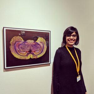
Today’s guest blog is written by Aparna Shah, a Post-Doc at Johns Hopkins University. Aparna visited the Promega campus in Madison, Wisconsin on January 18, 2019 and offered to share her experiences.


Aparna poses with her artwork at the Promega Employee Art Showcase

I donโ€™t recall ever having won a contest before, let alone the grand prize! In fact, I did a double take when I first read the email informing me that my SciArt submission had been selected as a winning entry for the Promega Art Contest for Creative Scientists. What did I win, you ask? A free trip to Madison, WI to meet with the team behind the contest and explore Promega’s headquarters!

I first heard about the contest on the HelloPhD podcast and considered participating primarily to support the SciArt movement. A couple of days later, I came across a perfectly-timed tweet about the contest that nudged me out of procrastination mode and reminded me to follow through with it. Iโ€™m going to take a second here to pitch both HelloPhD and Twitter to you. Regardless of whether youโ€™re an undergraduate student interning in a science lab or a senior postdoc, the HelloPhD podcast is incredible at calming you down while youโ€™re on the roller coaster ride called academia. As for Twitter, I can think of several pros for using it. But in the context of this post, it is one of the best resources for discovering opportunities that match your interests.

Continue reading “Oh, the Places You’ll Go, Thanks to Science!”

Meet Mฤ›nglร  Virus: the newest cousin in the Ebola and Marburg virus family tree

Ebola virus (EBOV) and Marburg virus (MARV) are two closely-related viruses in the family Filoviridae. Filoviruses are often pathogenic, causing hemorrhagic fever disease in human hosts. The Ebola outbreak of 2014 caught the world by surprise by spreading so quickly and severely that public health organizations were unprepared. The devastating outcome was a total of over 11,000 deaths by the time the outbreak ended in 2016. Research that provides further understanding of filoviruses and their potential for transmission is important in preventing future outbreaks from occurring. But what if the outbreak comes from a virus weโ€™ve never seen before?

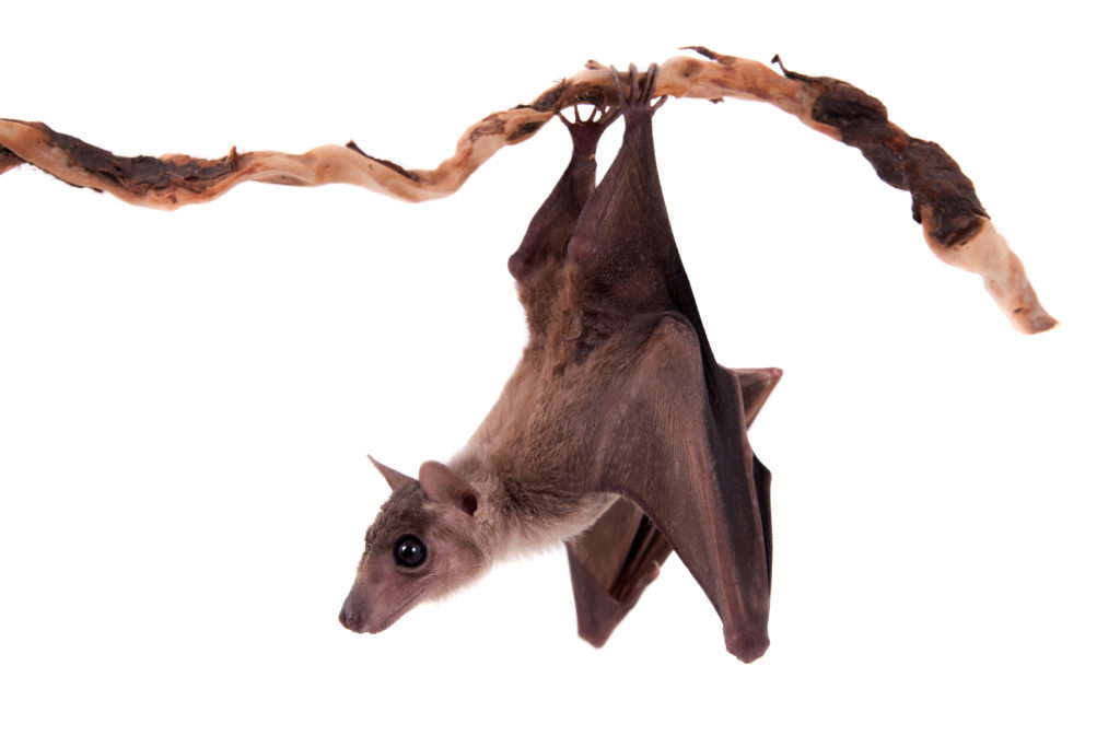
fruit_bat
Mฤ›nglร  virus was discovered among filoviruses isolated from Old World fruit bats (Rousettus)

All in the viral family

A recent study published in the journal Nature Microbiology provides evidence of a newly identified filovirus species. Using serum samples taken from bats, a well-known host for filoviruses, Yang et al. isolated and identified viral RNA for an unclassified viral genome sequence using next generation sequencing analysis. This new virus genome sequence was organized with the same open reading frames as other filoviruses, encoding for nucleoprotein (NP), viral protein 35 (VP35), VP40, glycoprotein (GP), VP30, VP24, and RNA-dependent RNA polymerase (L). This new genome sequence shared up to 54% of the nucleotide sequences for the filovirus species Lloviu virus (LLOV), EBOV and MARV, with MARV being the most similar. Their analysis suggested that this novel virus should be classified within the Filoviridae family tree as a separate genus, Dianlovirus, and was named Mฤ›nglร  virus (MLAV).

Continue reading “Meet Mฤ›nglร  Virus: the newest cousin in the Ebola and Marburg virus family tree”

Twisted CRISPR: A Novel Activation Strategy to Treat Genetically Driven Obesity

Two Is Better Than One

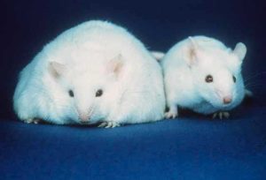
Obese and normal mouse

Redundancy equips us to survive. We have more than one lung or one kidney for a reasonโ€”if one organ in a pair gets damaged, we can still manage if the other is functional. At the cellular level, we have two copies of each chromosome in every non-germline cell. Each copy was inherited originally from a single sperm and ovum, which are โ€œhaploidโ€ cells. Consequently, there are two copies of any given gene in non-germline โ€œdiploidโ€ cells. In many cases, should one copy of a gene be damaged, the cell can still survive with the other, functional copy of a gene. In plants, this redundancy is common, and many plants exhibit polyploidy. In an extreme example of polyploidy, the large (by bacterial standards) but otherwise unassuming species Epulopiscium contains tens of thousands of copies of its genome.

Continue reading “Twisted CRISPR: A Novel Activation Strategy to Treat Genetically Driven Obesity”

Deep in the Jungle Something Is Happening: DNA Sequencing

This blog was written by guest blogger and 2018 Promega Social Media Intern Logan Godfrey.

Only 30 years ago, the polymerase chain reaction (PCR) was used for the first time, allowing the exponential amplification of a specific DNA segment. A small amount of DNA could now be replicated until there was enough of it to study accurately, even allowing sequencing of the amplified DNA. This was a massive breakthrough that produced immediate effects in the fields of forensics and life science research. Since these technologies were first introduced however, the molecular biology research laboratory has been the sole domain of PCR and DNA sequencing.

While an amazing revolution, application of a technology such as DNA sequencing is limited by the size and cost of DNA sequencers, which in turn restricts accessibility. However, recent breakthroughs are allowing DNA sequencing to take place in jungles, the arctic, and even spaceโ€”giving science the opportunity to reach further, faster than ever before. 

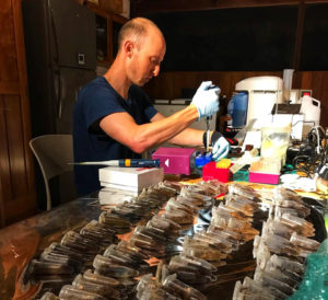
Gideon Erkenswick begins extractions on fecal samples collected from wild tamarins in 2017. Location: The GreenLab, Inkaterra.

Gideon Erkenswick begins extractions on fecal samples collected from wild tamarins in 2017. Location: The GreenLab, Inkaterra. Photo credit: Field Projects International.

The newfound accessibility of DNA sequencing means a marriage between fields of science that were previously largely unacquainted. The disciplines of genomics and wildlife biology/ecology have largely progressed independently. Wildlife biology is practiced in the field through observations and macro-level assessments, and genomics, largely, has developed in a lab setting. Leading the charge in the convergence of wildlife biology and genomics is Field Projects International. 

Continue reading “Deep in the Jungle Something Is Happening: DNA Sequencing”

How Cheese Helped Us Understand Microbial Interactions

In almost every environment on earth, such as soil, human skin and gut, there lives a whole community of microbesโ€”sometimes up to hundreds of species. It may seem like they all flourish in peace. But just like you may have friendly or hostile interactions with your neighbors, the different bacterial species interact in various ways. They may cooperate, compete or, sometimes, even kill each other. The interaction is complicated, and scientists have struggled to understand the nature of these microbiome interactions. How do microbiomes assemble and maintain stability? How do the interactions among different species affect gene expression?

Continue reading “How Cheese Helped Us Understand Microbial Interactions”

Combatting Gun Violence with Synthetic Biology

Imagine you are a high school student living in a community devastated by gun violence and death. In the U.S., this could be one of many communities, but it happens to be Baltimore which had 301 deaths due to gun violence in 2017 (with a per capita rate well above other large cities). Then imagine you were part of an organization within that community that helped you, along with other students, gain knowledge and skills to come up with a viable solution to the problem using synthetic biology.

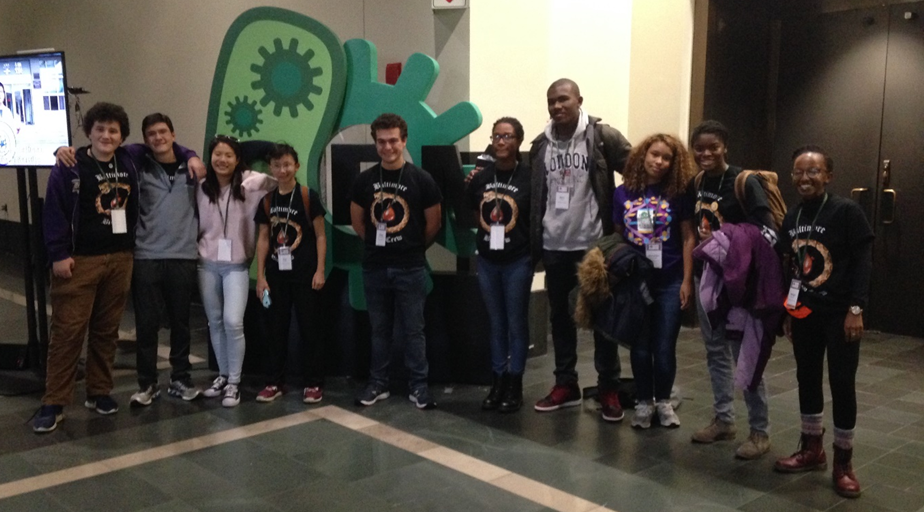
Baltimore Bio-Crew at the 2018 iGEM Giant Jamboree

This is exactly how the Baltimore Bio-Crew came up with their iGEM project, Coagulance Rx. The Baltimore Bio-Crew decided to tackle this community issue head-on. One team member, Mercedes Ferandes, reflected, โ€œLiving in Baltimore City, I have not only witnessed gun violence in front of me, but have had family members and friends die from it. I wanted to try to decrease the amount of deaths by gun violence using iGEM.โ€

After some research, they discovered that many of the gun violence deaths were due to blood loss and could have been prevented. The impoverished neighborhoods where this violence occurs lack the resources to provide timely emergency medical treatment. Many of these deaths can be attributed to delayed arrival of emergency response teamsโ€”wait times for an ambulance can be over an hour.

Although there were several contributing factors beyond their control, the team wanted to address this problem by focusing on blood clotting and how it could be helpful as a quick temporary treatment for open wounds. This solution could offer a reliable, cost efficient way to save lives by slowing or stopping blood loss until a victim could get medical attention. The team decided to pursue the use of snake venom after coming across some previous iGEM projects that had used it for clotting. Team member Henry Ryles pointed out that the need for snake venom powerful enough to clot blood quickly led them to choose the venom of the Russellโ€™s Viper
(Daboia russelii).

Continue reading “Combatting Gun Violence with Synthetic Biology”

A Roadmap for PROTAC Development

PROTACs or Proteolysis-Targeting Chimeras are an emerging tool in protein degradation studies, potentially suited to any need involving the removal of a specific protein. These small-molecule chimeras are exciting due to: 1) their target specificity; and 2) their ability to enable target destruction versus target inhibition. Here we discuss a paper that presents a roadmap for PROTAC development.

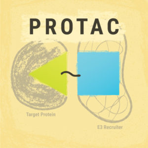
PROTAC components: target protein ligand, E3 ligase and linker, these are components in the described roadmap for PROTAC development.
PROTAC components: target protein ligand, E3 ligase and linker.

Destruction/Inhibition: Is There a Difference?
An analogy that microbiologists (and wrestlers or anyone that has ever spent time in a locker room shower) would understand, is fungicidal versus fungistatic compounds. A fungicidal compound kills fungus. A fungistatic compound just slows the fungus down a bit.

A small-molecule inhibitor attaches to its target protein, but for how long? What inhibitor testing must be done to determine how long the inhibition lasts?

On the other hand, a small-molecule agent that causes protein degradation first targets the protein of interest, then attaches ubiquitin to that target. Once a protein is marked with ubiquitin, it’s a dead man. E3 ligase must be involved, but if the ubiquitin is added by E3, the end is near. Next stop, Hades.

This ubiquitinated protein is headed to the proteasome and proteins that go there donโ€™t come back. Ubiquitination was called the โ€˜molecular kiss of deathโ€™ when this discovery was awarded the Nobel prize in Chemistry in 2004.

About PROTACs

PROTACs are degrader molecules composed of three parts: 1) a ligand that is specific for the target protein; 2) a ligand for E3 ligase; and 3) a linker molecule that connects the two ligands. The E3 ligase is one of three enzymes that can add ubiquitin to a cellular component, but only ubiquitins added by the E3 ligase cause targeting to the proteasome (Zoppi et al.).

Continue reading “A Roadmap for PROTAC Development”