Will Artificial Intelligence (AI) Transform the Future of Life Science Research?

Artificial intelligence (AI) is not a new technological development. The idea of intelligent machines has been popular for several centuries. The term “artificial intelligence” was coined by John McCarthy for a workshop at Dartmouth College in 1955 (1), and this workshop is considered the birthplace of AI research. Modern AI owes much of its existence to an earlier paper by Alan Turing (2), in which he proposed the famous Turing Test to determine whether a machine could exhibit intelligent behavior equivalent to—or indistinguishable from—that of a human.

The explosive growth in all things AI over the past few years has evoked strong reactions from the general public. At one end of the spectrum, some people fear AI and refuse to use it—even though they may have unwittingly been using a form of AI in their work for years. At the other extreme, advocates embrace all aspects of AI, regardless of potential ethical implications. Finding a middle ground is not always easy, but it’s the best path forward to take advantage of the improvements in efficiency that AI can bring, while still being cautious about widespread adoption. It’s worth noting that AI is a broad, general term that covers a wide range of technologies (see sidebar).
AI personified looking at a dna double helix against an abstract cosmic background
Image generated with Adobe Firefly v.2.

For life science researchers, AI has the potential to address many common challenges; a previous post on this blog discussed how AI can help develop a research proposal. AI can help with everyday tasks like literature searches, lab notebook management, and data analysis. It is already making strides on a larger scale in applications for lab automation, drug discovery and personalized medicine (reviewed in 3–5). Significant medical breakthroughs have resulted from AI-powered research, such as the discovery of novel antibiotic classes (6) and assessment of atherosclerotic plaques (7). A few examples of AI-driven tools and platforms covering various aspects of life science research are listed here.

Continue reading “Will Artificial Intelligence (AI) Transform the Future of Life Science Research?”
RAF Inhibitors: Quantifying Drug-Target Occupancy at Active RAS-RAF Complexes in Live Cells

Mitogen-activated protein kinases (MAPKs) are a large family of proteins that regulate diverse cellular functions in eukaryotes, including gene expression, proliferation, differentiation and apoptosis (1). MAPK signaling pathways typically include three sequentially activated kinases, and these pathways are triggered in response to extracellular stimuli, such as cytokines, mitogens, growth factors and oxidative stress (1). Ultimately, the signal is transmitted to the nucleus, with the activation of a specific transcription factor that modulates the expression of one or more genes.

Among MAPK pathways, the RAS-RAF-MEK-ERK signaling pathway has been studied extensively. Mutations in RAS family proteins and resultant dysregulation of the signaling pathway are implicated in a variety of cancers. Therefore, this pathway is a popular target for anticancer drug development.

An overview of the RAS-RAF-MEK-ERK signaling pathway.
Continue reading “RAF Inhibitors: Quantifying Drug-Target Occupancy at Active RAS-RAF Complexes in Live Cells”

Synthetic Biology: Minimal Cell, Maximal Opportunity

According to the National Human Genome Research Institute, synthetic biology is “a field of science that involves redesigning organisms for useful purposes by engineering them to have new abilities”. Synthetic biology has a broad range of applications, from manufacturing pharmaceuticals and other biologically active chemicals and biofuels, to accelerating the adoption of plant-based burgers (1).

At the heart of the synthetic biology revolution is the rapid technological advancement—and accompanying drop in costs—of DNA oligonucleotide synthesis. Typically, synthetic biology researchers use oligonucleotides as building blocks to assemble genes of interest that are then introduced into, and expressed by, a different organism. For example, to create the plant-based Impossible Burger, the soy leghemoglobin gene (normally found in the root nodules of leguminous plants) was synthesized and expressed in yeast cells (1). This component gives the burger its meaty flavor and appearance of “bleeding” when cooked.

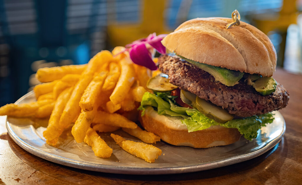
An Impossible Burger served with fries on the side

Continue reading “Synthetic Biology: Minimal Cell, Maximal Opportunity”

Phage Therapy: Meeting the Challenge of Drug-Resistant Bacterial Infections

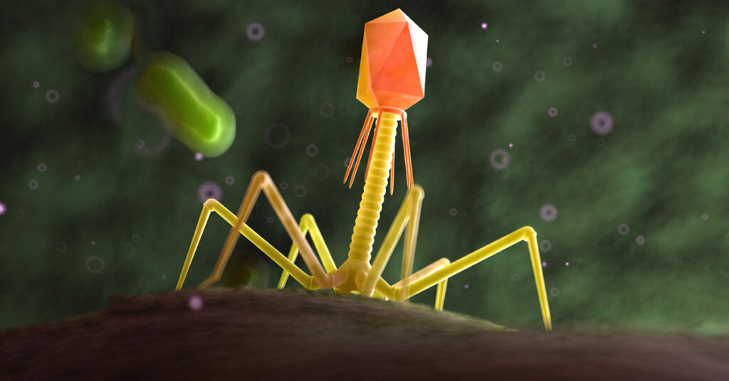
Global pandemics, such as COVID-19, have taught us to abhor viruses. The emergence of new, highly infectious viruses is—rightfully so—a cause for concern. However, despite the average human body harboring 380 trillion viruses, most of them simply coexist with us and are harmless. When it comes to an ancient lineage of viruses within the realm Duplodnaviria, researchers are even using them as weapons in the battle against infectious diseases.

In 1915, Frederick William Twort, an English bacteriologist at the University of London, reported the discovery of an unusual “ultramicroscopic virus” (1). Twort was culturing vaccinia virus as part of an experiment to determine if he could prepare smallpox vaccines in vitro. These vaccines, made in calves, were typically contaminated with Staphylococcus bacteria. When Twort plated the vaccines, he found small, clear areas on the agar plates where the bacteria would not grow, and these clear areas were the source of his ultramicroscopic virus. Two years later, a French-Canadian microbiologist, Félix d’Hérelle, independently discovered a similar phenomenon when culturing Shigella bacteria from fecal samples of patients with bacillary dysentery. He called the new virus “un bactériophage obligatoire” (2). Shortly after his discovery, he found that bacteriophages (phages) could be used as powerful agents to treat a variety of bacterial infections, and the field of phage therapy was born (3).

Continue reading “Phage Therapy: Meeting the Challenge of Drug-Resistant Bacterial Infections”

Genome-Wide CRISPR Screening: Putting Death on Hold

We share this planet with approximately 8.7 million species of plants and animals. Within such a diverse environment, it’s only natural that many complex relationships have developed among different species. Some relationships are mutually beneficial, some are parasitic—and some are lethal.

Genome wide - crisper screening to help with toxic compounds to humans

Natural toxins and venoms are biologically active compounds produced by normal metabolic processes in an organism but are harmful to other organisms. Typically, toxins are encountered passively or ingested by the affected organisms, and have a specific mode of action and binding site within a cell. In contrast, venoms are introduced directly into the victim through a specialized delivery mechanism, and they may consist of a mixture of compounds that affect a range of cell types and tissues (1). Both types of poisons are produced for predation, defense, or to offer a competitive advantage (1).

Continue reading “Genome-Wide CRISPR Screening: Putting Death on Hold”

PARP and DDR Pathways: Targeting the DNA Damage Response for Cancer Treatment

Our cells, and the DNA they contain, are under constant attack from external factors such as ionizing radiation, ultraviolet light and environmental toxins. Internal cellular processes can also generate metabolites, such as reactive oxygen species, that damage DNA. In most cases, DNA damage results in permanent changes to DNA molecules, including DNA mismatches, single-strand breaks (SSBs), double-strand breaks (DSBs), crosslinking, or chemical alteration of bases or sugars. If left unchecked, DNA damage can cause genome instability, mutations and aberrant transcription, and oncogenic transformation.

PARP DDR pathway for drug discovery

Fortunately, our cells have also evolved multiple pathways to repair damaged DNA, collectively known as the DNA damage response (DDR). The type of repair mechanism depends on the nature of the damage, and whether the damage occurs in mitochondrial or nuclear DNA. These mechanisms have been reviewed extensively (1,2). Recently, considerable attention has focused on pathways for repairing SSBs and DSBs, mediated by the ADP-ribosylating enzyme known as poly (ADP-ribose) polymerase 1, or PARP-1.

Continue reading “PARP and DDR Pathways: Targeting the DNA Damage Response for Cancer Treatment”

Monoclonal Antibody (mAb) Therapy to Delay the Onset of Type 1 Diabetes

On November 18, 2022, the US Food and Drug Administration (FDA) announced the approval of the first drug to delay the onset of stage 3 type 1 diabetes (T1D). The monoclonal antibody (mAb) drug, teplizumab, was approved for use in adults and pediatric patients 8 years and older.

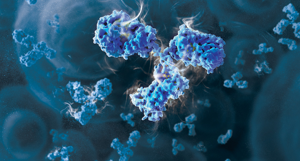
3D illustration of a monoclonal antibody

The road to approval has been a bumpy one for the manufacturer, Provention Bio. In 2020, the FDA rejected the application for teplizumab due to several concerns, including the small size of the clinical trial. With the current approval, based on new clinical trial results, Provention Bio confirmed a co-promotion agreement with Sanofi US. The agreement included a $35 million Sanofi equity investment in the company.

Continue reading “Monoclonal Antibody (mAb) Therapy to Delay the Onset of Type 1 Diabetes”

Left-Handed DNA: Is That Right?

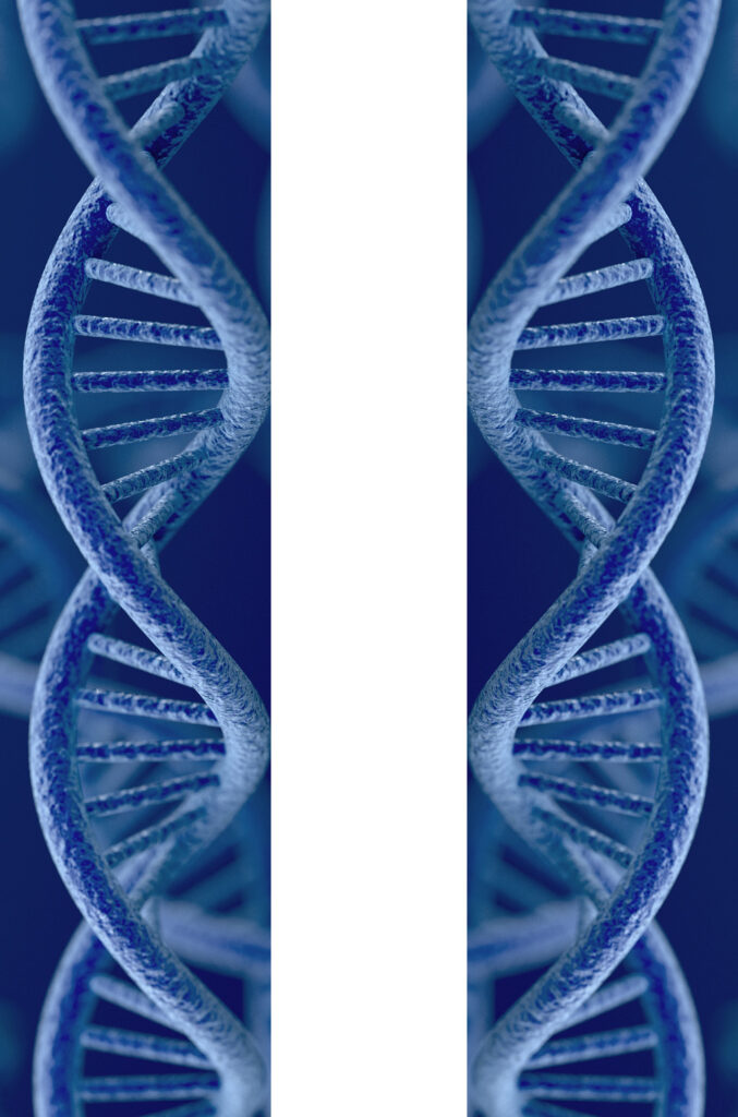
There’s a certain group of people (including this blog post author) who derive no small amount of amusement from seeing stock photos of DNA and pointing out flaws in the structure. It’s even more amusing when these photos are used in marketing by life science companies. The most common flaw: the DNA molecule is a left-handed double helix.

What does that even mean? DNA, like many organic chemicals in biology, is a chiral molecule. That is, it can exist in two structural forms that are mirror images of each other but are not superimposable (enantiomers). Just like your left and right hands are mirror images of each other, the two DNA structures are left-handed and right-handed double helices. The DNA double helix is chiral, because its building blocks (nucleotides) are chiral.

Two DNA helices that are mirror images

It can be challenging, at first glance, to tell whether an image of DNA is left-handed or right-handed. Various helpful hints are available; however, the one that I’ve found easiest to remember is described in a blog post by Professor Emeritus Larry Moran at the University of Toronto:

Imagine that the double helix is a spiral staircase, and you’re walking down the staircase. If you’re turning to the right as you descend, the DNA structure is right-handed; if turning to the left, it’s left-handed. In the image shown earlier, the DNA molecule on the right is a right-handed double helix, while its mirror image is left-handed.

Continue reading “Left-Handed DNA: Is That Right?”

Cellular Senescence and Cancer Therapy: Overcoming Immortality?

At the time of writing this post, no scientist had yet discovered the secret to immortality. In our world, we’ve come to accept that living things are born, grow old and die—the circle of life.

And yet, for many years, life scientists believed that the circle of life did not apply to our constituent cells when cultured in a laboratory. That is, cultured normal human cells were immortal, and they would continue to grow and proliferate forever, as long as they were provided with the necessary nutrients.

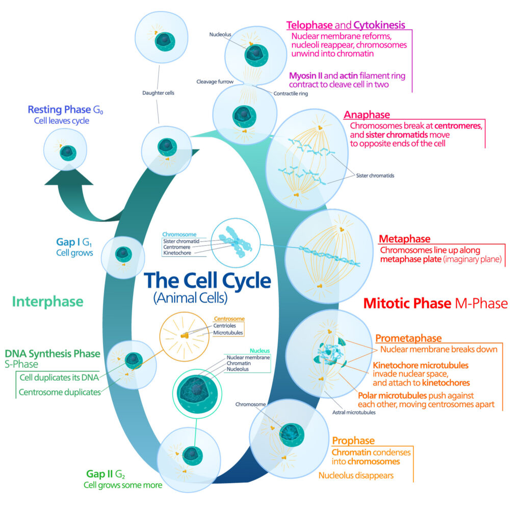
The animal cell cycle. Image by Kelvinsong; made available under the Creative Commons CC0 1.0 Universal Public Domain Dedication

Pioneering work published in 1961 by Leonard Hayflick and Paul Moorhead challenged that theory (reviewed in 1). Their research showed that normal cells in culture have a finite capacity to replicate. After they reach a certain number of replicative cycles, cells stop dividing. Hayflick and Moorhead made the important distinction between normal human cells and cultured cancer cells, which are truly immortal. In later years, the limit to the number of replicative cycles normal human cells can undergo became known as the Hayflick limit. Although some scientists still express skepticism about these findings, the Hayflick limit is widely recognized as a fundamental principle of cell biology.

Continue reading “Cellular Senescence and Cancer Therapy: Overcoming Immortality?”

Neanderthal DNA and Modern Humans: Svante Pääbo Receives the 2022 Nobel Prize in Physiology or Medicine

What makes humans “human”?

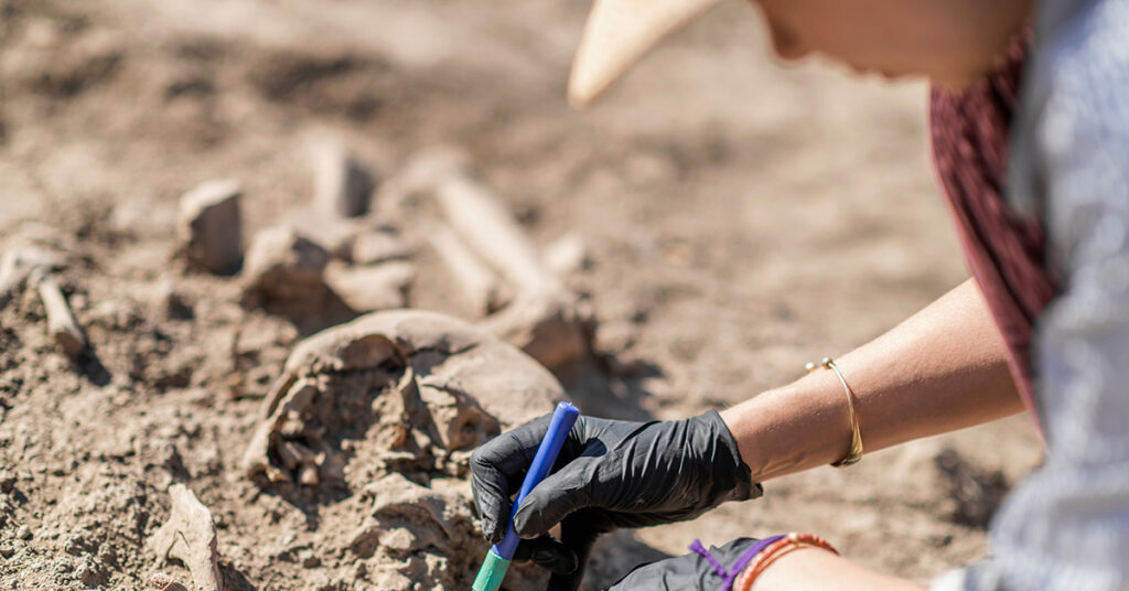
Neanderthal DNA sequencing from ancient bone samples

On October 3, 2022, the Nobel Assembly at Karolinska Institutet announced the 2022 Nobel Prize in Physiology or Medicine had been awarded to Svante Pääbo, director of the Department of Genetics at the Max Planck Institute for Evolutionary Anthropology in Leipzig, Germany. The Assembly cited his “discoveries concerning the genomes of extinct hominins and human evolution”. They mentioned the highlight of his research: the seemingly impossible task, at the time, of sequencing the Neanderthal genome. The discoveries that followed from this sequencing project continue to redefine our understanding of modern human origins.

The award showcases the technological advancements made in the analysis of ancient DNA. However, Pääbo’s research had an inauspicious beginning. In 1985, he published the results of his early work, cloning and sequencing DNA fragments from a 2,400-year-old Egyptian mummy (1). Unfortunately, later analysis revealed that the samples could have been contaminated by the researchers’ own DNA (2).

Continue reading “Neanderthal DNA and Modern Humans: Svante Pääbo Receives the 2022 Nobel Prize in Physiology or Medicine”