BTK (Bruton Tyrosine Kinase): Importance in Health and Disease
Bruton’s tyrosine kinase (BTK) was initially identified as a mediator of B-cell receptor signaling in the development and functioning of adaptive immunity. More recent and growing evidence supports an additional role for BTK in mononuclear cells of the innate immune system, especially dendritic cells and macrophages. For example, BTK functions in receptor-mediated recognition of infectious agents, cellular maturation and recruitment processes, and Fc receptor signaling. BTK has recently been identified as a direct regulator of a key innate inflammatory machinery, the NLRP3 inflammasome (2). Continue reading “Kinase Drug R & D: Helping Your Inhibitor Make the Cut”
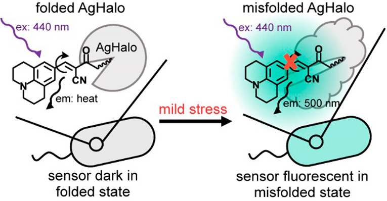
Cellular stress is associated with global misfolding and aggregation of the endogenous proteome. Monitoring stress-induced abnormalities remains one of the major technical challenges facing established sensors. Misfolded monomers induced by mild stresses, however, remain largely invisible with current sensors.
In a recent publication (1) Fares and colleagues describe a new sensor based upon a fluorescent molecular rotor that is conjugated to a Halo mutant (AgHalo). In non-stressed cells, the AgHalo sensor remains largely folded, and is fluorescent when misfolded. The fluorescent molecular rotor, when conjugated to purified AgHalo to form the proteome stress sensor, is able to report on urea-induced partially unfolded (misfolded) conformations with a higher fluorescent increase than the previously reported fluorophore-based sensors. Heat-induced misfolding is also effectively monitored by the fluorescence change of the sensor that is based on fluorescent molecular rotor, but not the solvatochromic fluorophore. The unique feature of the fluorescent molecular rotor makes the new generation of the AgHalo proteome sensor more sensitive to misfolded conformations that are primarily induced by mild proteome stress. Further, the new sensor exhibits a higher fluorescence signal when detecting soluble and insoluble protein aggregates that are induced by more severe proteome stress. These data collectively suggest that thermo-labile Halo conjugated with a fluorescent molecular rotor serves as a suitable sensor to detect a wide range of proteome stress conditions.
Cancer has been studied for decades by scientists trying to find a vulnerability to exploit and testing compounds to develop as potential drugs. As the “Emperor of All Maladies”, cancer has proven itself to be a wily beast with many varieties of genetic mutations for eluding cellular control, tireless in its ability to divide and spread. In the end, a cancer cell is still a cell and subject to its environment even though cancer does not play by the same rules as the normal cells that exist around it. To be able to grow, a cell needs access to metabolites, molecules needed for building the materials and machinery needed by the cell to function and divide. These requirements also offer potential pathways to target for halting cancer growth and spread.
All cells use glucose to generate ATP, but normal and cancer cells differ in how glucose is converted to ATP. Most cells use glucose in oxidative phosphorylation, but cancer cells use aerobic glycolysis, converting glucose to lactate without oxygen. This Warburg effect (glucose converted to lactate) is a hallmark of cancer cells as they take up glucose at a much higher rate than normal cells. Blocking glucose uptake is one way to target cancer cells. While 2-deoxyglucose (2DG) has been shown to slow glucose uptake in vitro, the compound proved toxic in clinical trials and lower dosages do not seem to be an effective treatment against cancer. While not an ideal drug target, glucose uptake has been helpful in monitoring cancer response to therapies via fluorodeoxyglucose positron emission tomography (FDG-PET).
Today is the start of Teacher Appreciation Week in the United States, punctuated tomorrow by National Thank a Teacher Day. I used to be on the receiving end of the various expressions of gratitude bestowed upon our educators: platters of brownies or cookies from the Board of Education, free meals from restaurants, discounts at retail stores and, if you were really lucky, maybe a student or two (likely initiated by their parents) would bring a gift card or note.
I would also reflect on the teachers that I was personally thankful for: my elementary teachers through graduate school professors (I still remember most of them by name and, with few exceptions, I received what I needed from all of them to learn and grow), my colleagues (who provided mentorship, support and comradery to me and so much more to their students) and my parents (who taught my earliest and most important lessons).
But now I find myself looking at this annual celebration of teachers from the other side—it has been two years since I became a science writer after nearly a decade of teaching high school science. The transition has completely changed my life in ways I could not have imagined and has also impacted the way I think about educators.
The main impetus for this career change was burnout. I had spent countless early mornings, late nights and weekends grading, planning lessons, completing professional development requirements and simply worrying about what challenges I would face the next morning, week or class period. The pressures of each school year would crescendo to a near breaking point every May, and then be swiftly wiped away by the arrival of summer break.
This cycle seemed inevitable, but I had been conditioned by the cultural narrative about teachers to consider it a tolerable tradeoff to the enviable benefits of teaching: holidays and summers “off”, ending the workday before 4 (even I groaned while typing that), great (read: better than average American, worse than someone with similar level of education and experience) benefits & retirement.
Unfortunately, this wasn’t sustainable for me. Moreover, legislative changes and budget cuts exacerbated the ever-present stress to new levels during my last few years as an educator. The strain was taking a toll on my mental health and my ability to be present with family and friends, especially my children.
In my new position, I have been met with intellectual challenges equal to those I encountered as a teacher but face a manageable amount of stress and few threats to work-life balance. Ending my teaching career was probably one of the best decisions I have ever made for my personal well-being. But despite this newfound joie de vivre, I am left with a feeling of guilt that resurfaces whenever issues I used to be so connected with make their way to the national spotlight.
Two of these have been in the news a lot this year—repercussions from budget cuts to education and gun violence in schools. I shouldered the burden of helping my students’ process school shootings and personally dealing with the reality that I could be in the middle of such a tragedy. Similar to the recent wave of teacher walkouts, budget measures that targeted educators brought me to the state capitol in protest.
Yet, I don’t have to face these issues with the sense of urgency I used to. My guilt is rooted in the fact that being a good teacher required selflessness and I chose to be selfish and leave because I couldn’t meet that expectation. It is perhaps because of this nagging feeling that I now feel a gratitude toward teachers that I didn’t before. I am still thankful to all the teachers in my past, but now my appreciation also extends to those that are and will become the future of education.
This year for Teacher Appreciation Week I want to express special gratitude for all of the teachers who feel the same pressures I did and are able to persist. I admire those of you already in the classroom and know you are putting your students’ needs ahead of your own. I’m grateful for all of you who are studying to become teachers, looking past all of the reasons you shouldn’t go into education and focusing instead on the impact you’ll have on future generations.
At a time when it is increasingly difficult to be optimistic about the future, knowing that there are still teachers willing to fight for themselves and their students gives me all the hope I need. Thank you teachers, this week and every week, for all you do!
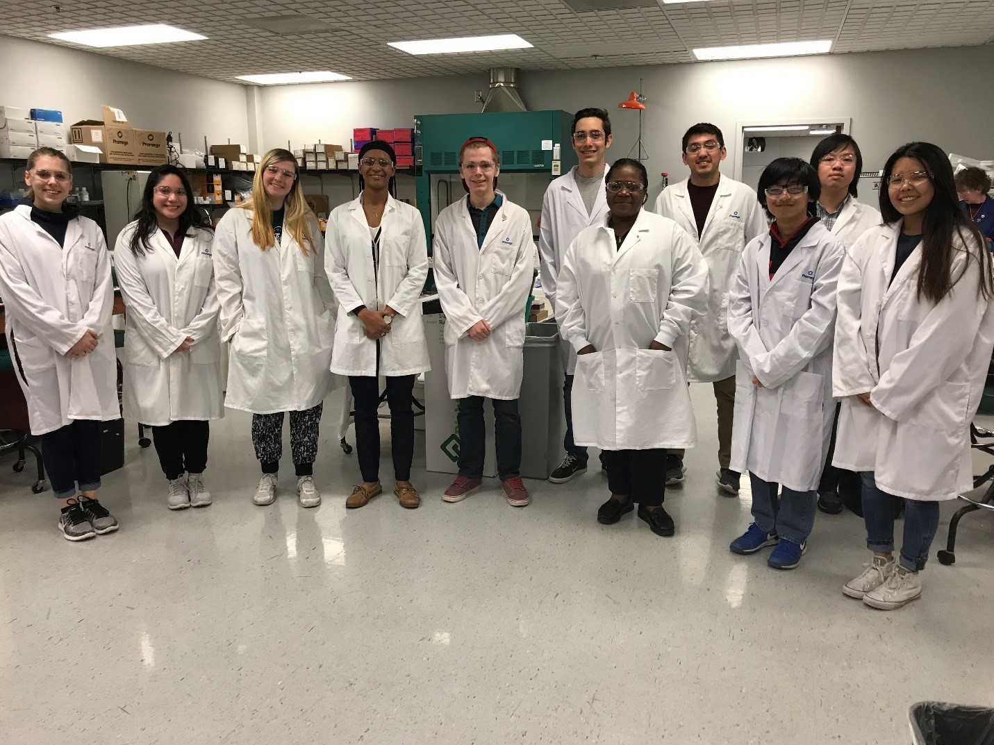
During the week of March 26, 2018, while many students were having fun and relaxing during Spring Break, others were busy doing extra lab work at the BTC Institute. This four-day workshop was designed to provide an introduction to the molecular biology laboratory for students affiliated with the Center for Educational Opportunity (CeO) on the UW-Madison campus. As noted on its website: “CeO promotes access to resources, academic achievement and personal growth for students whose parents have not received a four-year degree, students who meet specific federal family income guidelines, and students with documented disabilities.”
It is well known that first-generation college students, women and students of color persist in STEM fields at lower rates than the general population. This interferes with the creation of a diverse STEM talent pool, in turn needed to ensure diverse problem-solving perspectives.
Further, STEM fields are often seen as being stressful, given their competitive learning environments. This may be especially discouraging for students from racial/ethnic minorities who may not have as many mentors and role models to turn to.
Introduction to the Laboratory attendees
This workshop aimed to give students an experience that would strengthen their skills and confidence as they continue to pursue scientific paths. In addition to laboratory work, students discussed the importance of clear communication in written and oral presentations, were required to work as partners to experience teamwork, and were encouraged to use reflection and lab reporting as ways to internalize what they learned throughout the week.
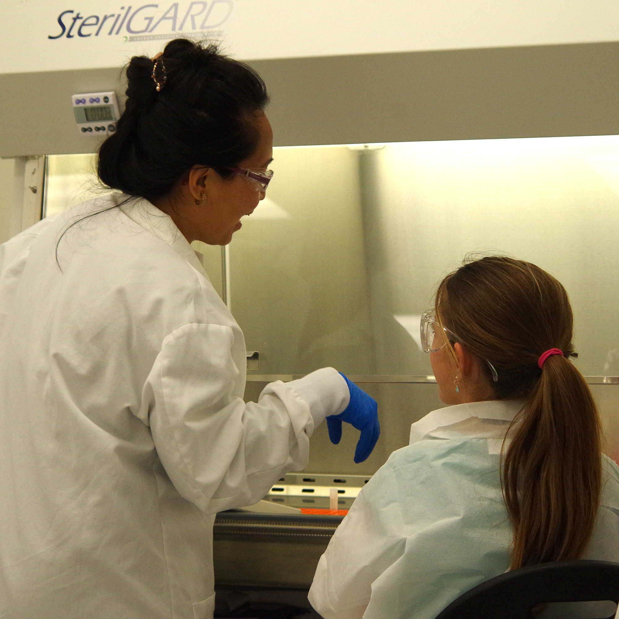
Local girls scouts worked with scientists at Promega to learn how a cell culture facility operates.
My twin daughters are finishing up their 10th-grade year next month, finding themselves smack in the middle of their high school experience, and discussions of classes, colleges and careers are increasing in frequency in my household. (It’s cliché, but I have to say it… Where does the time go?) As the girls begin to ponder their future, my husband and I are encouraging them to gain real-life insight from adults who work in fields they’re curious about. It’s never too early to get a first-hand perspective.
One of my girls has known from a pretty young age that she wants to pursue something in STEM, and likely the “S” in the acronym. Her schedule happened to be open the night a few months ago that one of my Promega colleagues, Senior R&D Scientist Danette Daniels, was speaking on a panel sponsored by the University of Wisconsin – Madison chapter of Graduate Women in Science. My daughter wasn’t sure about how she’d be received as the only high school student in the room, but she agreed to go with me anyway. Besides, I told her, they’re serving pie.
The six women on the panel represented a huge variety of avenues (academic to industry), specialties (biophysics to geology) and professional styles. During introductions, one panelist declared, “I had a job in a lab and was depressed. When I was stuck in a library all day, I was totally excited.” She now works with an organization to recruit more women into STEM fields. The woman sitting beside her runs a research lab and declared, “I love the bench quite a bit, and I don’t want to be in an office reading!” Continue reading “Inspiring the Next Generation of Scientists”
Recently I wrote about the completion of the human genome sequencing project and the promise, problems and questions that the project has generated in the last decade and a half. One of the biggest realizations that I had from researching and writing that post is that our human genome makes us more alike than different at the molecular level, yet there is incredible variability in the human species around the globe.
I started to think about other things where the basic building blocks were the same, yet the final products were so very different—and I landed in the middle of a symphony orchestra.
Orchestras, if we look at the instruments that they have at their disposal, are very similar: dare I say 99% identical? For instance the instruments listed in the February 2017 roster for the New York Philharmonic Orchestra on Wikipedia (1) are very similar to the lists of instruments listed for the musicians of the Atlanta Symphony Orchestra on its web site (2). Numbers and groupings might vary, but the instruments are the same.
However no one would argue that the New York Philharmonic Orchestra and the Atlanta Symphony Orchestra and Chorus are interchangeable. Experiencing one is not the same as experiencing the other, and two separate experiences of either are often completely different.
The orchestral “DNA” is the same: highly trained musicians playing essentially the same set of instruments, and quite often the same piece of music. What makes each experience of these organizations unique is the when, the where and the how of the expression of that DNA.
Pearl Jam, a popular alternative rock band in the 1990s (and still pretty awesome!). Photo credit: Rolling Stone Magazine.
This post could easily start out as an ode to ’90s alternative music (of which I’m a huge fan). That new and totally different sound (a la Pearl Jam, Smashing Pumpkins, Soundgarden, Nirvana, etc.) in the 1990s eventually made its way into the mainstream as it gained popularity. (I have to say that I got a shock when I recently heard some Pearl Jam on “classic rock” radio stations. But I digress…)
Why isn’t the same true for science career paths? Science careers outside of academia are still referred to as “alternative.” In a previous post, I highlighted statistics from a 2012 NIH report that found that only 20% of recent life sciences Ph.D.’s go on to become faculty members1. That means that 80% of recent life sciences Ph.D.’s took the “alternative” path. It seems like the academic path could now be viewed as the alternative to the mainstream, but somehow there’s an underlying stigma associated with straying from a path which few can travel down successfully.
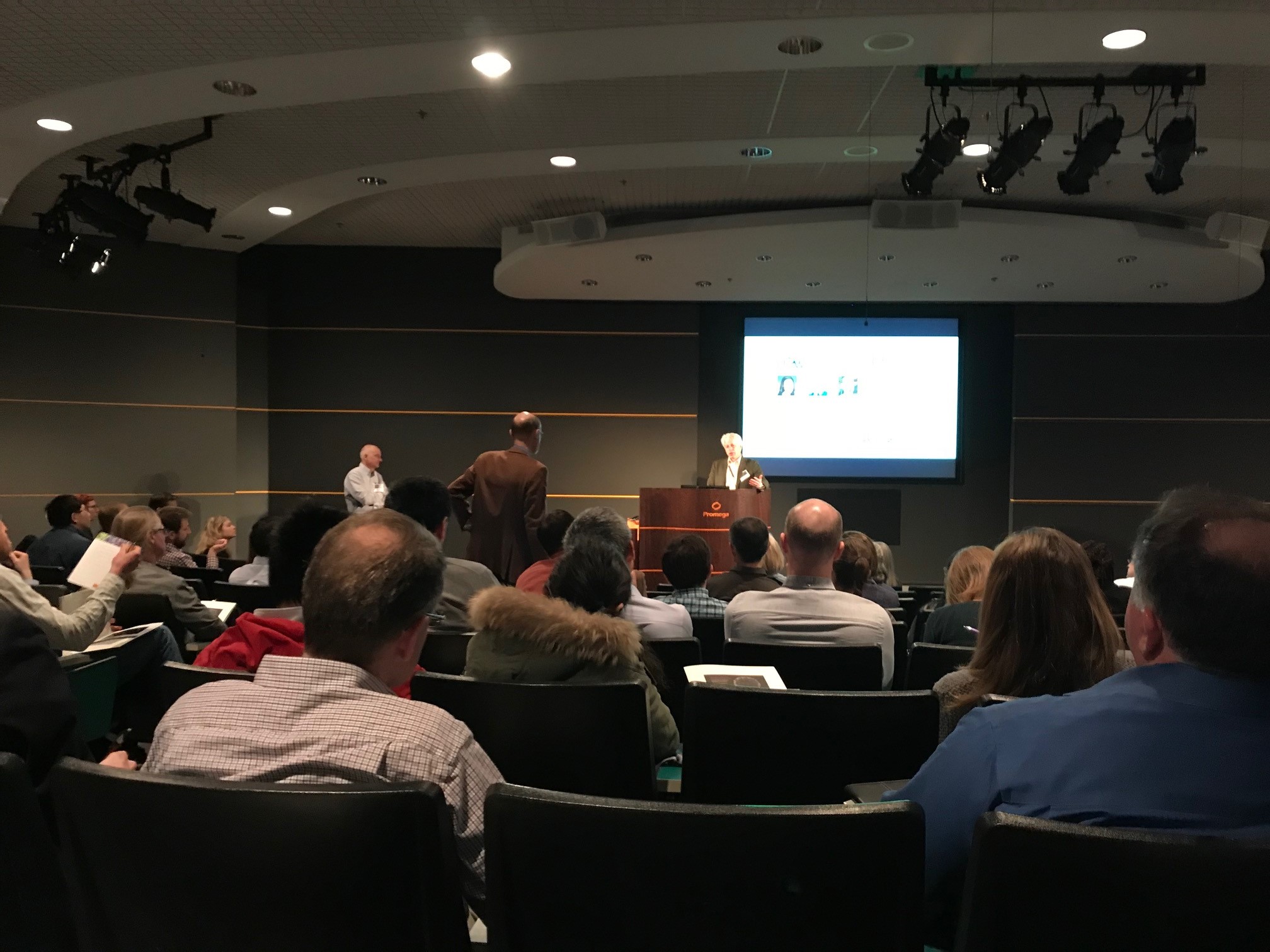
Dr. David Russell presenting at the 13th Wisconsin Stem Cell Symposium. The session was moderated by Dr. James Thomson.
“20 years ago, when I first heard about the creation of human embryonic stem cells, I knew that this was the future. I immediately requested the cells from Dr. Thomson and dropped almost everything else we were doing in our lab. It has been my focus to this day.” The person presenting is Dr. David Russell, a professor at the University of Washington. He is just one of the hundreds of researchers gathered at the BioPharmaceutical Technology Center Institute (a nonprofit supported by Promega) in Madison, Wisconsin for the 13th Annual Wisconsin Stem Cell Symposium that happened this week. This year, it’s not just a symposium, but also a celebration—it’s the 20-year anniversary of the first-ever isolation and culture of human embryonic stem cells (ES cells).
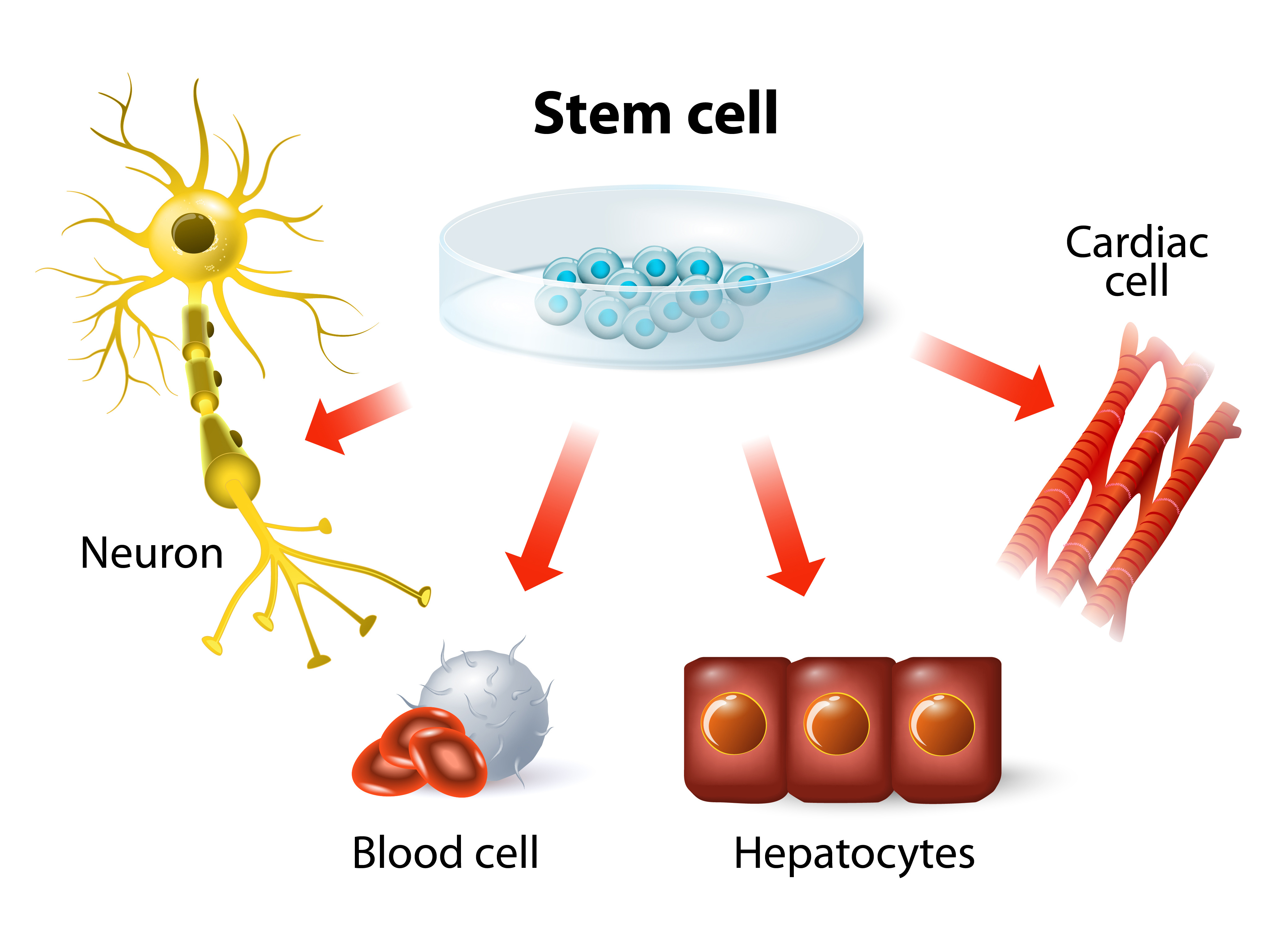
In 1998, Dr. James Thomson, at the University of Wisconsin-Madison, created the first ES-cell line using donated (unused) embryos from a fertility clinic. The study sent a shockwave through the scientific community and general public. We now had the technology to grow human pluripotent ES cells—with the potential to develop into every cell type in the human body—in a dish! Thomson quickly became a celebrity scientist. (Thomson’s headshot was on the cover of the August 20, 2001 issue of Time Magazine, next to big text that read: “The Man Who Brought You Stem Cells”.)
However, not all were excited about the news. Backlash from conservative communities, who opposed the use of human embryos, resulted in a temporary ban on developing new ES cell lines with government funding. Nonetheless, the ban did not deter researchers from studying ES cells using private or state funding. By 2001, human ES cells have been successfully derived into neural, cardiac, hematopoietic, endothelial, and insulin-producing cells. In 2010, the first in-human clinical trial was initiated; which used human ES cell-derived materials to treat spinal cord injury.
2006 marked another milestone in stem cell research: the discovery of induced pluripotent stem (iPS) cells. Dr. Shinya Yamanaka at Kyoto University successfully reprogrammed adult fibroblasts (common cells in connective tissue that form the extracellular matrix and collagen) to revert back into an embryonic-like pluripotent state—simply by expressing four specific genes. He named these reprogrammed cells “induced pluripotent stem cells” or iPS cells. A year later, human iPS cells were made in a similar fashion by both Thomson and Yamanaka. Yamanaka later received the 2012 Nobel Prize (some argue that Thomson deserved to share the prize).
Photo credit: 123rf stock photos.
The ability to reprogram adult cells back into a pluripotent state suggested we could create an unlimited supply of pluripotent cells that genetically matched a specific individual—without the ethical baggage of using human embryos. This meant, in theory, you could take fibroblasts from a patient with a neurological disorder, such as Parkinson’s disease, revert the fibroblasts into iPS cells, edit the “faulty genes” in those cells, then redifferentiate the healthy iPS cells into neural stem cells that can be introduced back into the same patient to produce healthy neurons. Of course, this is easier said than done. The technical difficulties and high cost of generating and editing iPS cells from individual patients have complicated the development of iPS-based treatments. Currently, there is only one human clinical trial using cells derived from iPS cells, which treats macular degeneration (an incurable eye disease that leads to blindness).
Despite the emergence of iPS cells, ES cells have continued to dominate in the clinical realm. To this date, there are 18 clinical trials using ES cells to treat various disorders, including macular degeneration, Parkinson’s disease, spinal cord injury, heart disease and diabetes. The future is bright, but there is still one major problem in ES cell-based therapies. Because ES cell treatments use donor cells from other healthy individuals—not the patients’ own cells—there is a high risk of immune rejection. But no fear, scientists have a plan.
In 2017, Dr. David Russell (mentioned in the beginning of this blog) re-engineered human embryonic stem cells to remove specific proteins—human leukocyte antigens (HLA)—from the cell surface. HLA proteins allow the immune system to determine whether the presenting cell is “self” or “foreign”. Removing HLA proteins is like wrapping the foreign cell with an invisible cloak, rendering it unnoticeable by the immune system. In his talk at the Stem Cell Symposium, Russell discussed the many advantages of using these “universal donor cells (UDCs)” to treat diseases. Only one cell line is needed, which reduces the cost, complexity and time required for clinical trials. Also, it does not require immunosuppression, which weakens the patient’s immune system. Russell and many others believe that UDCs are the future of regenerative medicine. In fact, UDC-based therapies to treat cancer, macular degeneration, skin wounds and type 1 diabetes are already being developed.
It is amazing to see how far we have come over the last 20 years. Thanks to visionary scientists like James Thomson, Shinya Yamanaka, David Russell—and countless other principal investigators, post-docs and grad students who work tirelessly in the lab every day—treatments for many life-threatening diseases may be available in the near future. Nonetheless, there is still much more to learn and many more challenges to overcome. Who knows where the next 20 years will take us?
Transfection can sometimes seem more like an art than a science—the perfect transfection experiment being dependent on optimization of conditions, including cell density, transfection reagent and DNA:reagent ratio. No one reagent is perfect for every cell type, so there is the added challenge of optimizing performance in your cell line of choice—which may fall into the well-populated “difficult-to-transfect” category that includes many primary cells.
Among transfection reagents, Lipofectamine® (Thermofisher), and FuGENE® (Promega) are popular and widely used choices. Viafect™ Transfection Reagent is newer and less well-known, but gaining popularity as a high-performance, low-toxicity reagent that performs well across a wide range of cell lines. In head-to-head comparisons with FuGENE and Lipofectamine, Viafect outperformed or equaled the others for expression of transfected reporter genes and resulting cell viability (see the data in this article).
The story of ViaFect begins with Promega Custom Assay Services (CAS), a group that uses Promega technologies to construct made-to-order assays, typically in a cell line. Many projects from the CAS group involve transfecting cells with expression vectors and reporter vectors. In some instances, customers contact CAS to have an assay constructed in a difficult cell line, after attempting and failing, or experiencing difficulty building the assay themselves.
CAS projects start with a proof-of-concept experiment using transient transfection before moving on to production of a clonal, stable cell line. For difficult cell lines, the CAS group previously turned to electroporation after exhausting lipid-based transfection options. Electroporation often worked, but success came with a price—cytotoxicity. The CAS group challenged R&D to find a better solution—better transfection with low toxicity for difficult-to-use cells. The result of that challenge is the ViaFect™ Transfection Reagent. Continue reading “ViaFect™ Reagent: Building Assays in Difficult Cells”
XWe use cookies and similar technologies to make our website work, run analytics, improve our website, and show you personalized content and advertising. Some of these cookies are essential for our website to work. For others, we won’t set them unless you accept them. To learn more about our approach to Privacy we invite you to Read More
By clicking “Accept All”, you consent to the use of ALL the cookies. However you may visit Cookie Settings to provide a controlled consent.
We use cookies and similar technologies to make our website work, run analytics, improve our website, and show you personalized content and advertising. Some of these cookies are essential for our website to work. For others, we won’t set them unless you accept them. To find out more about cookies and how to manage cookies, read our Cookie Policy.
If you are located in the EEA, the United Kingdom, or Switzerland, you can change your settings at any time by clicking Manage Cookie Consent in the footer of our website.
Necessary cookies are absolutely essential for the website to function properly. These cookies ensure basic functionalities and security features of the website, anonymously.
Cookie
Duration
Description
cookielawinfo-checbox-analytics
11 months
This cookie is set by GDPR Cookie Consent plugin. The cookie is used to store the user consent for the cookies in the category "Analytics".
cookielawinfo-checbox-functional
11 months
The cookie is set by GDPR cookie consent to record the user consent for the cookies in the category "Functional".
cookielawinfo-checbox-others
11 months
This cookie is set by GDPR Cookie Consent plugin. The cookie is used to store the user consent for the cookies in the category "Other.
cookielawinfo-checkbox-advertisement
1 year
The cookie is set by GDPR cookie consent to record the user consent for the cookies in the category "Advertisement".
cookielawinfo-checkbox-necessary
11 months
This cookie is set by GDPR Cookie Consent plugin. The cookies is used to store the user consent for the cookies in the category "Necessary".
cookielawinfo-checkbox-performance
11 months
This cookie is set by GDPR Cookie Consent plugin. The cookie is used to store the user consent for the cookies in the category "Performance".
gdpr_status
6 months 2 days
This cookie is set by the provider Media.net. This cookie is used to check the status whether the user has accepted the cookie consent box. It also helps in not showing the cookie consent box upon re-entry to the website.
lang
This cookie is used to store the language preferences of a user to serve up content in that stored language the next time user visit the website.
viewed_cookie_policy
11 months
The cookie is set by the GDPR Cookie Consent plugin and is used to store whether or not user has consented to the use of cookies. It does not store any personal data.
Analytical cookies are used to understand how visitors interact with the website. These cookies help provide information on metrics the number of visitors, bounce rate, traffic source, etc.
Cookie
Duration
Description
SC_ANALYTICS_GLOBAL_COOKIE
10 years
This cookie is associated with Sitecore content and personalization. This cookie is used to identify the repeat visit from a single user. Sitecore will send a persistent session cookie to the web client.
vuid
2 years
This domain of this cookie is owned by Vimeo. This cookie is used by vimeo to collect tracking information. It sets a unique ID to embed videos to the website.
WMF-Last-Access
1 month 18 hours 24 minutes
This cookie is used to calculate unique devices accessing the website.
_ga
2 years
This cookie is installed by Google Analytics. The cookie is used to calculate visitor, session, campaign data and keep track of site usage for the site's analytics report. The cookies store information anonymously and assign a randomly generated number to identify unique visitors.
_gid
1 day
This cookie is installed by Google Analytics. The cookie is used to store information of how visitors use a website and helps in creating an analytics report of how the website is doing. The data collected including the number visitors, the source where they have come from, and the pages visted in an anonymous form.
Advertisement cookies are used to provide visitors with relevant ads and marketing campaigns. These cookies track visitors across websites and collect information to provide customized ads.
Cookie
Duration
Description
IDE
1 year 24 days
Used by Google DoubleClick and stores information about how the user uses the website and any other advertisement before visiting the website. This is used to present users with ads that are relevant to them according to the user profile.
test_cookie
15 minutes
This cookie is set by doubleclick.net. The purpose of the cookie is to determine if the user's browser supports cookies.
VISITOR_INFO1_LIVE
5 months 27 days
This cookie is set by Youtube. Used to track the information of the embedded YouTube videos on a website.
Performance cookies are used to understand and analyze the key performance indexes of the website which helps in delivering a better user experience for the visitors.
Cookie
Duration
Description
YSC
session
This cookies is set by Youtube and is used to track the views of embedded videos.
_gat_UA-62336821-1
1 minute
This is a pattern type cookie set by Google Analytics, where the pattern element on the name contains the unique identity number of the account or website it relates to. It appears to be a variation of the _gat cookie which is used to limit the amount of data recorded by Google on high traffic volume websites.
 Today is the start of Teacher Appreciation Week in the United States, punctuated tomorrow by National Thank a Teacher Day. I used to be on the receiving end of the various expressions of gratitude bestowed upon our educators: platters of brownies or cookies from the Board of Education, free meals from restaurants, discounts at retail stores and, if you were really lucky, maybe a student or two (likely initiated by their parents) would bring a gift card or note.