Directed Targeted Protein Degradation with Pre-Built HiBiT Cell Lines

Recently, selectively targeting proteins for degradation using the cell’s natural ubiquitin proteasome pathway (UPS) has surfaced as an effective strategy to bypass difficult-to-drug proteins related to diseases like cancer. Using sensitive bioluminescence technology, CRISPR-edited cell lines can facilitate studying popular protein degradation targets.

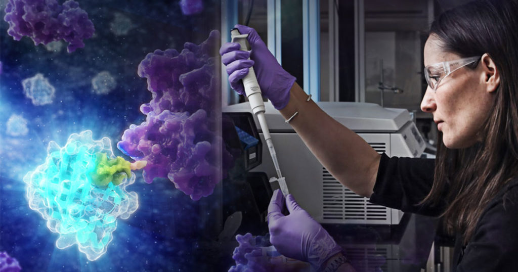
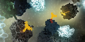
Woman at lab bench and artist 3D rendering of directed targeted protein degradation in a HiBiT cell line

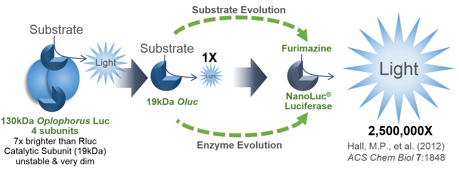
NanoLuc® Luciferase (NLuc) has made biology more accessible than ever (1). Further experimentation with NLuc led to creation of a protein complementation system (2) and the discovery of the HiBiT bioluminescent peptide. HiBiT combines spontaneously with the engineered complementary subunit LgBiT to yield an active luciferase called NanoBiT® Luciferase.

Continue reading “Directed Targeted Protein Degradation with Pre-Built HiBiT Cell Lines”

NanoBRET™ Assays to Analyze Virus:Host Protein:Protein Interactions in Detail

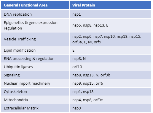
Recently, Gordon et al. published an atlas of protein:protein interactions of all proposed SARS-CoV-2 proteins expressed individually in HEK 293 cells (Table 1). The study tagged each of the viral proteins with an epitope tag and performed a pull-down of the expressed protein followed by trypsin digestion and mass spec analysis, a process referred to as affinity purification–mass spec analysis. The group identified 332 human proteins interacting with 27 SARS-CoV-2 proteins.

The interactions identified in the HEK 293 cells helped Appelberg et al. analyze interactions over time in SARS-CoV-2-infected Huh7 cells. Gordon et al. used the PPI data to identify FDA-approved drugs, drugs in clinical trials, and pre-clinical compounds that bound to the identified human proteins and labs in New York and Paris tested some of these drugs for antiviral effects.   

Table 1. The general functional area of human proteins identified to interact with individually expressed SARS-CoV-2 proteins as reported by Gordon, et al. (1). The SARS-CoV-2 proteins are classified as non-structural proteins (nsp#), structural proteins (E, M, and N) and accessory proteins (orf#).  
Continue reading “NanoBRET™ Assays to Analyze Virus:Host Protein:Protein Interactions in Detail”

Choices for Measuring Luciferase-Tagged Reporter Pseudotyped Viral Particles in Coronavirus Research

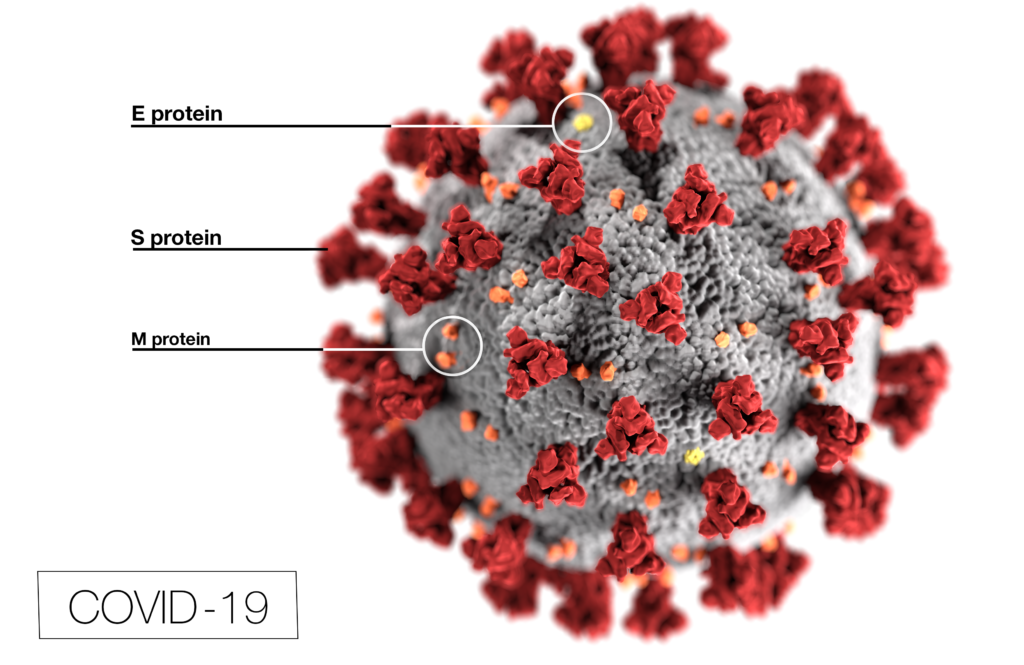
Coronavirus (CoV) researchers are working quickly to understand the entry of SARS-CoV-2 into cells. The Spike or S proteins on the surface of a CoV is trimer. The monomer is composed of an S1 and S2 domain. The division of S1 and S2 happens in the virus producing cell through a furin cleavage site between the two domains. The trimer binds to cell surface proteins. In the case of the SARS-CoV, the receptor is angiotensin converting enzyme 2. (ACE2). The MERS-CoV utilizes the cell-surface dipeptidyl peptidase IV protein. SARS-CoV-2 uses ACE2 as well. Internalized S protein goes though a second cleavage by a host cell protease, near the S1/S2 cleavage site called S2′, which leads to a drastic change in conformation thought to facilitate membrane fusion and entry of the virus into the cell (1).  

CDC / Alissa Eckert, MS; Dan Higgins, MAMS

Rather than work directly with the virus, researchers have chosen to make pseudotyped viral particles. Pseudotyped viral particles contain the envelope proteins of a well-known parent virus (e.g., vesicular stomatitis virus) with the native host cell binding protein (e.g., glycoprotein G) exchanged for the host cell binding protein (S protein) of the virus under investigation. The pseudotyped viral particle typically carries a reporter plasmid, most commonly firefly luciferase (FLuc), with the necessary genetic elements to be packaged in the particle. 

To create the pseudotyped viral particle, plasmids or RNA alone are transfected into cells and the pseudotyped viruses work their way through the endoplasmic reticulum and golgi to bud from the cells into the culture medium. The pseudoviruses are used to study the process of viral entry via the exchanged protein from the virus of interest. Entry is monitored through assay of the reporter. The reporter could be a luciferase or a fluorescent protein.   

Continue reading “Choices for Measuring Luciferase-Tagged Reporter Pseudotyped Viral Particles in Coronavirus Research”

Investigation of Remdesivir as a Possible Treatment for SARS-2-CoV (2019-nCoV)

Remdesivir (RDV or GS-5734) was used in the treatment of the first case of the SARS-CoV-2 (formerly 2019-nCoV ) in the United States (1). RDV is not an approved drug in any country but has been requested by a number of agencies worldwide to help combat the SARS-CoV-2 virus (2). RDV is an adenine nucleotide monophosphate analog demonstrated to inhibit Ebola virus replication (3). RDV is bioactivated to the triphosphate form within cells and acts as an alternative substrate for the replication-necessary RNA dependent RNA polymerase (RdRp). Incorporation of the analog results in early termination of the primer extension product resulting in the inhibition.

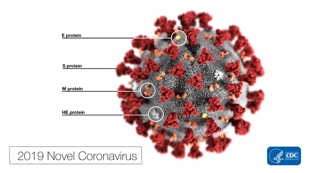
 Note the spikes that adorn the outer surface of the virus, which impart the look of a corona surrounding the virion, when viewed electron microscopically. In this view, the protein particles E, S, M, and HE, also located on the outer surface of the particle, have all been labeled as well. A novel coronavirus virus was identified as the cause of an outbreak of respiratory illness first detected in Wuhan, China in 2019.
This illustration, created at the Centers for Disease Control and Prevention (CDC), reveals ultrastructural morphology exhibited by coronaviruses. Photo Credit: Alissa Eckert, MS; Dan Higgins, MAM CDC

Why all the interest in RDV as a treatment for SARS-CoV-2 ? Much of the interest in RDV is due to a series of studies performed by collaborating groups at the University of North Carolina Chapel Hill (Ralph S. Baric’s lab) and Vanderbilit University Medical Center (Mark R. Denison’s lab) in collaboration with Gilead Sciences. 

Continue reading “Investigation of Remdesivir as a Possible Treatment for SARS-2-CoV (2019-nCoV)”

ViaFect™ Reagent: Building Assays in Difficult Cells

Transfection can sometimes seem more like an art than a science—the perfect transfection experiment being dependent on optimization of conditions, including cell density, transfection reagent and DNA:reagent ratio. No one reagent is perfect for every cell type, so there is the added challenge of optimizing performance in your cell line of choice—which may fall into the well-populated “difficult-to-transfect” category  that includes many primary cells.

Among transfection reagents, Lipofectamine® (Thermofisher), and FuGENE® (Promega) are popular and widely used choices. Viafect™ Transfection Reagent is newer and less well-known, but gaining popularity as a high-performance, low-toxicity reagent that performs well across a wide range of cell lines. In head-to-head comparisons with FuGENE and Lipofectamine, Viafect outperformed or equaled the others for expression of transfected reporter genes and resulting cell viability (see the data in this article).

The story of ViaFect begins with Promega Custom Assay Services (CAS), a group that uses Promega technologies to construct made-to-order assays, typically in a cell line. Many projects from the CAS group involve transfecting cells with expression vectors and reporter vectors. In some instances, customers contact CAS to have an assay constructed in a difficult cell line, after attempting and failing, or experiencing difficulty building the assay themselves.

CAS projects start with a proof-of-concept experiment using transient transfection before moving on to production of a clonal, stable cell line. For difficult cell lines, the CAS group previously turned to electroporation after exhausting lipid-based transfection options. Electroporation often worked, but success came with a price—cytotoxicity. The CAS group challenged R&D to find a better solution—better transfection with low toxicity for difficult-to-use cells. The result of that challenge is the ViaFect™ Transfection Reagent. Continue reading “ViaFect™ Reagent: Building Assays in Difficult Cells”

Shining Stars: Cool NanoLuc® Plasmid Constructs Available Through the Addgene Repository

Researchers having been sharing plasmids ever since there were plasmids to share. Back when I was in the lab, if you read a paper and saw an interesting construct you wished to use, you could either make it yourself or you could “clone by phone”.  One of my professors was excellent at phone cloning with labs around the world and had specific strategies and tactics for getting the plasmids he wanted. Addgene makes this so much easier to share your constructs from lab to lab. Promega supports the Addgene mission statement: Accelerate research and discovery by improving access to useful research materials and information.  Many of our technology platforms like HaloTag® Fusion Protein, codon-optimized Firefly luciferase genes (e.g., luc2), and NanoLuc® Luciferase are present in the repository. We encourage people to go to Addgene to get new innovative tools. Afterall, isn’t science better when we share?

I’d like to focus on some tools in the Addgene collection based on NanoLuc® Luciferase (NLuc).  The creation of NanoLuc® Luciferase and the optimal substrate furimazine is a good story (1).  From a deep sea shrimp to a compact powerhouse of bioluminescence, NLuc is 100-fold brighter than our more common luciferases like firefly (FLuc) and Renilla (RLuc) luciferase.  This is important not so much for how bright you can make a reaction but for how sensitive you can make a reaction.  NLuc requires 100-fold less protein to produce the same amount of light from a Fluc or RLuc reaction.  NLuc lets you work at physiological concentrations.  NLuc is bright enough to detect endogenous tagged genes generated through the CRISPR/Cas9 knock-in.  NLuc is very inviting for endogenous tagging as it is only 19kDa.  An example is the CRISPaint-NLuc construct (Plasmid #67178) for use in the system outlined in Schmid-Burgk, J.L. et al (2).

Two applications of NanoLuc® Technology have caught my attention through coupling the luciferase with fluorescent proteins to make better imaging reporters and biosensors. Continue reading “Shining Stars: Cool NanoLuc® Plasmid Constructs Available Through the Addgene Repository”

Making BRET the Bright Choice for In vivo Imaging: Use of NanoLuc® Luciferase with Fluorescent Protein Acceptors

13305818-cr-da-nanoluc-application_ligund

Live animal in vivo imaging is a common and useful tool for research, but current tools could be better. Two recent papers discuss adaptations of BRET technology combining the brightness of fluorescence with the low background of a bioluminescence reaction to create enhanced in vivo imaging capabilities.

The key is to image photons at wavelengths above 600nm, as lower wavelengths are absorbed by heme-containing proteins (Chu, J., et al., 2016 ). Fluorescent protein use in vivo is limited because the proteins must be excited by an external light source, which generates autofluorescence and has limited penetration due to absorption by tissues. Bioluminescence imaging continues to be a solution, especially firefly luciferase (612nm emission at 37°C), but its use typically requires long image acquisition times. Other luciferases, like NanoLuc, Renilla, and Gaussia, etc. either do not produce enough light or the wavelengths are readily absorbed by tissues, limiting their use to near-surface imaging.

The two papers discussed here illustrate how researchers have combined NanoLuc® luciferase with a fluorescent protein to harness bioluminescent resonance energy transfer (BRET) for brighter in vivo imaging reporters.

Continue reading “Making BRET the Bright Choice for In vivo Imaging: Use of NanoLuc® Luciferase with Fluorescent Protein Acceptors”

Making the Switch from FRET to BRET: Applications of NanoLuc® Luciferase with Fluorescent Protein Acceptors for Sensing Cellular Events

A Bioluminescent Alternative

Fluorescence resonance energy transfer (FRET) probes or sensors are commonly used to measure cellular events. The probes typically have a matched pair of fluorescent proteins joined by a ligand-binding or responsive protein domain. Changes in the responsive domain are reflected in conformational changes that either bring the two fluorescent proteins together or drive them apart. The sensors are measured by hitting the most blue-shifted fluorescent protein with its excitation wavelength (donor). The resulting emission is transferred to the most red-shifted fluorescent protein in the pair, and the result is ultimately emission from the red-shifted protein (acceptor).

As pointed out by Aper, S.J.A. et al. below, FRET sensors face challenges of photobleaching, autofluorescence, and, in the case of exciting cyan-excitable donors, phototoxicity. Another challenge to using FRET sensors comes when employing optogenetic regulators to initiate the event you wish to monitor. Optogenetic regulators respond to specific wavelengths and initiate signaling. The challenge comes when the FRET donor excitation overlaps with the optogenetic initiation wavelengths. Researchers have sought to alleviate many of these challenges by exchanging the fluorescent donor for a bioluminescent donor, making bioluminescence resonance energy transfer (BRET) probes. In the three papers described below, the authors chose NanoLuc® Luciferase as the BRET donor due to its extremely bright signal.

Continue reading “Making the Switch from FRET to BRET: Applications of NanoLuc® Luciferase with Fluorescent Protein Acceptors for Sensing Cellular Events”

Probing RGS:Gα Protein Interactions with NanoBiT Assays

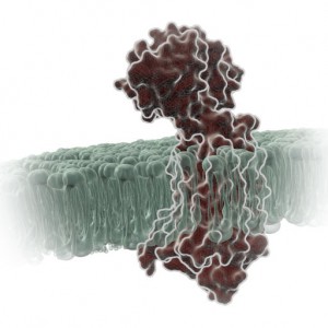
gpcr_in_membrane_on_white2When I was a post-doc at UT Southwestern, I was fortunate to interact with two Nobel prize winners, Johann Deisenhofer and Fred Gilman.  Johann once helped me move a -80°C freezer into his lab when we lost power in my building. I once replaced my boss at small faculty mixer with a guest speaker and had a drink with Fred Gilman and several other faculty members from around the university. Among the faculty, one professor had a cell phone on his belt, an odd sight in 1995. Fred Gilman asked him what it was and why he had it. It was so his lab could notify him of good results anytime of the day. Fred laughed and told him to get rid of it – if it’s good data, it will survive until morning.

I was reminded of this story when I read a recent paper by Bodle, C.R. et al (1) about the development of a NanoBiT® Complementation Assay (2) to measure interactions of Regulators of G Protein Signaling (RGS) with Gα proteins in cells. (Fred Gilman was the first to isolate a G protein and that led to him being a co-recipient of the Nobel Prize in 1994). The authors created over a dozen NanoBiT Gα:RGS domain pairs and found they could classify different RGS proteins by the speed of the interaction in a cellular context. The interactions were readily reversible with known inhibitors and were suitable for high-throughput screening due to Z’ factors exceeding 0.5. The study paves the way for future work to identify broad spectrum RGS domain:Gα inhibitors and even RGS domain-specific inhibitors. This is the second paper applying NanoBiT Technology to GPCR studies (3).

A Little Background…
A primary function of GPCRs is transmission of extracellular signals across the plasma membrane via coupling with intracellular heterotrimeric G proteins. Upon receptor stimulation, the Gα subunit dissociates from the βγ subunit, initiating the cascade of downstream second messenger pathways that alter transcription (4). The Gα subunits are actually slow GTPases that propagate signals when GTP is bound but shutdown and reassociate with the βγ subunit when GTP is cleaved to GDP. This activation process is known as the GTPase cycle. G proteins are extremely slow GTPases. Continue reading “Probing RGS:Gα Protein Interactions with NanoBiT Assays”

Bioassay for Cannabinoid Receptor Agonists Designed with NanoBiT™ Techology

Cannabinoids. What are they? Sometimes, Wikipedia can give a nice definition:

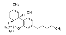
Tetrahydrocannabinol (THC), a partial agonist of the CB1 and CB2 cannabinoid receptors. Wikipedia Commons

A cannabinoid is one of a class of diverse chemical compounds that acts on cannabinoid receptors in cells that alter neurotransmitter release in the brain. Ligands for these receptor proteins include the endocannabinoids (produced naturally in the body by animals), the phytocannabinoids (found in Cannabis and some other plants), and synthetic cannabinoids (manufactured artificially).

Synthetic cannabinoids (SCs) were originally created for the scientific investigation of two cannabinoid receptors, CB1 and CB2, but have made their way to the streets as “safe” and “legal” alternatives to marijuana.

The problem is that these SCs engage the cannabinoid receptors more completely and with higher affinity than anything derived from marijuana. As a result, SCs can produce serious side effects that often require medical attention. In fact, you are 30 times more likely to seek emergency medical attention following the use of an SC than with natural cannabinoid sources like marijuana. Continue reading “Bioassay for Cannabinoid Receptor Agonists Designed with NanoBiT™ Techology”