Harnessing the Power of Massively Parallel Sequencing in Forensic Analysis

The rapid advancement of next-generation sequencing technology, also known as massively parallel sequencing (MPS), has revolutionized many areas of applied research. One such area, the analysis of mitochondrial DNA (mtDNA) in forensic applications, has traditionally used another methodโ€”Sanger sequencing followed by capillary electrophoresis (CE).

Although MPS can provide a wealth of information, its initial adoption in forensic workflows continues to be slow. However, the barriers to adoption of the technology have been lowered in recent years, as exemplified by the number of abstracts discussing the use of MPS presented at the 29th International Symposium for Human Identification (ISHI 29), held in September 2018. Compared to Sanger sequencing, MPS can provide more data on minute variations in the human genome, particularly for the analysis of mtDNA and single-nucleotide polymorphisms (SNPs). It is especially powerful for analyzing mixture samples or those where the DNA is highly degraded, such as in human remains. 

Continue reading “Harnessing the Power of Massively Parallel Sequencing in Forensic Analysis”

Control Samples: 3 Terrifying Tales for Scientists

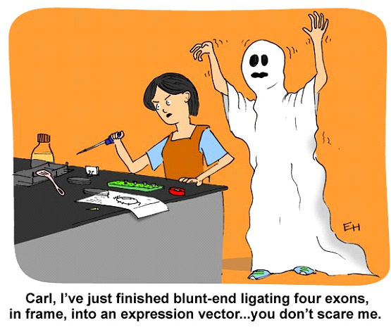
Lab science cartoon
Carl may not scare her…but did she remember the controls?

Warning: This blog contains stories about phantom serial killers, frankenfoods, mysteriously phosphorylated bands and unrequited ligations that may be disturbing to some people. Children or scientists prone to anxiety over irreproducible results should read this with their eyes shut.

I

Clouds hung low in the sky, and the late October wind howled between the buildings, rattling the window panes of the basement laboratory. The grackles cawed in desperate warning, their flocks changing the evening color palette from gray to black. I was as unsettled as the weather, watching my blot slosh back and forth.

Continue reading “Control Samples: 3 Terrifying Tales for Scientists”

Overcoming 5 Bottlenecks in Communicating Life Sciences Research

As a first-year grad student, I was so excited to start my thesis work. I brainstormed to make a list of experiments to try and then discussed them with one of the senior grad students in the lab. As I enthusiastically explained the goals of my experiments and what I was planning, he gave me a strange look. Puzzled, I asked for some feedback. He told me that, while these were good research ideas, almost all of them had been published. Hence, my first lesson learned from grad school: immerse yourself in the field by reading relevant papers and then plan some innovative experiments to move forward. Itโ€™s critical to have a deep knowledge of your field of studyโ€”not just to be a good grad student, but to see what is being done and then build on it, or take a totally different approach to innovate.

Reading papers is a big part of keeping up with the latest research. And attending conferences can give you a sense of current work before itโ€™s published. However, Iโ€™m sure that, at least once, youโ€™ve heard a cool talk at a conference and then quite a while later, havenโ€™t seen the corresponding paper (so that you can read about all the ins and outs of what they did!). Why would this be?  They may have been discussing the data early on in their project. Or perhaps they submitted a manuscript and the review/publishing process is taking a long time. Maybe the data were so surprising that they felt they needed to do a lot of follow-up work to support their conclusions. Or maybe their PI takes forever to write/comment on manuscripts. Etc.

The sooner that you can find out what is going on in a field, the sooner you can design smart, relevant experiments. What can be done to get cutting edge work out there to facilitate the progression of a field as a whole?

Bottlenecks in communicating research can occur at 5 different points in the process. Here are some tips to try to alleviate these delays.

Continue reading “Overcoming 5 Bottlenecks in Communicating Life Sciences Research”

An Ode to Packaging Engineers

Comic by Ed Himelblau.

In my second or third year as a graduate student, I had to ship some microfluidic masters to a collaborator in Kenya. The masters were extremely fragile and took me several days in a cleanroom gown to make. I was horrified at having to send them on a perilous journey overseas, and somewhat flabbergasted that they made it to Nairobi whole and well. And yet, every day thousands of delicate items zoom around the world and arrive at their destinations in one piece. How?

A couple months ago, I visited the lab where our packaging engineers (yes, that’s a thing) do their work. Here’s what I learned. Continue reading “An Ode to Packaging Engineers”

Hey, iGEMers! We’re talking to you!

The 2018 iGEM Giant Jamboree is upon us! This Wednesday, October 24th, thousands of you will flood into Boston, weighed down by posters and presentation materials, but energized by the excitement of a non-stop science-packed conference. Promega will also be attending, with a booth full of helpful giveaways and staff standing by to answer all your questions about science, Promega or future careers. As you make your final plans for the Jamboree, here are a few helpful tips for making the most of this incredible opportunity.

Continue reading “Hey, iGEMers! We’re talking to you!”

Nano, Nano: Tiny Lipid Particles with Big Therapeutic Potential

cell-transfection-viafect-luciferase-assay

Getting DNA or RNA into cells can be a tricky business, and a variety of transfection reagents have been developed over the years to make the process easier. Lipid-based reagents are especially popular because they combine efficient transfection with relatively low toxicity.

When it comes to transfection, it pays to think small. Human cells range in volume from 20โ€“40 ยตm3 (sperm cells) to as large as 4 million ยตm3 (mature egg cells, or oocytes). For several decades, transfection reagents have targeted this size range. However, breakthrough research involves leaving the โ€œmicroโ€ realm and entering a world that was once the domain only of science fiction: nanotechnology.

Continue reading “Nano, Nano: Tiny Lipid Particles with Big Therapeutic Potential”

A Tale of Two Toxins: the mechanisms of cell death in Clostridium difficile infections

When someone is admitted to a hospital for an illness, the hope is that medical care and treatment will help them them feel better. However, nosocomial infectionsโ€”infections acquired in a health-care settingโ€”are becoming more prevalent and are associated with an increased mortality rate worldwide. This is largely due to the misuse of antibiotics, allowing some bacteria to become resistant. Furthermore, when an antibiotic wipes out the โ€œgoodโ€ bacteria that comprise the human microbiome, it leaves a patient vulnerable to opportunistic infections that take advantage of disruptions to the gut microbiota.

One such bacteria, Clostridium difficile, is of growing concern world-wide since it is resistant to many different antibiotics. When a patient is treated with an antibiotic, C. difficile can thrive in the intestinal tract without other bacteria populating the gut. C. difficile infection is the leading cause of antibiotic-associated diarrhea. While symptoms can be mild, aggressive infection can lead to pseudomembranous colitisโ€”a severe inflammation of the colon which can be life-threatening.

C. difficile causes disease by releasing two large toxins, TcdA and TcdB. Understanding the role these toxins play in colonic disease is important for treatment strategies. However, most published research data only report the effects of the toxins independently. A 2016 study demonstrated a method of comparing the toxins side-by-side using the same time points and cell assays to investigate the role each toxin plays in the cell death that leads to disease of the colon. Continue reading “A Tale of Two Toxins: the mechanisms of cell death in Clostridium difficile infections”

MSI Analysis and the Application of Therapies Based on 2018 Nobel Immuno-Oncology Work

The 2018 Nobel Prize in Physiology and Medicine was awarded to James P. Allison of the United States and Tasuku Honjo of Japan for their work to identify pathways in the immune system that can be used to attack cancer cells (1). Although immunotherapy for cancer has been a goal for many decades, Dr. Allison and Dr. Honjo succeeded through their manipulation of โ€œcheckpoint inhibitorโ€ pathways to target cancer cells.

Immune checkpoint inhibitor drugs have been effective in cancers such as aggressive metastatic melanoma, some lung cancers, kidney, bladder and head and neck cancers. These therapies have succeeded in pushing many aggressive cancers below detectable limits, though these cases are notably not relapse-free or necessarily โ€œcuredโ€ (2,3).

One challenge in implementing immunotherapy in a cancer treatment regime is the need to understand the genetic makeup of the tumor. Certain tumors, with specific genetic features, are far more likely to respond to immune checkpoint therapy than others. For this reason, Microsatellite Instability (MSI) analysis has become an increasingly relevant tool in genetic and immuno-oncology research.

What is MSI Analysis?

Continue reading “MSI Analysis and the Application of Therapies Based on 2018 Nobel Immuno-Oncology Work”

Meeting the Needs of Scientists at All Levels

One of the best things about the BTC Institute is that we have programs for all levels of learners. It is as rewarding to introduce the concept of how bioluminescence is used by different organisms in the natural world to middle-school students as it is to have top-level scientists use reporter genes to track their knock-in genome edits.

We spend a lot of time working over our curricula to determine whether the content meets the learner where they are to allow our students to achieve their goals. We develop activities that let students who comes to us โ€”via field trips, high school courses, non-scientist sessions and graduate level programsโ€”to test ideas and evaluate strategies for problem solving as they learn techniques and concepts central to biotechnology. Continue reading “Meeting the Needs of Scientists at All Levels”

How Can a ADCC Reporter Bioassay Help You Save Time and Reduce Variability?

Fc receptor-mediated antibody-dependent cell-mediated cytotoxicity (ADCC) is an important mechanism of action (MOA) by which antibodies target diseased cells for elimination. Traditional methods for measuring ADCC require primary donor peripheral blood mononuclear cells (PBMCs) or purified natural killer (NK) cells that express Fc receptors on the cell surface. However, primary cultures of PBMCs and NK cells introduce variability, high background, and can be tedious to prepare. Using a commercially available ADCC reporter bioassay can overcome many of the limitations of these primary cell assays.

Our ADCC and ADCP Reporter Bioassays are biologically relevant, MOA-based assays that can be used to measure the potency and stability of antibodies and other biologics that specifically bind and activate Fcฮณ receptors. The ADCC Reporter Bioassays use an alternative readout from traditional primary cell-based assays: the FcฮณR and NFAT-mediated activation of luciferase activity in the effector cells. Primary cells are replaced with a Jurkat cell line stably expressing human FcฮณR variant and NFAT-induced luciferase.

The thaw-and-use cell format of the ADCC Reporter Bioassay saves time and labor of primary cell assays, while reducing variability. While a primary cell assay can take 1-2 weeks from culturing cells to results, ADCC reporter bioassay can be performed in 3โ€“24 hours. The bioassays include all of the required reagents and are easily amenable to high-throughput workflows, enabling you to have precisely the right throughput for your workflow needs.

Check out the full Promega portfolio of Fc effector reporter bioassays to discover the best tool for your research and read more about how these assays