The review “Kinase Inhibitors: the road ahead” was recently published in Nature Reviews Drug Discovery. In it, authors Fleur Ferguson and Nathanael Gray provide an up-to-date look at the “biological processes and disease areas that kinase-targeting small molecules are being developed against”. They note the related challenges and the strategies and technologies being used to efficiently generate highly-optimized kinase inhibitors.
This review describes the state of the art for kinase inhibitor therapeutics. To understand why kinase inhibitors are so important in the development of cancer (and other) therapeutics research, let’s start with the role of kinases in cellular physiology.
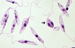
The Medicinal Chemistry Center (CQMED), headquartered at Campinas State University in Brazil, recently started a project in partnership with Promega to develop drugs that can be used against Leishmania. This genus of protozoans is the etiological agent of leishmaniasis, transmitted to humans by sandflies.
Microscopic image of Leishmania tropica. Credit: Brian E. Keas at Michigan State University.
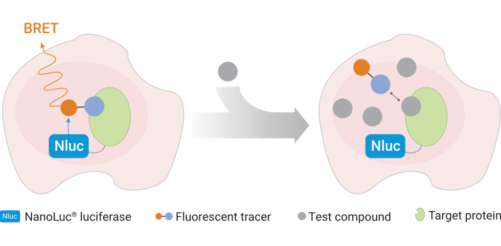
Leishmaniasis is classified as a neglected tropical disease that mainly affects poor communities. Symptoms include large skin sores and an enlarged spleen. The challenge in developing drugs to treat Leishmania is finding appropriate therapeutic targets. These targets are normally proteins whose inhibition leads to death of the parasite. In addition to pharmaceutical company Eurofarma, whose goal is to develop drugs for Leishmania, Promega was chosen to help solve this problem because of our NanoBRET™ Target Engagement (TE) assay*, a well-established technique for measuring protein interactions. In this assay, NanoLuc® luciferase is attached to the protein of interest, and a fluorescent NanoBRET™ tracer molecule is added to the cells. This produces a BRET signal. When a competing ligand is added, it will displace the tracer molecule, enabling quantification of the strength of the interaction compared to the tracer molecule..
A challenge that researchers will face will be ensuring that the NanoBRET™ tracer reaches the inside of the parasite cells; because Leishmania is an intracellular parasite, molecules need to cross the host cell membrane, the membrane of the vacuole containing the parasites, and the membrane of the parasite itself. Another challenge the slow reproduction of Leishmania within macrophages. On top of that is the fact that the parasite’s metabolism varies depending on its biological cycle, meaning that there could be long periods of time during which a drug’s therapeutic target is not expressed in the cell, during which time the drug would have no effect. The ideal target would be expressed at high levels throughout the cell cycle.
The project is being led by Rafael Couñago, a researcher at CQMED, and Promega scientists Matt Robers and Jean-Luc Vaillaud.
*An earlier version of this blog incorrectly said that these experiments are based on the NanoBRET™ assay using HaloTag® protein.
Have you read last week’s breaking story about the microbiome of the human placenta? Wait, stop, don’t run away to Google it! I’ll tell you all about it – this is a science blog, remember?
I’m asking because as I started reading about the topic in preparation for writing this blog post, I noticed two things. First, as a science writer who tries to stay well-connected with what’s going on in the world of biology research, it would have been nearly impossible for me to avoid this story. I get eight or nine daily digest emails from scientific publications every day, and I think over the course of last week, every single one came with a headline related to the placenta study. (Of course, I read them all. And the Nature study they were based on.)
Second, I noticed that each story I read had a slightly different angle on covering the research. As scientists, we like to believe that science is, well, just science. It’s factual. We pore over the data and reach a conclusion. If we aren’t sure of something, we search the journals. The story, if there is one, is about methods and controls, protocols and reagent quality. However, when information about that research is communicated broadly, outside of the journals, we can get a different impression based on how the author frames their article.
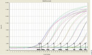
qPCR monitors amplification in real and allows you to measure starting material.
For those of us well versed in traditional, end-point PCR, wrapping our minds and methods around real-time or quantitative (qPCR) can be challenging. Here at Promega Connections, we are beginning a series of blogs designed to explain how qPCR works, things to consider when setting up and performing qPCR experiments, and what to look for in your results.
First, to get our bearings, let’s contrast traditional end-point PCR with qPCR.
End-Point PCR
qPCR
Visualizes by agarose gel the amplified product AFTER it is produced (the end-point)
Visualizes amplification as it happens (in real time) via a detection instrument
Does not precisely measure the starting DNA or RNA
Measures how many copies of DNA or RNA you started with (quantitative = qPCR)
Less expensive; no special instruments required
More expensive; requires special instrumentation
Basic molecular biology technique
Requires slightly more technical prowess
Quantitative PCR (qPCR) can be used to answer the same experimental questions as traditional end-point PCR: Detecting polymorphisms in DNA, amplifying low-abundance sequences for cloning or analysis, pathogen detection and others. However, the ability to observe amplification in real-time and detect the number of copies in the starting material can quantitate gene expression, measure DNA damage, and quantitate viral load in a sample and other applications.
Anytime that you are performing a reaction where something is copied over and over in an exponential fashion, contaminants are just as likely to be copied as the desired input. Quantitative PCR is subject to the same contamination concerns as end-point PCR, but those concerns are magnified because the technique is so sensitive. Avoiding contamination is paramount for generating qPCR results that you can trust.
Use aerosol-resistant pipette tips, and have designated pipettors and tips for pre- and post-amplification steps.
Wear gloves and change them frequently.
Have designated areas for pre- and post-amplification work.
Use reaction “master mixes” to minimize variability. A master mix is a ready-to-use mixture of your reaction components (excluding primers and sample) that you create for multiple reactions. Because you are pipetting larger volumes to make the reaction master mix, and all of your reactions are getting their components from the same master mix, you are reducing variability from reaction to reaction.
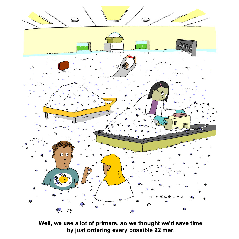
Dispense your primers into aliquots to minimize freeze-thaw cycles and the opportunity to introduce contaminants into a primer stock.
These are very basic tips that are common to both end-point and qPCR, but if you get these right, you are off to a good start no matter what your experimental goals are.
If you are looking for more information regarding qPCR, watch this supplementary video below.
Are you looking for more in-depth information about qPCR? Check out our qPCR and RT-qPCR Guide!
The first time I performed PCR was in 1992. I was finishing my Bachelors in Genetics and had an independent study project in a population genetics laboratory. My task was to try using a new technique, RAPD PCR, to distinguish clonal populations of the sea anemone, Metridium senile. These creatures can reproduce both sexually and asexually, which can make population genetics studies challenging. My professor was looking for a relatively simple method to identify individuals who were genetically identical (i.e., potential clones).
PCR was still in its infancy. No one in my lab had ever tried it before, and the department had one thermal cycler, which was located in a building across the street. We had a paper describing RAPD PCR for population work, so we ordered primers and Taq DNA polymerase and set about grinding up bits of frozen sea anemone to isolate the DNA. [The grinding process had to be done using a mortar and pestle seated in a bath of liquid nitrogen because the tissue had to remain frozen. If it thawed it became a disgusting mass of goo that was useless—but that is a topic for a different blog.] Since I had never done any of the procedures before, my professor and I assembled the first set of reactions together. When we ran our results on a gel, we had all sorts of bands—just what he was hoping to see. Unfortunately, we realized that we had added 10X more Taq DNA polymerase than we should have used. I repeated the amplification with the correct amount of Taq polymerase, and I saw nothing. Continue reading “Optimizing PCR: One Scientist’s Not So Fond Memories”
Ten years ago, I wrote about the distressing news of lack of genetic diversity in the wild Amur tiger population. International Tiger Day seemed like a good time to check in on what progress has been made to both sustain and establish wild tiger populations worldwide. In 2010, 13 tiger range countries (TRC) committed to a goal of doubling the world’s tiger population by 2022.
Amur Tiger.
That timeline was an ambitious goal, as highlighted by a report published in PLOS One in November of 2018 (1). The authors assessed the recovery potential of 18 sites identified under the World Wide Fund for Nature’s (WWF) Tigers alive initiative. The recovery system has several parts: A source site with higher density of tigers that the area around it and has a legal framework that does (or will) protect the tiger population; a recovery site that has a lower density of tigers than the surrounding regions, has the ability to support more tigers but is not as supported as a source site; and a support region that connects a source and recovery site. These different site types all require different levels of management, available resources and legal protections, but they need to be managed in a coordinated way.
Aside from what is needed to manage these recovery sites, there are also other things that need to exist to support recovery of tiger populations. Some of these include support from local populations and governments, as well as environmental requirements such as breeding habitats and prey populations. For 15 of the 18 sites it is the prey population that is the sticking point. Recovery of prey populations is a slow process. The authors concluded that there need to be a commitment to achieving a realistic recovery of tiger populations, even if we miss the 2022 goal.
The fate of the wild tiger is still tenuous. Only time will tell if the interventions that are being implemented can be realized in time.
Reference
Abishek, H. et al. (2018) Recovery planning towards doubling wild tigers Panthera tigris numbers: Detailing 18 recovery sites from across the range. PLOS One13. e0207114. published online
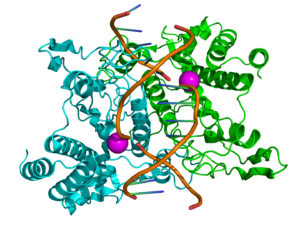
Ribbon diagram of EcoRI homodimer bound to doublestranded DNA
Restriction enzymes sometimes get a lot of flak. In the not-so-distant past, they were the workhorses of molecular biology. Restriction enzymes played a huge role in developing early DNA sequencing techniques. They chop DNA in a predictable manner, which makes cutting and pasting genes of interest manageable and relatively easy, enabling the development of genetic engineering and recombination technologies. These technologies are now moving beyond restriction enzymes toward more modern methods, with the most talked-about method being CRISPR /Cas9. As technology continues to advance at such a rapid pace, restriction analysis and other “ancient” technologies feel antiquated. But this is not necessarily the case.
G protein-coupled receptors (GPCRs) are a large family of receptors that traverse the cell membrane seven times. Functionally, GPCRs are extremely diverse, yet they contain highly conserved structural regions. GPCRs respond to a variety of signals, from small molecules to peptides and large proteins. Many GPCRs are involved in disease pathways and, not surprisingly, they present attractive targets for both small-molecule and biologic drugs.
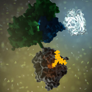
In response to a signal, GPCRs undergo a conformational change, triggering an interaction with a G protein—a specialized protein that binds GDP in its inactive state or GTP when activated. Typically, the GPCR exchanges the G protein-bound GDP molecule for a GTP molecule, causing the activated G protein to dissociate into two subunits that remain anchored to the cell membrane. These subunits relay the signal to various other proteins that interact with or produce second-messenger molecules. Activation of a single G protein can result, ultimately, in the generation of thousands of second messengers.
Given the complexity of GPCR signaling pathways and their importance to human health, a considerable amount of research has been devoted to GPCR interactions, both with specific ligands and G proteins.
Today’s blog brought to you by Julia Nepper, a Promega science writer guest blogging for the BioPharmaceutical Technology Center Institute (BTC Institute)!
“We all benefit from STEM role models. When students from underrepresented populations meet and learn about STEM professionals of color, they can see themselves as the scientists and engineers of the future. Fun, engaging science programming for children is also essential to light the spark for the next generation. A Celebration of Life, the partnership between the BTC Institute and the African American Ethnic Academy, two community nonprofits, has combined these 2 objectives for over twenty years.” according to Barbara Bielec, K-12 Program Director.
This year, the theme of the program is Sunsational!, with a number of activities related to the sun, solar energy, and STEM careers. As part of the program, students heard talks from several STEM professionals of color about their work. Mehrdad Arjmand, co-founder of solar energy company NovoMoto, was one of those speakers.
Dr. Arjmand was born and raised in Iran. His path to becoming a mechanical engineer began as a child, with him “destroying a lot of equipment” in his house. After completing his undergraduate education, he came to the States to pursue a PhD at the University of Wisconsin-Madison, where he met Aaron Olson, a student who was born in the Democratic Republic of Congo. These two discovered a shared passion for starting a business and helping their communities, which led directly to the founding of NovoMoto. The name derives from Portuguese for “new” (novo) and Lingala—a language spoken in Congo—for “fire” (moto). Continue reading “Empowering Communities with the Light of the Sun”
Restriction enzymes recognize short DNA sequences and cleave double-stranded DNA at specific sites within or adjacent to these sequences. These enzymes are the workhorse in many molecular biology applications such as cloning, RFLP, methylation-specific restriction enzyme analysis of DNA, etc. Restriction enzymes with enhanced capabilities can help you streamline and shorten these workflows and improve success of restriction enzyme digestion.
A subset of Promega restriction enzymes offer capabilities that include rapid digestion of DNA in 15 minutes or less, ability to completely digest DNA directly in the GoTaq® Green Master Mix, and Blue/White Cloning Qualification which allows for rapid, reliable detection of transformants.
To learn more about restriction enzymes and applications, check out Restriction Enzyme Resource on the web. The resource provides everything from information on restriction enzyme biology to practical information on how to set up and design a restriction enzyme digestion. This resource also contains useful online tools, including the Restriction Enzyme Tool, to help you use enzymes more effectively. It helps you choose the best reaction buffer for double digests, find the commercially available enzyme that cuts your sequence of interest, find compatible ends, and search for specific information on cut site, overhang isoschizomers and neoschizomers by enzyme name.
For added convenience, you can download the mobile app available for iOS devices and use the Restriction Enzyme Tool to plan your next digest.
For additional information regarding Restriction Enzyme Digest, reference the supplementary video below.
XWe use cookies and similar technologies to make our website work, run analytics, improve our website, and show you personalized content and advertising. Some of these cookies are essential for our website to work. For others, we won’t set them unless you accept them. To learn more about our approach to Privacy we invite you to Read More
By clicking “Accept All”, you consent to the use of ALL the cookies. However you may visit Cookie Settings to provide a controlled consent.
We use cookies and similar technologies to make our website work, run analytics, improve our website, and show you personalized content and advertising. Some of these cookies are essential for our website to work. For others, we won’t set them unless you accept them. To find out more about cookies and how to manage cookies, read our Cookie Policy.
If you are located in the EEA, the United Kingdom, or Switzerland, you can change your settings at any time by clicking Manage Cookie Consent in the footer of our website.
Necessary cookies are absolutely essential for the website to function properly. These cookies ensure basic functionalities and security features of the website, anonymously.
Cookie
Duration
Description
cookielawinfo-checbox-analytics
11 months
This cookie is set by GDPR Cookie Consent plugin. The cookie is used to store the user consent for the cookies in the category "Analytics".
cookielawinfo-checbox-functional
11 months
The cookie is set by GDPR cookie consent to record the user consent for the cookies in the category "Functional".
cookielawinfo-checbox-others
11 months
This cookie is set by GDPR Cookie Consent plugin. The cookie is used to store the user consent for the cookies in the category "Other.
cookielawinfo-checkbox-advertisement
1 year
The cookie is set by GDPR cookie consent to record the user consent for the cookies in the category "Advertisement".
cookielawinfo-checkbox-necessary
11 months
This cookie is set by GDPR Cookie Consent plugin. The cookies is used to store the user consent for the cookies in the category "Necessary".
cookielawinfo-checkbox-performance
11 months
This cookie is set by GDPR Cookie Consent plugin. The cookie is used to store the user consent for the cookies in the category "Performance".
gdpr_status
6 months 2 days
This cookie is set by the provider Media.net. This cookie is used to check the status whether the user has accepted the cookie consent box. It also helps in not showing the cookie consent box upon re-entry to the website.
lang
This cookie is used to store the language preferences of a user to serve up content in that stored language the next time user visit the website.
viewed_cookie_policy
11 months
The cookie is set by the GDPR Cookie Consent plugin and is used to store whether or not user has consented to the use of cookies. It does not store any personal data.
Analytical cookies are used to understand how visitors interact with the website. These cookies help provide information on metrics the number of visitors, bounce rate, traffic source, etc.
Cookie
Duration
Description
SC_ANALYTICS_GLOBAL_COOKIE
10 years
This cookie is associated with Sitecore content and personalization. This cookie is used to identify the repeat visit from a single user. Sitecore will send a persistent session cookie to the web client.
vuid
2 years
This domain of this cookie is owned by Vimeo. This cookie is used by vimeo to collect tracking information. It sets a unique ID to embed videos to the website.
WMF-Last-Access
1 month 18 hours 24 minutes
This cookie is used to calculate unique devices accessing the website.
_ga
2 years
This cookie is installed by Google Analytics. The cookie is used to calculate visitor, session, campaign data and keep track of site usage for the site's analytics report. The cookies store information anonymously and assign a randomly generated number to identify unique visitors.
_gid
1 day
This cookie is installed by Google Analytics. The cookie is used to store information of how visitors use a website and helps in creating an analytics report of how the website is doing. The data collected including the number visitors, the source where they have come from, and the pages visted in an anonymous form.
Advertisement cookies are used to provide visitors with relevant ads and marketing campaigns. These cookies track visitors across websites and collect information to provide customized ads.
Cookie
Duration
Description
IDE
1 year 24 days
Used by Google DoubleClick and stores information about how the user uses the website and any other advertisement before visiting the website. This is used to present users with ads that are relevant to them according to the user profile.
test_cookie
15 minutes
This cookie is set by doubleclick.net. The purpose of the cookie is to determine if the user's browser supports cookies.
VISITOR_INFO1_LIVE
5 months 27 days
This cookie is set by Youtube. Used to track the information of the embedded YouTube videos on a website.
Performance cookies are used to understand and analyze the key performance indexes of the website which helps in delivering a better user experience for the visitors.
Cookie
Duration
Description
YSC
session
This cookies is set by Youtube and is used to track the views of embedded videos.
_gat_UA-62336821-1
1 minute
This is a pattern type cookie set by Google Analytics, where the pattern element on the name contains the unique identity number of the account or website it relates to. It appears to be a variation of the _gat cookie which is used to limit the amount of data recorded by Google on high traffic volume websites.


 GPCRs
GPCRs