Mass Spec for Glycosylation Analysis of SARS-CoV-2 Proteins Implicated in Host-Cell Entry

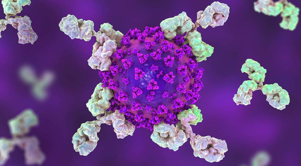
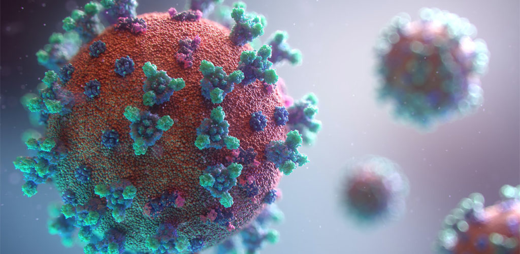
The spike protein of the SARS-CoV-2 virus is a very commonly researched target in COVID-19 vaccine and therapeutic studies because it is an integral part of host cell entry through interactions between the S1 subunit of the spike protein with the ACE2 protein on the target cell surface. Viral proteins important in host cell entry are typically highly glycosylated. Looking at the sequence of the SARS-CoV-2 virus, researchers predict that the spike protein is highly glycosylated. In a recent study, researchers conducted a glycosylation analysis of SARS-CoV-2 proteins using mass spec analysis to determine the N-glycosylation profile of the subunits that make up the spike protein.

3d model of coronavirus covid-19 showing the spike protein. A recent study performed a glycosylation analysis of SARS-CoV-2 protein.

Glycans assist in protein folding and help the virus avoid immune recognition by the host. Glycosylation can also have an impact on the antigenicity of the virus, as well as potential effects on vaccine safety and efficacy. Mass spectrometry is widely used for viral characterization studies of influenza viruses. Specifically, mass spec has been used to study influenza protein glycosylation, antigen quantification, and determination of vaccine potency.

Continue reading “Mass Spec for Glycosylation Analysis of SARS-CoV-2 Proteins Implicated in Host-Cell Entry”

Proteomics from a Different Point of View: Introducing ProAlanase, the Newest Mass-Spec Grade Protease from Promega

Sometimes, when using trypsin to study a protein sequence or protein modifications, sequence coverage just isn’t quite as complete as you’d like. Looking for a protease with novel cleavage specificity or a protease that functions well in a low pH environment? Promega has a protease for that.

ProAlanase is a new site-specific endoprotease that preferentially cleaves proteins on the C-terminal side of proline and alanine amino acids. The unique cleavage specificity of ProAlanase (also known as An-PEP or EndoPro; 1–3) can help to uncover parts of the proteome not previously accessible with proteases typically used in proteomic studies.

Continue reading “Proteomics from a Different Point of View: Introducing ProAlanase, the Newest Mass-Spec Grade Protease from Promega”

Go fISH! Using in situ Hybridization to Search for Expression of a SARS-CoV-2 Viral Entry Protein

Loss of smell (olfaction) is a commonly reported symptom of COVID-19 infection. Recently, Bilinska, et al. set out to better understand the underlying mechanisms for loss of smell resulting from SARS-CoV-2 infection. In their research, they used in situ hybridization to investigate the expression of TMPRSS2, a SARS-CoV-2 viral entry protein in olfactory epithelium tissues of mice.

Continue reading “Go fISH! Using in situ Hybridization to Search for Expression of a SARS-CoV-2 Viral Entry Protein”

In Vitro Transcription and the Use of Modified Nucleotides

In vitro transcription
RNA polymerase unwinds DNA strands for transcription.

Transcription is the production of RNA from a DNA sequence. It’s a necessary life process in most cells. Transcription performed in vitro is also a valuable technique for research applications—from gene expression studies to the development of RNA virus vaccines.

During transcription, the DNA sequence is read by RNA polymerase to produce a complimentary, antiparallel RNA strand. This RNA strand is called a primary transcript, often referred to as an RNA transcript. In vitro transcription is a convenient method for generating RNA in a controlled environment outside of a cell.

In vitro transcription offers flexibility when choosing a DNA template, with a few requirements. The template must be purified, linear, and include a double stranded promoter region. Acceptable template types are plasmids or cloning vectors, PCR products, synthetic oligos (oligonucleotides), and cDNA (complimentary DNA). 

In vitro transcription is used for production of large amounts of RNA transcripts for use in many applications including gene expression studies, RNA interference studies (RNAi), generation of guide RNA (gRNA) for use in CRISPR, creation of RNA standards for quantification of results in reverse-transcription quantitative PCR (RT-qPCR), studies of RNA structure and function, labeling of RNA probes for blotting and hybridization or for RNA:protein interaction studies, and preparation of specific cDNA libraries, just to name a few!

In vitro transcription can also be applied in general virology to study the effects of an RNA virus on a cell or an organism, and in development and production of RNA therapeutics and RNA virus vaccines. The large quantity of viral RNA produced through in vitro transcription can be used as inoculation material for viral infection studies. Viral mRNA transcripts, typically coding for a disease-specific antigen, can be quickly created through in vitro transcription, and used in the production of vaccines and therapeutics.

Continue reading “In Vitro Transcription and the Use of Modified Nucleotides”

Cloning with pGEM®-T Vectors: Ligation

T-Vector Cloning

One of the easiest methods for cloning blunt-ended DNA fragments including PCR products is T-vector cloning, such as with pGEM®-T or pGEM®-T Easy Vector Systems. This method takes advantage of the “A” overhang added by a PCR enzyme like Taq DNA Polymerase. T vectors are linearized plasmids that have been treated to add 3′ T overhangs to match the A overhangs of the insert. The insert is directly ligated to the T-tailed plasmid vector with T4 DNA ligase. The insert can then be easily transferred from the T vector to other plasmids using the restriction sites present in the multiple cloning region of the T vector.

Proofreading polymerases like Pfu do not add “A” overhangs so PCR products generated with these polymerases are blunt-ended. In a previous blog, we discussed a simple method for adding an A-tail to any blunt-ended DNA fragment to enable T-vector cloning. Below, we think about the next step: Ligation.

Continue reading “Cloning with pGEM®-T Vectors: Ligation”

Cloning Blunt-Ended DNA Fragments is Hard: pGEM®-T Vectors Can Make It Easier.

PCR amplification with a proofreading polymerase, like Pfu DNA polymerase, will leave you with a blunt end. However, another thermostable DNA polymerase, like Taq DNA Polymerase, adds a single nucleotide base to the 3’ end of the DNA fragment, usually an adenine, creating an “A” overhang. This “A” overhang can create difficulties when cloning the fragment is your end goal. You might consider creating a blunt end with Klenow or adding restriction sites to the ends of your PCR fragment by designing them in your primers. But why go through all those extra steps, when that “A” overhang allows efficient cloning of these fragments into T-Vectors such as the pGEM®-T Vectors? Fewer steps? Who can argue with that?

Continue reading “Cloning Blunt-Ended DNA Fragments is Hard: pGEM®-T Vectors Can Make It Easier.”

Targeted Gene Modification in Prairie Voles Using CRISPR and pGEM®-T Easy Vectors

As the number of children diagnosed with autism spectrum disorder (ASD) continues to rise, the search for a cause continues. Scientists have been studying genetically modified oxytocin receptors, which have shown promise as a target for studying ASD-related behaviors. One of the obstacles to designing robust scientific experiments for investigating potential ASD causes or treatments is the lack of a truly appropriate model organism for social behaviors in humans (1). Sure, there are the traditional lab rats and lab mice that demonstrate a certain level of social behaviors. However, there has been a loss of natural social behaviors in common lab mice strains because of the reduction in genetic complexity from inbreeding and adaptation to captivity (2). These animals cannot fully represent the depth of human social behaviors, including the ability of humans to form lasting social bonds (1).

Enter: The prairie vole (Microtus ochrogaster).

Continue reading “Targeted Gene Modification in Prairie Voles Using CRISPR and pGEM®-T Easy Vectors”

T-Vector Cloning: Questions, Answers and Tips

Blue/White colony screening helps you pick only the colonies that have your insert.

Q: Can PCR products generated with GoTaq DNA Polymerase be used to for T- vector cloning?

A: Yes. GoTaq® DNA Polymerase is a robust formulation of unmodified Taq Polymerase. GoTaq® DNA Polymerase lacks 3’ →5’ exonuclease activity and displays terminal transferase activity that adds a 3′ deoxyadenosine (dA) to product ends. As a result, PCR products amplified using GoTaq® DNA Polymerases (including the GoTaq® Flexi and GoTaq® G2 polymerases) will contain A-overhangs which makes them suitable for T-vector cloning with the pGEM®-T (Cat.# A3600), pGEM®-T Easy (Cat.# A1360) and pTARGET™ (Cat.# A1410) Vectors.

Continue reading “T-Vector Cloning: Questions, Answers and Tips”

A Diamond in the Rough: New Applications of Diamond Nucleic Acid Dye

Diamond™ Nucleic Acid Dye (Cat# H1181) is a safe, inexpensive and sensitive fluorescent dye option that binds to single-stranded and double-stranded DNA and RNA. Diamond™ Dye typically is used for staining electrophoresis gels to visualize nucleic acids in a similar to its carcinogenic counterpart, ethidium bromide. However Diamond™ Dye has several advantages: gels stained with Diamond™ Dye can be visualized using either UV or blue-light transilluminators. Also, a wash step after staining is not necessary when using Diamond™ Dye, unlike what is typically recommended for ethidium bromide.

Besides staining electrophoresis gels, there are other applications for this diamond in the rough. Highlighted below are two fascinating uses of this multifaceted tool: touch DNA localization and qPCR detection.

Continue reading “A Diamond in the Rough: New Applications of Diamond Nucleic Acid Dye”

Think Restriction Enzymes are so last decade? Not so fast!

Ribbon diagram of EcoRI homodimer bound to doublestranded DNA

Restriction enzymes sometimes get a lot of flak. In the not-so-distant past, they were the workhorses of molecular biology. Restriction enzymes played a huge role in developing early DNA sequencing techniques. They chop DNA in a predictable manner, which makes cutting and pasting genes of interest manageable and relatively easy, enabling the development of  genetic engineering and recombination technologies. These technologies are now moving beyond restriction enzymes toward more modern methods, with the most talked-about method being CRISPR /Cas9. As technology continues to advance at such a rapid pace, restriction analysis  and other “ancient” technologies feel antiquated. But this is not necessarily the case.

Continue reading “Think Restriction Enzymes are so last decade? Not so fast!”