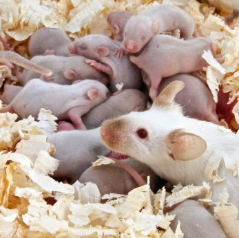
 Imagine that you are sitting in your room when you smell cherries and you are suddenly, inexplicably afraid. Although odors can elicit strong emotional responses, you have no bad memories of cherries. What you don’t know is that your father did, and you have inherited his fear. Sound far fetched? Maybe not. A paper published in Nature Neuroscience found just such an inherited association in mice (1). Continue reading “Inheriting Fear: Mice Haunted by Parent’s Fears”
ADCC Reporter Bioassay Makes Top 10 Innovator List
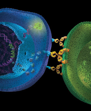
For the second year running a Promega technology has made The Scientist Magazine’s list of Top 10 Innovations. In 2012 NanoLuc® luciferase technology was in the spotlight; in 2013 the ADCC Reporter Bioassay took center stage.
Antibody-dependent cell-mediated cytotoxicity (ADCC) is the main mechanism of action (MOA) of antibodies through which virus-infected or other diseased cells are targeted for destruction by components of the cell-mediated immune system. These assays are often used to assess the effectiveness of monoclonal antibody therapies during the manufacture and development of biologic drugs. The bioluminescent assays use an alternative readout at an earlier point in ADCC MOA pathway for the quantification of Fc effector function of antibody-based molecules: the activation of gene transcription through the NFAT (nuclear factor of activated T-cells) pathway in the effector cell.
The bioassay uses engineered Jurkat cells stably expressing the FcγRIIIa receptor, V158 (high affinity) variant, and an NFAT response element driving expression of firefly luciferase. The assay is MOA-based and features frozen, thaw-and-use effector cells and optimized reagents and protocol to perform a reporter-based ADCC bioassay in a single day. The bioassay correlates with classic cytotoxic ADCC assays and is a suitable replacement for these cumbersome and highly variable assays.
The novel bioassay is linear, accurate, precise and stability indicating. Moreover, the bioassay shows good linear correlation between levels of glycosylation or fucosylation and ADCC activity. All of these features indicate the assay is suitable for use across biologic drug development programs.
NanoLuc® Luciferase: A Good Thing for Small Packages
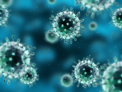
A paper published on October 2 in the Journal of Virology describes an exciting development in the world of influenza research—the construction of a luciferase reporter virus that does not affect virulence and can be used to track development and spread of infection in mice.
Insertion of luciferase reporter genes into viruses has been accomplished before for several viruses, but has not been successful for influenza. Construction of influenza reporter viruses is complicated because the viral genome is small and all the viral genes are critical for infection. Therefore, replacement of an existing gene with a reporter gene or insertion of additional reporter sequences without affecting the virus’s ability to replicate and cause infection has proven difficult. To be successful, a reporter gene needs to be small enough to insert into the viral genome without eliminating any other vital functionality.
Continue reading “NanoLuc® Luciferase: A Good Thing for Small Packages”Piecing the Puzzle Together: Using Multiple Assays to Better Understand What Is Happening with Your Cells
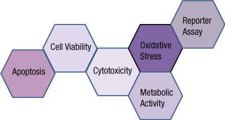
You often need several pieces of information to really understand what is happening within a cell or population of cells. If your cells are not proliferating, are they dying? Or, are you seeing cytostasis? If they are dying, what is the mechanism? Is it apoptosis or necrosis? If you are seeing apoptosis, what is the pathway: intrinsic or extrinsic?
If you are measuring expression of a reporter gene and you see a decrease in expression, is that decrease due to transfection inefficiencies, cytotoxicity, or true down regulation of your reporter gene?
To investigate these multiple parameters, you can run assays in parallel, but that requires more sample, and sample isn’t always abundant.
Multiplexing assays allows you to obtain information about multiple parameters or events (e.g., reporter gene expression and cell viability; caspase-3 activity and cell viability) from a single sample. Multiplexing saves sample, saves time and gives you a more complete picture of the biology that is happening with your experimental sample.
Multiplexing assay reagents to measure biomarkers in the same sample has often been considered an application only accomplished with antibodies or dyes and sophisticated detection instrumentation. However, Promega has developed microwell plate based assays for cells in culture that allow multiplexed detection of biomarkers in the same sample well using standard multimode multiwell plate readers. Continue reading “Piecing the Puzzle Together: Using Multiple Assays to Better Understand What Is Happening with Your Cells”
Remembering Frederick Sanger and Sanger Sequencing
It is with sadness that we recognize the passing of Dr. Frederick Sanger. Sanger is known to molecular biologists and biochemists worldwide for his DNA sequencing technique, which won for him the 1980 Nobel prize in Chemistry.
Also noteworthy, Sanger’s laboratory accomplished the first complete genome sequence, that of a viral DNA genome more than 5,000 base pairs in length.
The 1980 prize was Sanger’s second Nobel award, his first awarded in 1958 for determining the chemical structure of proteins. In this work, Sanger elucidated not only the amino acids that comprised insulin but also the order in which the amino acids occurred.
About Sanger Sequencing
Sanger DNA sequencing is also known as the chain-termination method of sequencing. The Sanger technique uses dideoxynucleotides or ddNTPs in addition to typical deoxynucleotides (dNTPs) in the reaction. ddNTPs result in termination of the DNA strand because ddNTPs lack the 3’-OH group required for phosphodiester bond formation between nucleotides. Without this bond, the chain of nucleotides being formed is terminated.
Sanger sequencing requires a single-stranded DNA, a DNA primer (either radiolabeled or with a fluorescent tag), DNA polymerase, dNTPs and ddNTPs. Four reactions are set up, one for each nucleotide, G, A, T and C. In each reaction all four dNTPs are included, but only one ddNTP (ddATP, ddCTP, ddGTP or ddTTP) is added. The sequencing reactions are performed and the products denatured and separated by size using polyacrylamide gel electrophoresis.
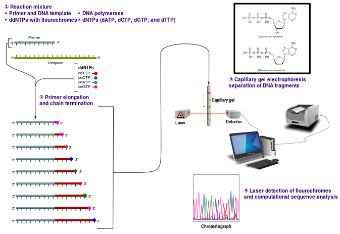
This reaction mix results in various lengths of fragments representing, for instance, the location of each A nucleotide in the sequence, because while there is more dATP than ddATP in the reaction, there is enough ddATP that each ATP ultimately instead is replaced with a ddATP, resulting in chain termination. Separation by gel electrophoresis reveals the size of these ddATP-containing fragments, and thus the locations of all A nucleotide in the sequence. Similar information is provided for GTP, CTP and TTP.
The Maxam and Gilbert DNA sequencing method had the advantage at the time of being used with double-stranded DNA. However, this method required DNA strand separation or fractionation of the restriction enzyme fragments, resulting in a somewhat more time-consuming technique, compared to the 1977 method published by Sanger et al.
Dr. Sanger was born in Gloucestershire, U.K. in 1918, the son of a physician. Though he initially planned to follow his father into medicine, biochemistry became his life-long passion and area of research endeavor. Sanger retired at age 65, to spend more time at hobbies of gardening and boating.
References
Sanger, F. , Nicklen, S. and Coulson, A.R. (1977) DNA sequencing with chain-terminating inhibitors. Proc. Natl. Acad. Sci. USA 74, 5463-7.
Maxam, A.M. and Gilbert, W. (1977) A New Method for Sequencing DNA. Proc. Natl. Acad. Sci. USA
There is something special about seeing the original Sanger publication from 1977, available here as a scan.
ProTEV Protease Compound Compatibility Analysis
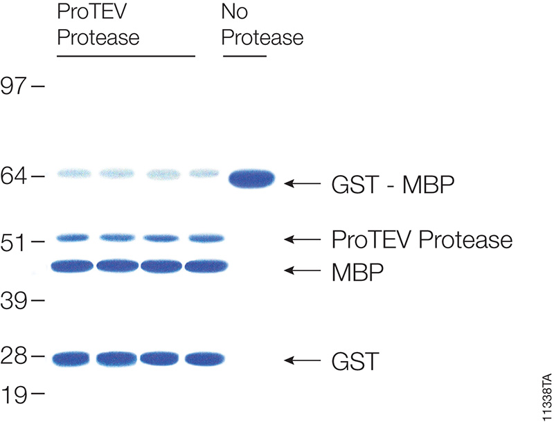
Many proteins are expressed as fusion partners with affinity tags, such as the HaloTag® fusion, glutathione-S-transferase (GST) or maltose binding protein (MBP), to selectively bind the proteins using affinity purification resins. While such resins yield high-purity protein quickly, the large affinity tags are undesirable for some downstream applications. Most expression vectors are designed with a specific protein cleavage site between the two fusion partners to remove the affinity tag after purification. ProTEV Protease recognizes a rare amino acid sequence, EXXYXQ, where X is any amino acid, and cleaves after the glutamine residue.
ProTEV Plus functions over a broad pH and temperature range. In a recent study the enzymatic activity of ProTEV Plus in the presence of various compounds (Table 1) commonly found in protein purification protocols were evaluated.
Continue reading “ProTEV Protease Compound Compatibility Analysis”Update: Is It the Blood of Louis XVI?

In an earlier blog entry, I wrote about the ill-fated Louis XVI, the French king who was famously beheaded along with his wife, Marie-Antoinette, during the French Revolution in 1793. Witnesses to the execution dipped handkerchiefs in the king’s blood and kept them as souvenirs of the common people’s rebellion. In 2010, scientists published the presumptive DNA profile of the king, obtained from one of these bloody handkerchiefs (1). Shortly after this profile was published, doubters surfaced, arguing that scientists could not say with certainty that the blood was that of Louis XVI. Clearly, more work was needed to identify the source of the blood. Recently, additional work was published (2,3). The most recent data (3) were presented at the 2013 International Symposium on Human Identification; these newest data cast doubt on the identification of the remains of not one king, but two.
Continue reading “Update: Is It the Blood of Louis XVI?”Methods for Quantitating Your Nucleic Acid Sample
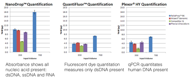
For most molecular biology applications, knowing the amount of nucleic acid present in your purified sample is important. However, one quantitation method might serve better than another, depending on your situation, or you may need to weigh the benefits of a second method to assess the information from the first. Our webinar “To NanoDrop® or Not to NanoDrop®: Choosing the Most Appropriate Method for Nucleic Acid Quantitation” given by Doug Wieczorek, one of our Applications Scientists, discussed three methods for quantitating nucleic acid and outlined their strengths and weaknesses.
Continue reading “Methods for Quantitating Your Nucleic Acid Sample”Articles, Blog Posts, Tweets, New Products and One Page to See Them All
If you are like me, there are just not enough hours in the day. The list of things that I need to get done regularly out distances the time I have to do them in. Keeping up with my favorite blogs, staying in tune with things on twitter and staying on top of new product and features often fall by the wayside because it takes so much time to go to all those pages and find the content I want.
Recently we updated the Promega PubHub page on our website with the hopes that it will help you use the time you spend visiting the PubHub page more efficiently. In addition to latest technical articles from Promega, useful lab facts and the ever-popular cartoons, we now offer a live feed of our Promega Connections Blog posts, tweets from @Promega and a list of new products.

We know that your time is valuable, and if you are interested in the articles and more from Promega, there is now one page to see it all.
Top Ten Tips for Successful PCR
We decided to revisit a popular blog from our Promega Connections past for those of you in the amplification world. Enjoy:
-
- Modify reaction buffer composition to adjust pH and salt concentration.
- Titrate the amount of DNA polymerase.
- Add PCR enhancers such as BSA, betaine, DMSO, nonionic detergents, formamide or (NH4)2SO4.
- Switch to hot-start PCR.
- Optimize cycle number and cycling parameters, including denaturation and extension times.
- Choose PCR primer sequences wisely.
- Determine optimal DNA template quantity.
- Clean up your DNA template to remove PCR inhibitors.
- Determine the optimal annealing temperature of your PCR primer pair.
[Drum roll please]…and the most important thing you can do to improve your PCR results is:
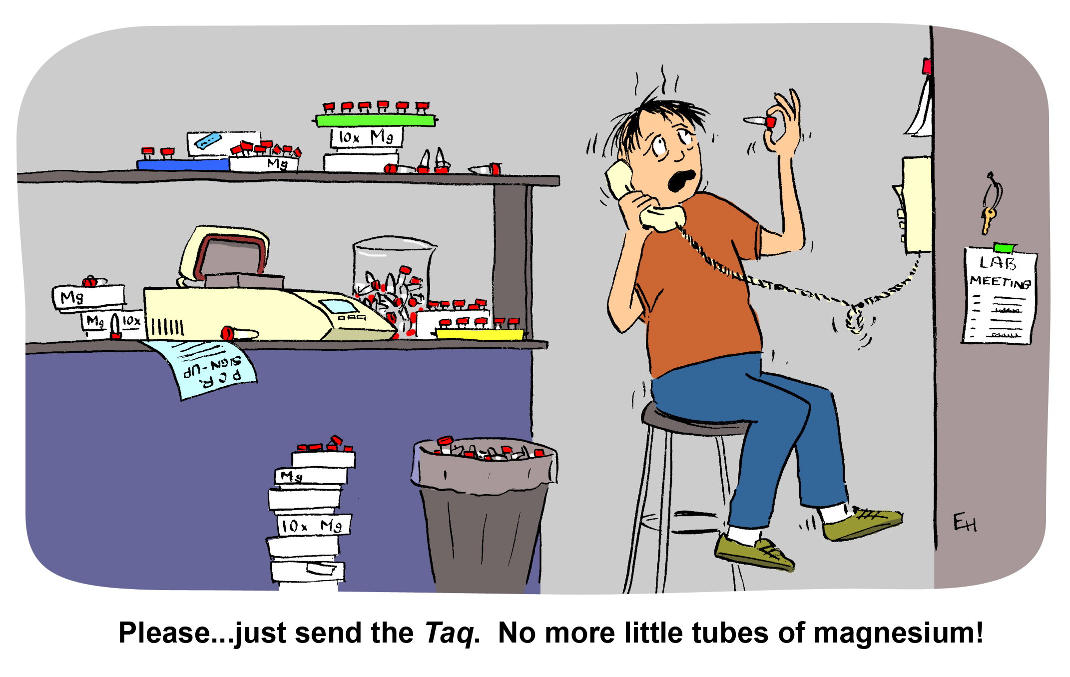
- Titrate the magnesium concentration.