Can pre-digestion with trypsin improve mass spec analysis?
The trypsin protease cleaves proteins on the carboxyterminus of Arginine (Arg) and Lysine (Lys). This cleavage reaction leaves a positive charge on the C-terminus of the resulting peptide, which enhances mass spectrometry analysis (1,2). Because of this advantage, trypsin has become the most commonly used protease for mass spectrometry analysis. Other proteases which cleave differently from trypsin, yielding complementary data are also used in mass spec analysis: these include Asp-N and Glu-C , which cleave acidic residues, and chymotrypsin which cleaves at aromatic residues. The broad spectrum protease, proteinase K is also used for some proteomic analyses. In a recent study, Dau and colleagues investigated whether sequential digestion with trypsin followed by the complementary proteases could improve protein digests for mass spectrometry analysis.
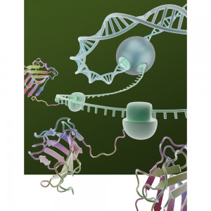
Cell-free gene expression systems are a staple tool for the researcher seeking to understand the regulation of transcription and translation. Many factors can affect the efficiency of cell-free gene expression including vector sequence, reaction components and the template DNA concentration. One factor that has not been extensively studied is how DNA template length influences gene expression.
A tiny worm called Onchocerca lupi can make life uncomfortable for both humans and their best friends. This thread-like nematode is found in the eyes or under the skin of infected animals. Historically, diagnosis required skin biopsy or surgical removal of ocular tissue, but a recent study demonstrates a new non-invasive diagnostic tool for infection by Onchocerca lupi in dogs.
Mass spectrometry depends on the successful digestion of proteins using proteases. Many commercially available proteomic-grade trypsins contain natural contaminants that produce non-specific cleavages. Trypsin Platinum, a new protease from Promega provides maximum specificity, giving you cleaner and more conclusive data from mass spec.
Trypsin is typically extracted from bovine or porcine pancreas. In addition to trypsin, both of these sources also contain chymotrypsin. To suppress chymotryptic activity, trypsin is treated with tosyl phenylalanyl chloromethyl ketone, or TPCK, to irreversibly inhibit the chymotrypsin. However, trace amounts of chymotrypsin appear to escape this inhibition and produce non-specific cleavages, as seen in the figure below.
A new article in Nature Scientific Reports answers open questions about TOPBP1, a protein involved in repairing DNA double-strand breaks (DSBs). The study used cell-free protein expression and a unique DSB system to identify domains that were important for activation of a protein kinase.
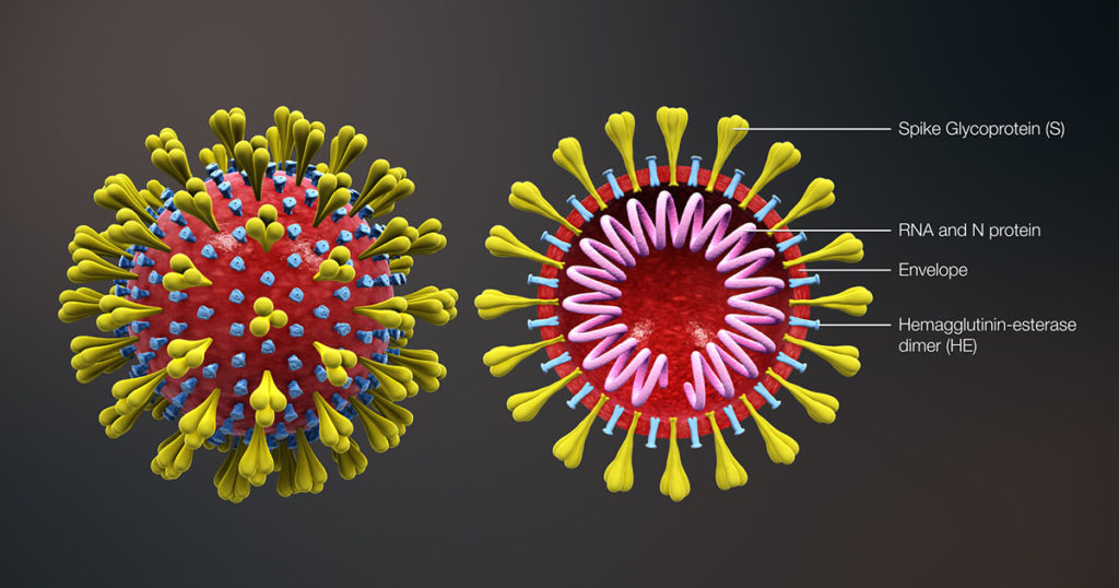
The SARS-CoV-2 nucleocapsid protein accounts for the largest proportion of viral structural proteins and is the most abundant protein in infected cells. Nucleocapsid proteins have the job of “packaging” the viral nucleic acid (in this case, RNA). Viral nucleocapsid proteins can also enter the host nucleus and interact with a variety of host proteins to interfere with critical processes of the host cell, including protein degradation. Here we review a study that used an in vitro protein degradation assay to investigate the interaction of the SARS-CoV-2 nucleocapsid protein and the proteasome activator PA28γ.
In SARS-CoV-2 infections, the nucleocapsid protein is critical for infection, replication and packaging. The SARS-CoV-2 nucleocapsid protein is not only localized in the cytosol of the host cell but also is translocated into the nucleus. There, it interacts with various cellular proteins that modulate cellular functions, such as the degradation of unneeded or damaged proteins by proteolysis. Researchers have proposed that the protein degradation system plays an important part in coronavirus infection (1).
In older people, low muscle mass is strongly associated with reduced functional capacity and an increased risk of disability. Myostatin is a negative regulator of muscle growth and has become an important target for pharmaceutical companies designing therapeutics to address age-associated muscle loss.
Anti-myostatin drugs increase muscle size and strength in preclinical studies. Fortetropin is a proteo-lipid complex made from fertilized egg yolk and shows anti-myostatin activity. When Fortetropin is provided as a supplement, lowered circulating myostatin levels are observed both in rodents and in young men. Fortetropin in combination with resistance exercise also lowers myostatin and increased lean body mass.
Glycosylation is the process by which a carbohydrate is covalently attached to target macromolecules, typically proteins. This modification serves various functions including guiding protein folding (1,2), promoting protein stability (2), and participating signaling functions (3).
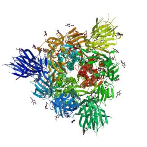
Ribbon Structure of SARS-CoV-2 Spike Protein
SARS-CoV-2 utilizes an extensively glycosylated spike (S) protein that protrudes from the viral surface to bind to angiotensin-converting enzyme 2 (ACE2) to mediate host-cell entry. Vaccine development has been focused on this protein, which is the focus of the humoral immune response. Understanding the glycan structure of the SARS-CoV-2 virus spike (S) protein will be critical in the development of glycoprotine-based vaccine candidates.
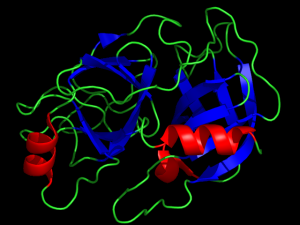
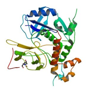
Structure of a HIF-1a-pVHL-ElonginB-ElonginC complex
William G. Kaelin Jr., Sir Peter J. Ratcliffe and Gregg L. Semenza were awarded the 2019 Nobel Prize in Physiology or Medicine for their discoveries of how cells sense and adapt to oxygen availability.
Kaelin and Ratcliffe’s labs focused their efforts on the transcription factor HIF (hypoxia-inducible factor). This transcription factor is critical in the cellular adaptation of to changes in oxygen availability.
When oxygen levels are elevated cells contain very little HIF. Ubiquitin is added to the HIF protein via the VHL complex and it is degraded in the proteasome. When oxygen levels are low (hypoxia) the amount of HIF increases.
In 2001 both groups published articles characterizing the interaction between VHL and HIF, and these articles were referenced by the Nobel Prize Organization in their press release about this year’s award. (1,2). Both studies demonstrated that under the normal oxygen conditions hydroxylation of proline residue P564 enabled VHL to recognize and bind to HIF.
The use of cell free expression (i.e., TNT Coupled Transcription/Translation System) by both labs was key in the characterization of the VHL:HIF interaction The labs utilized HIF and VHL 35-S labeled proteins generated via the TNT system under both normal or in a hypoxic work station to:
Determine the affect of ferrous chloride and cobaltous chloride on the interaction
Map the specific region of HIF required for the interaction to occur (556-574)
Determine the effect of HIF point mutations on the interaction
Use synthetic peptides to block the interaction
Conclude that a factor in mammalian cells was necessary for the interaction to occur.
XWe use cookies and similar technologies to make our website work, run analytics, improve our website, and show you personalized content and advertising. Some of these cookies are essential for our website to work. For others, we won’t set them unless you accept them. To learn more about our approach to Privacy we invite you to Read More
By clicking “Accept All”, you consent to the use of ALL the cookies. However you may visit Cookie Settings to provide a controlled consent.
We use cookies and similar technologies to make our website work, run analytics, improve our website, and show you personalized content and advertising. Some of these cookies are essential for our website to work. For others, we won’t set them unless you accept them. To find out more about cookies and how to manage cookies, read our Cookie Policy.
If you are located in the EEA, the United Kingdom, or Switzerland, you can change your settings at any time by clicking Manage Cookie Consent in the footer of our website.
Necessary cookies are absolutely essential for the website to function properly. These cookies ensure basic functionalities and security features of the website, anonymously.
Cookie
Duration
Description
cookielawinfo-checbox-analytics
11 months
This cookie is set by GDPR Cookie Consent plugin. The cookie is used to store the user consent for the cookies in the category "Analytics".
cookielawinfo-checbox-functional
11 months
The cookie is set by GDPR cookie consent to record the user consent for the cookies in the category "Functional".
cookielawinfo-checbox-others
11 months
This cookie is set by GDPR Cookie Consent plugin. The cookie is used to store the user consent for the cookies in the category "Other.
cookielawinfo-checkbox-advertisement
1 year
The cookie is set by GDPR cookie consent to record the user consent for the cookies in the category "Advertisement".
cookielawinfo-checkbox-necessary
11 months
This cookie is set by GDPR Cookie Consent plugin. The cookies is used to store the user consent for the cookies in the category "Necessary".
cookielawinfo-checkbox-performance
11 months
This cookie is set by GDPR Cookie Consent plugin. The cookie is used to store the user consent for the cookies in the category "Performance".
gdpr_status
6 months 2 days
This cookie is set by the provider Media.net. This cookie is used to check the status whether the user has accepted the cookie consent box. It also helps in not showing the cookie consent box upon re-entry to the website.
lang
This cookie is used to store the language preferences of a user to serve up content in that stored language the next time user visit the website.
viewed_cookie_policy
11 months
The cookie is set by the GDPR Cookie Consent plugin and is used to store whether or not user has consented to the use of cookies. It does not store any personal data.
Analytical cookies are used to understand how visitors interact with the website. These cookies help provide information on metrics the number of visitors, bounce rate, traffic source, etc.
Cookie
Duration
Description
SC_ANALYTICS_GLOBAL_COOKIE
10 years
This cookie is associated with Sitecore content and personalization. This cookie is used to identify the repeat visit from a single user. Sitecore will send a persistent session cookie to the web client.
vuid
2 years
This domain of this cookie is owned by Vimeo. This cookie is used by vimeo to collect tracking information. It sets a unique ID to embed videos to the website.
WMF-Last-Access
1 month 18 hours 24 minutes
This cookie is used to calculate unique devices accessing the website.
_ga
2 years
This cookie is installed by Google Analytics. The cookie is used to calculate visitor, session, campaign data and keep track of site usage for the site's analytics report. The cookies store information anonymously and assign a randomly generated number to identify unique visitors.
_gid
1 day
This cookie is installed by Google Analytics. The cookie is used to store information of how visitors use a website and helps in creating an analytics report of how the website is doing. The data collected including the number visitors, the source where they have come from, and the pages visted in an anonymous form.
Advertisement cookies are used to provide visitors with relevant ads and marketing campaigns. These cookies track visitors across websites and collect information to provide customized ads.
Cookie
Duration
Description
IDE
1 year 24 days
Used by Google DoubleClick and stores information about how the user uses the website and any other advertisement before visiting the website. This is used to present users with ads that are relevant to them according to the user profile.
test_cookie
15 minutes
This cookie is set by doubleclick.net. The purpose of the cookie is to determine if the user's browser supports cookies.
VISITOR_INFO1_LIVE
5 months 27 days
This cookie is set by Youtube. Used to track the information of the embedded YouTube videos on a website.
Performance cookies are used to understand and analyze the key performance indexes of the website which helps in delivering a better user experience for the visitors.
Cookie
Duration
Description
YSC
session
This cookies is set by Youtube and is used to track the views of embedded videos.
_gat_UA-62336821-1
1 minute
This is a pattern type cookie set by Google Analytics, where the pattern element on the name contains the unique identity number of the account or website it relates to. It appears to be a variation of the _gat cookie which is used to limit the amount of data recorded by Google on high traffic volume websites.