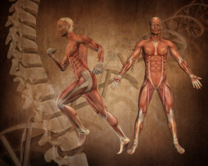
Mass Spec for Glycosylation Analysis of SARS-CoV-2 Proteins Implicated in Host-Cell Entry

The spike protein of the SARS-CoV-2 virus is a very commonly researched target in COVID-19 vaccine and therapeutic studies because it is an integral part of host cell entry through interactions between the S1 subunit of the spike protein with the ACE2 protein on the target cell surface. Viral proteins important in host cell entry are typically highly glycosylated. Looking at the sequence of the SARS-CoV-2 virus, researchers predict that the spike protein is highly glycosylated. In a recent study, researchers conducted a glycosylation analysis of SARS-CoV-2 proteins using mass spec analysis to determine the N-glycosylation profile of the subunits that make up the spike protein.

3d model of coronavirus covid-19 showing the spike protein. A recent study performed a glycosylation analysis of SARS-CoV-2 protein.

Glycans assist in protein folding and help the virus avoid immune recognition by the host. Glycosylation can also have an impact on the antigenicity of the virus, as well as potential effects on vaccine safety and efficacy. Mass spectrometry is widely used for viral characterization studies of influenza viruses. Specifically, mass spec has been used to study influenza protein glycosylation, antigen quantification, and determination of vaccine potency.

Continue reading “Mass Spec for Glycosylation Analysis of SARS-CoV-2 Proteins Implicated in Host-Cell Entry”

More muscle from eggs? Proteo-lipid complex may help prevent age-associated loss of muscle-mass

In older people, low muscle mass is strongly associated with reduced functional capacity and an increased risk of disability. Myostatin is a negative regulator of muscle growth and has become an important target for pharmaceutical companies designing therapeutics to address age-associated muscle loss.

Anti-myostatin drugs increase muscle size and strength in preclinical studies. Fortetropin is a proteo-lipid complex made from fertilized egg yolk and shows anti-myostatin activity. When Fortetropin is provided as a supplement, lowered circulating myostatin levels are observed both in rodents and in young men. Fortetropin in combination with resistance exercise also lowers myostatin and increased lean body mass.

Continue reading “More muscle from eggs? Proteo-lipid complex may help prevent age-associated loss of muscle-mass”

Proteomics from a Different Point of View: Introducing ProAlanase, the Newest Mass-Spec Grade Protease from Promega

Sometimes, when using trypsin to study a protein sequence or protein modifications, sequence coverage just isn’t quite as complete as you’d like. Looking for a protease with novel cleavage specificity or a protease that functions well in a low pH environment? Promega has a protease for that.

ProAlanase is a new site-specific endoprotease that preferentially cleaves proteins on the C-terminal side of proline and alanine amino acids. The unique cleavage specificity of ProAlanase (also known as An-PEP or EndoPro; 1–3) can help to uncover parts of the proteome not previously accessible with proteases typically used in proteomic studies.

Continue reading “Proteomics from a Different Point of View: Introducing ProAlanase, the Newest Mass-Spec Grade Protease from Promega”

CRISPR/Cas9 HiBiT Knock-In: A Scalable Approach for Studying Endogenous Protein Dynamics

Studying protein function in live cells is limited by the tools available to analyze the expression and interactions of those proteins. Although mass spectrometry and antibody-based protein detection are valuable technologies for protein analysis, both methods have drawbacks that limit the range of targets and contexts in which proteins can be investigated.

Mass spectrometry is often poor at detecting low-abundance proteins. Antibody-based techniques require high quality, specific antibodies, which can be difficult to impossible to acquire. Both methods require cell lysis, preventing real-time analysis and limiting the physiological relevance, and both methods can be limiting for higher-throughput analysis. While plasmid-based overexpression of tagged target proteins simplifies detection and can allow for real time analysis, protein levels don’t typically resemble endogenous levels. Overexpression also has the potential to create experimental artifacts or limit the dynamic range of an observed response.

In 2018, Promega R&D scientists published a paper in ACS Chemical Biology demonstrating the use of CRISPR/Cas9 to integrate the 11 amino acid, bioluminescent HiBiT tag directly into the genome to serve as an easily measured reporter for endogenous proteins. This provides a highly quantitative method for investigating cellular protein dynamics that sidesteps the need for cloning and other drawbacks to conventional methods, including the ability to measure changing protein dynamics in real-time. (For more details about CRISPR/Cas9 knock-in tagging and other applications, read this blog.)

While their findings showed that this method provides efficient and specific tagging of endogenous proteins, the research was limited to just five different proteins within a single signaling pathway in two cell lines. This left unanswered questions about whether this approach was scalable, had broader applications and how accurately the natural biology of the cells was represented.

Continue reading “CRISPR/Cas9 HiBiT Knock-In: A Scalable Approach for Studying Endogenous Protein Dynamics”

Designing BET(ter) Inhibitors to Guide Therapy for Cancer and Inflammatory Diseases

bet proteins brd nanoluc

Transcriptional activation of genes within the nucleus of eukaryotic cells occurs by a variety of mechanisms. Typically, these mechanisms rely on the interaction of regulatory proteins (transcriptional activators or repressors) with specific DNA sequences that control gene expression. Upon DNA binding, regulatory proteins also interact with other proteins that are part of the RNA polymerase II transcriptional complex.

One type of transcriptional activation relies on inducing a conformational change in chromatin, the DNA-protein complex that makes up each chromosome within a cell. In a broad sense, “extended” or loosely wound chromatin is more accessible to transcription factors and can signify an actively transcribed gene. In contrast, “condensed” chromatin hinders access to transcription factors and is characteristic of a transcriptionally inactive state. Acetylation of lysine residues in histones—the primary constituents of the chromatin backbone—results in opening up the chromatin and consequent gene activation. Disruption of histone acetylation pathways is implicated in many types of cancer (1).

Continue reading “Designing BET(ter) Inhibitors to Guide Therapy for Cancer and Inflammatory Diseases”

NanoBRET™ Assays to Analyze Virus:Host Protein:Protein Interactions in Detail

Recently, Gordon et al. published an atlas of protein:protein interactions of all proposed SARS-CoV-2 proteins expressed individually in HEK 293 cells (Table 1). The study tagged each of the viral proteins with an epitope tag and performed a pull-down of the expressed protein followed by trypsin digestion and mass spec analysis, a process referred to as affinity purification–mass spec analysis. The group identified 332 human proteins interacting with 27 SARS-CoV-2 proteins.

The interactions identified in the HEK 293 cells helped Appelberg et al. analyze interactions over time in SARS-CoV-2-infected Huh7 cells. Gordon et al. used the PPI data to identify FDA-approved drugs, drugs in clinical trials, and pre-clinical compounds that bound to the identified human proteins and labs in New York and Paris tested some of these drugs for antiviral effects.   

Table 1. The general functional area of human proteins identified to interact with individually expressed SARS-CoV-2 proteins as reported by Gordon, et al. (1). The SARS-CoV-2 proteins are classified as non-structural proteins (nsp#), structural proteins (E, M, and N) and accessory proteins (orf#).  
Continue reading “NanoBRET™ Assays to Analyze Virus:Host Protein:Protein Interactions in Detail”

NanoLuc® Luciferase Powers More than Reporter Assays

Bright NanoLuc® Luciferase

What can you do with a small, super bright luciferase? Amazing things. We’ve highlighted many of the papers and new applications that NanoLuc® luciferase has enabled on this blog. While NanoLuc® luciferase was first introduced as a reporter enzyme to assess promoter activity, its capabilities have expanded far beyond a genetic reporter, creating bioluminescent tools used to study endogeneous protein dynamics, target engagement, protein degradation, immunodetection and more. So where did the NanoLuc luciferase come from and how does one enzyme power so many research capabilities? Read further for a primer on the various technologies and applications developed from this enzyme over the last 10 years.

Continue reading “NanoLuc® Luciferase Powers More than Reporter Assays”

From Live Cells to Lysates: Adapting NanoBiT to a Biochemical Assay Format

The ability to target protein interactions with low solubility or weak binding affinities can present a significant challenge when it comes to drug screening. The beauty of these types of challenges we often face in the lab is that finding solutions to these problems doesn’t necessarily require brand new tools. Sometimes we already have the right tools in our arsenal and, with just a little creativity and collaboration, they can be adapted to address the challenge at hand.

In the following video, Dr. Mohamed (Soly) Ismail, a Postdoctoral Fellow at the Downward Lab of the Francis Crick Institute, presents the perfect example of this with his novel approach to the NanoBiT® Protein:Protein Interaction Assay. Through a collaboration with Promega R&D Scientists, Dr. Ismail has translated the assay into a cell-free, biochemical format, termed the NanoBiT Biochemical Assay (NBBA).

Watch the subtitled version of the video here >

Continue reading “From Live Cells to Lysates: Adapting NanoBiT to a Biochemical Assay Format”

Giant Rodent, Lowered Cancer Rates: What Genetic Analysis Reveals about the Capybara and Cancer

Do you find the thought of a giant rodent off-putting? Do your thoughts go to huge rats running amuck in dark allies, threatening unsuspecting passers by?

I personally hold rodents in low esteem. Rats, mice…who needs them? With the exception of cavies. I spent countless hours as a child playing with guinea pigs. We had as many as 16 of these little rodents at one time (the males are very capable of chewing or climbing out of cardboard boxes to reach a female in the next box). The baby guinea pigs were very cute and the adults had quite pronounced personalities, and a lot of attitude.

It was this history with guinea pigs that made me interested in learning more about the largest rodent in the world, the South American capybara (Hydrochoerus hydrochaeris). These family-oriented herbivores are found in savannas and forested areas, living in groups of as many as 100 members. They are excellent swimmers and can remain underwater for as long as 5 minutes. In fact, capybara mate only in the water. (Perhaps it’s not surprising then that the South American alligator, the caiman, is one of the capybara’s greatest predators.)

Photos of an adult male capybara.
A male capybara, with a scent gland (called a morillo) on his head. Photo by: Charles J Sharp – Own work, from Sharp Photography, sharpphotography, CC BY-SA 4.0.

With their squared-off nose and lack of tail, capybaras actually resemble guinea pigs. However, these oversized cavies weigh as much as 40 pounds. and can reach 24” at the shoulder, the size of an average standard poodle. Guinea pigs, on the other hand, weigh in at 2–3 pounds, and are 3–4” tall.

Their proportions make capybaras 60 times more massive than their closest relatives, rock cavies (Kerodon sp.) and 2,000 times more massive than the common mouse (Mus musculus). This tremendous size difference is why Herrera-Álvarez et al. took a closer look at the capybara, studying its propensity to develop cancer and other tradeoffs that would seem to coincide with its exceptional size.

Continue reading “Giant Rodent, Lowered Cancer Rates: What Genetic Analysis Reveals about the Capybara and Cancer”

Targeted Protein Degradation: A Bright Future for Drug Discovery

targeted protein degradation and protacs

Our cells have evolved multiple mechanisms for “taking out the trash”—breaking down and disposing of cellular components that are defective, damaged or no longer required. Within a cell, these processes are balanced by the synthesis of new components, so that DNA, RNA and proteins are constantly undergoing turnover.

Proteins are degraded by two major components of the cellular machinery. The discovery of the lysosome in the mid-1950s provided considerable insight into the first of these degradation mechanisms for extracellular and cytosolic proteins. Over the next several decades, details of a second protein degradation mechanism emerged: the ubiquitin-proteasome system (UPS). Ubiquitin is a small, highly conserved polypeptide that is used to selectively tag proteins for degradation within the cell. Multiple ubiquitin tags are generally attached to a single targeted protein. This ill-fated, ubiquitinated protein is then recognized by the proteasome, a large protein complex with proteolytic activity. Ubiquitination is a multistep process, involving several specialized enzymes. The final step in the process is mediated by a family of ubiquitin ligases, known as E3.

Continue reading “Targeted Protein Degradation: A Bright Future for Drug Discovery”