The Wnt/β-catenin pathway, long studied in the context of developmental biology, has become increasingly recognized for its role in a wide range of human diseases. Its dysregulation has been implicated in cancer, fibrosis, immune modulation, and neurodegenerative conditions—making it a clinically actionable target across diverse therapeutic areas1. In this blog, we cover the fundamentals of Wnt/β-catenin signaling, highlight ongoing research efforts to understand its role in disease, and show how combining live-cell imaging with luminescent assays complements functional studies.
Wnt/β-Catenin Signaling in Development, Homeostasis, and Disease
Wnt/β-catenin signaling is one of the key pathways our cells use to manage growth, repair, and renewal. Wnt, named as a portmanteau of the genes Wingless and Int-1, refers to a family of secreted signaling proteins that trigger cellular responses like proliferation, migration, and polarity2. β-catenin is a dual-function protein that helps anchor cells together at their membranes and acts as a transcriptional co-activator in the nucleus3. The Wnt/β-catenin pathway is active during early embryonic development and continues to play a steady role throughout life—especially in rapidly renewing tissues like the skin, intestine, and hair follicles1.
At the molecular level, this pathway is governed by a destruction complex that tightly controls β-catenin stability. In the absence of Wnt signals, β-catenin is targeted for degradation by a group of proteins that includes APC, Axin, and the kinases CK1α and GSK3β4. Axin acts as a scaffold that brings the complex components together, facilitating β-catenin phosphorylation and subsequent degradation4. But when Wnt ligands are present, the destruction complex is disrupted. β-catenin escapes degradation, accumulates in the cytoplasm, and translocates to the nucleus, where it activates genes like c-MYC, cyclin D1, and CD44—many of which are associated with cell proliferation and cancer progression5.
The movement of β-catenin from the cytoplasm to the nucleus serves as a regulatory signal that determines what a cell will do next. In healthy tissues, this is tightly regulated. But in cancer—especially colorectal cancer—mutations in genes like APC or CTNNB1 (which encodes β-catenin) can tip the balance toward unchecked signaling6. In fact, around 90% of colorectal cancers involve mutations that disrupt this pathway7, making it both a major driver of disease and an important therapeutic target.
Researchers are starting to use this pathway to understand mechanisms of disease and how to treat them. For example, mutations in the CTNNB1 gene help classify Wnt-driven medulloblastoma subtypes1, while methylation of Wnt inhibitors like DKK1 and SFRP1 have been linked to prognosis in leukemia and colorectal cancer. Even in non-cancer contexts, proteins like serum DKK1 are being investigated as predictors of cardiovascular risk and cognitive decline1. Taken together, these findings position Wnt/β-catenin as a clinically actionable target in a wide range of diseases.
Using Reporter Assays to Identify Wnt Pathway Inhibitors
Luciferase-based reporter assays have played a key role in identifying small molecules that disrupt Wnt/β-catenin signaling—particularly transcriptional activity in the nucleus where β-catenin drives gene transcription. In one study, researchers used a β-catenin–responsive luciferase reporter to screen nearly 15,000 compounds for their ability to inhibit canonical Wnt signaling downstream of Axin. This high-throughput screen led to the discovery of the iCRT class of compounds, which disrupt the interaction between β-catenin and TCF—key partners in activating Wnt target genes. Importantly, iCRT compounds (iCRT3, iCRT5, and iCRT14) selectively impaired survival of colon cancer cells with constitutive Wnt activity and reduced tumor growth in mouse models, highlighting the therapeutic potential of targeting nuclear β-catenin function8.
Studies like this one underscore how valuable functional assays can be for discovering Wnt pathway inhibitors. While luciferase reporters, like those used in the RNAi-based study above, provide a population-level view of pathway activity, they don’t reveal where within a cell these interactions are happening—or how uniformly they occur across a cell population. Nuclear translocation of β-catenin is a hallmark of Wnt activation, and tracking this process in real time can help confirm whether candidate compounds are disrupting the pathway as intended.
Visualizing β-Catenin Dynamics in Real Time to Validate Biology
Traditionally, reporter assays are measured with a microplate reader (such as a GloMax® Discover Plate Reader) to collect Relative Luminescence Units (RLU) from a population of cells. While this can tell us whether β-catenin is active in the nucleus, it doesn’t reveal how individual cells respond over time—or how consistently they respond across a population. Fluorescence-based imaging can provide some of this spatial and temporal information, but often requires overexpression, fixation, or staining that limits live-cell resolution. Bioluminescent imaging offers a complementary approach, enabling researchers to visualize β-catenin dynamics in live cells at the single-cell level using the same reporter from their workflow. This added layer of information can strengthen the connection between molecular interactions and cellular outcomes, ultimately revealing how pathway modulation translates to phenotype.
Using bioluminescent imaging, we can observe β-catenin localization dynamically in live cells, without the need for overexpression or harsh processing. In a recent experiment, researchers at Promega used the HiBiT Protein Tagging System to tag endogenous β-catenin with a small, luminescent tag. When paired with LgBiT and Nano-Glo® Live Cell substrate, the luminescent signal acts as a proxy for β-catenin abundance and location. Captured in the animations below, we can see that in untreated cells, β-catenin remains mostly in the cytoplasm. But after treatment with AZD2858—an inhibitor that blocks β-catenin degradation—cells showed clear nuclear accumulation of β-catenin over a five-hour time course.
By combining endogenously tagged β-catenin with live-cell imaging using the GloMax® Galaxy Bioluminescence Imager, researchers can validate not only that a pathway is active, but how individual cells respond to specific perturbations over time. This can be especially useful when screening for modulators like iCRTs or GSK3β inhibitors, confirming their mechanism of action visually—and in some cases, catching off-target effects early. In a pathway as clinically relevant and tightly regulated as Wnt/β-catenin, that kind of resolution is hard to ignore.
A Flexible Platform for Visualizing Assays
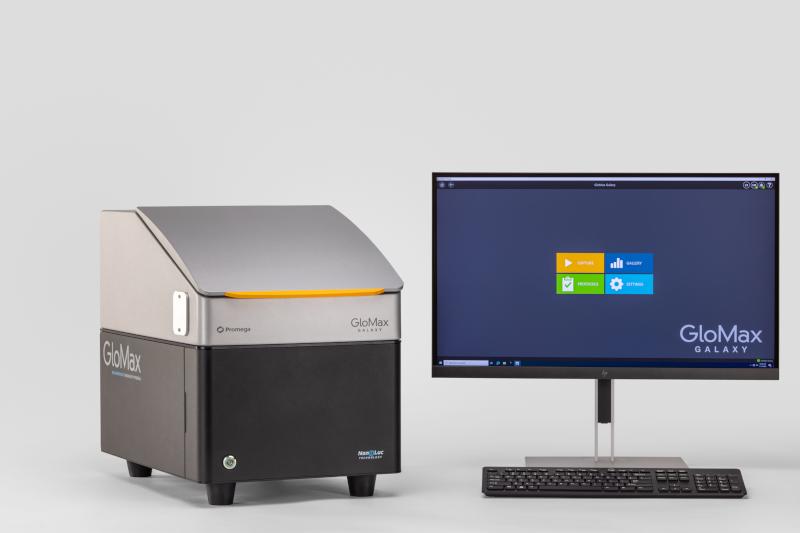
The GloMax® Galaxy Bioluminescence Imager was built to visualize the NanoLuc® luciferase-based assays we engineered, including HiBiT, NanoBiT, and NanoBRET, in both live-cell and end-point formats. It’s fully equipped for luminescence, fluorescence, and brightfield imaging, so you can explore protein localization, degradation, and interactions in real time. See Your Research in a New Light With the GloMax® Galaxy.
The GloMax® Galaxy Bioluminescence Imager is for Research Use Only.
References
- Liu, J., et al. (2022). Wnt/β-catenin signalling: function, biological mechanisms, and therapeutic opportunities. Signal Transduction and Targeted Therapy, 7, Article 3. https://doi.org/10.1038/s41392-021-00762-6 ↩︎
- Nusse, R., & Varmus, H. (1982). Many tumors induced by the mouse mammary tumor virus contain a provirus integrated in the same region of the host genome. Cell, 31(1), 99–109. https://doi.org/10.1016/0092-8674(82)90409-3
↩︎ - Clevers, H. (2006). Wnt/β-catenin signaling in development and disease. Cell, 127(3), 469–480. https://doi.org/10.1016/j.cell.2006.10.018 ↩︎
- MacDonald, B. T., Tamai, K., & He, X. (2009). Wnt/β-Catenin Signaling: Components, Mechanisms, and Diseases. Developmental Cell, 17(1), 9–26. https://doi.org/10.1016/j.devcel.2009.06.016 ↩︎
- Jeong, W.-J., Ro, E. J., & Choi, K.-Y. (2018). Interaction between Wnt/β-catenin and RAS-ERK pathways. NPJ Precision Oncology, 2, Article 5. https://doi.org/10.1038/s41698-018-0049-y ↩︎
- Kim, G., et al. (2018). Nuclear β-catenin localization and mutation of the CTNNB1 gene. Modern Pathology, 31(10), 1553–1559. https://doi.org/10.1038/s41379-018-0080-0 ↩︎
- The Cancer Genome Atlas Research Network et al. (2013). The Cancer Genome Atlas Pan-Cancer analysis project. Nature Genetics, 45(10), 1113–1120. https://doi.org/10.1038/ng.2764 ↩︎
- Gonsalves, F. C., et al. (2011). An RNAi-based chemical genetic screen identifies three small-molecule inhibitors of the Wnt/Wingless signaling pathway. PNAS, 108(15), 5954–5963. https://doi.org/10.1073/pnas.1017496108 ↩︎
Anna Bennett
Latest posts by Anna Bennett (see all)
- The Molecular Blueprint for Virus-Resistant Cowpea - June 9, 2026
- Piecing Together the Primate Gut Microbiome: Known Residents and Novel Species - April 28, 2026
- Down the Rabbit Hole: The Search for New England’s Disappearing Cottontail - March 31, 2026