Today’s blog written by guest author Kendra Hanslik.
In the intricate dance of cellular processes that sustain life, pyruvate emerges as a central figure. It plays a crucial role in the energy production saga. This small molecule is the linchpin between glycolysis and the citric acid cycle, linking the breakdown of glucose to the production of adenosine triphosphate (ATP). In this article, we explore pyruvate’s origins, multifaceted roles, and its association with various diseases.
Public awareness of mental disorders has increased over the past decade. Post-traumatic stress disorder (PTSD), anxiety and depression are both debilitating and complex to approach therapeutically. Recent research has begun exploring monoamine oxidase (MAO) enzymes as potential treatment options. MAO enzymes are responsible for the metabolism of monoamine neurotransmitters in the central nervous system, such as serotonin and dopamine (Jones & Raghanti, 2021). Abnormal levels of these neurotransmitters within the nervous system are a key characteristic of several neurological conditions. Thus, exploring MAO regulation may help our understanding of these complex clinical conditions.
Model organisms are essential tools in the pursuit of understanding biological processes, elucidating the mechanisms of diseases, and developing potential treatments and therapies. Use of these organisms in scientific research has paved the way for groundbreaking discoveries across various fields of biology. In particular, non-mammalian models can be valuable due to characteristics such as rapid life cycles, low cost, and amenability to use with advanced genetic tools, including bioluminescent reporters such as NanoLuc® Luciferase.
NanoLuc® is a small (19.1 kDa) luciferase enzyme originating from deep sea shrimp that is 100x brighter than firefly or Renilla luciferase. It utilizes a furimazine substrate to produce its bright glow-type luminescence. In the decade following its development, the NanoLuc® toolbox has expanded to include NanoBiT® complementation, NanoBRET™ energy transfer methods, and new reagents such as the Nano-Glo® Fluorofurimazine In Vivo Substrate (FFz) which was designed for in vivo detection of NanoLuc® Luciferase, NanoLuc® fusion proteins or reconstituted NanoBiT® Luciferase. In addition to the aqueous-soluble reagents increased substrate bioavailability in vivo, with fluorofurimazine, NanoLuc® and firefly luciferase can be used together in dual-luciferase molecular imaging studies.
Here we spotlight some recent research that demonstrates how the expanded NanoLuc® toolbox can be adapted to use in non-mammalian models, shedding new light on fundamental biological processes and advancing our understanding of complex mechanisms in these diverse organisms.
Bioluminescence imaging is a powerful tool for non-invasive studies of the effect of treatments on cells and tissues. The luminescent signal is strong, and can be used in vivo, enabling repeated observations over time, allowing longitudinal study of cellular changes for hours or days. Bioluminescence imaging can be used in live animals over varying periods of time, without interfering with normal cellular processes.
Fluorescence imaging is also used in cellular studies. Although it can provide a stronger signal than luminescence, fluorescence requires light for excitation, and thus its in vivo use is limited at a tissue or cell depth greater than 1mm.
NanoLuc® Luciferase. Small, bright and now useful in brain bioluminescence imaging.
In addition, autofluorescence can be an issue with fluorescence imaging, as cellular components and surrounding proteins and cells can fluoresce when exposed to light. Autofluorescence can result in high background signals, making it difficult to distinguish true fluorescence from background.
Traditional approaches for protein degrader compound screening like Western blotting can be laborious, time consuming and cannot be streamlined with automation. By implementing a high-throughput, automated workflow that uses our CRISPER/Cas9 knock-in cell lines, live-cell bioluminescent assays and sensitive GloMax® Discover microplate readers, our custom assay services offer protein degradation profiling at an accelerated rate.
To do this, we collaborated with HighRes® Biosolutions, to develop an automated system that can screen up to 100 384-well plates each day, generating roughly 40,000 data points with minimal hands-on work.
An important step of building this system is to integrate four GloMax® Discover microplate readers into the automated system using instrument’s built-in SiLA2 communication driver. The driver software makes it easy to connect the microplate readers with HighRes® Biosolution’s robotic components.
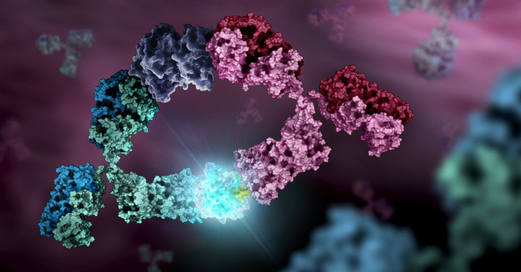
The first monoclonal antibody (mAb) was produced in a lab 1975, and the first therapeutic mAb was introduced in the United States to prevent kidney transplant rejection in 1986. The first mAb used in cancer treatment the anti-CD20 mAb, rituximab, was used to treat non-Hodgkin’s lymphoma and chronic lymphocytic leukemia. Today therapeutic mAbs have become a mainstay of cancer, autoimmune disease, and metabolic disease therapies and include HERCEPTIN® used to treat certain forms of breast cancer, Prolia used to treat bone loss in post-menopausal women, and Stelara used to treat autoimmune diseases like psoriatic arthritis and severe Crohn disease, among many others. Therapeutic mAbs bind targets with high specificity and affinity and they can recruit effector cells to drive target elimination through mechanisms such as antibody-dependent cellular cytotoxicity (ADCC) or antibody-dependent cellular phagocytosis (ADCP), making them highly specific, effective therapies.
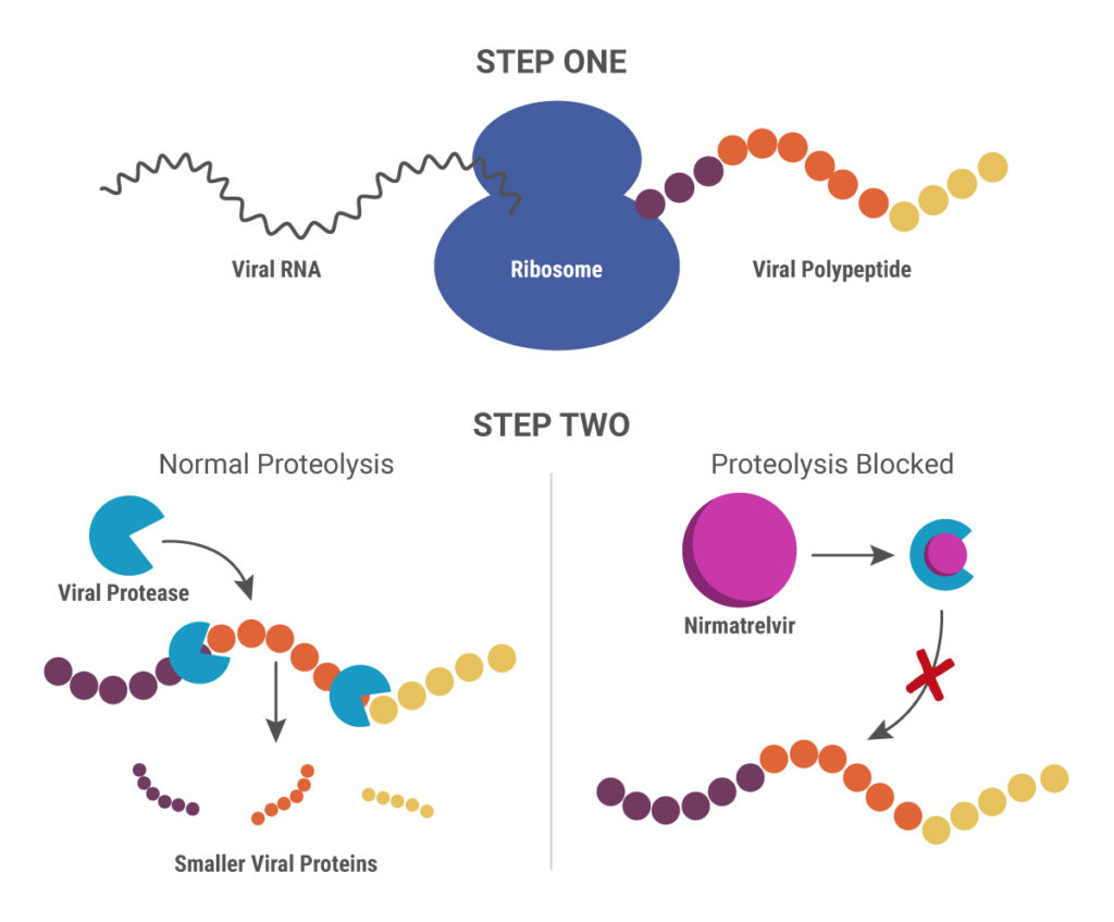
Cytochrome P450 (CYP) inhibitors are often used as boosting agents in combination with other drugs. This drug development strategy is front and center for Paxlovid, the new anti-SARS-CoV-2 treatment from Pfizer. Paxlovid is a combination therapy, comprised of two protease inhibitors, nirmatrelvir and ritonavir. It significantly reduces the risk of COVID-19 hospitalization in high-risk adults and is ingested orally rather than injected, which is an advantage over other SARS-CoV-2 treatments, such as Remdesivir.
Nirmatrelvir was originally developed by Pfizer almost 20 years ago to treat HIV and works by blocking enzymes that help viruses replicate. Pfizer created another version of this drug to combat SARS in 2003, but, once that outbreak ended, further development was put on pause until the advent of the COVID-19 pandemic. After developing an intravenous form of nirmatrelvir early in the pandemic, Pfizer created another version that can be taken orally and combined it with ritonavir.
When ritonavir was originally developed, it wasn’t considered particularly useful because it metabolized so quickly in the body. Now it is recognized as a pharmacokinetic enhancer in combination with other drugs. Ritonivir inhibits CYP3A4, an enzyme which plays a key role in the metabolism of drugs and xenobiotics. By inhibiting CYP3A4, ritonivir slows the metabolism of other drugs. In the case of Paxlovid, this allows nirmatrelvir to stay in the body longer at a high enough concentration to be effective against the virus. This ultimately means that patients can be given lower doses of the drug with reducing efficacy.
Nirmatrelvir inhibits the viral 3CL protease, so that functional, smaller viral proteins cannot be produced.
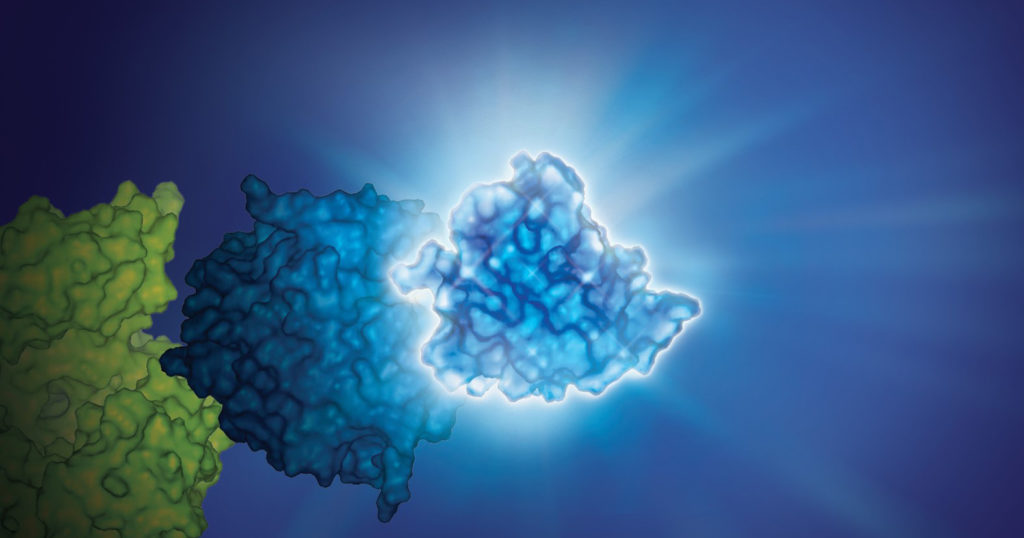
A new study published in Nature Chemical Biology shows that the most commonly mutated protein in cancer might not be as “undruggable” as previously believed. Promega R&D scientists collaborated with the research group led by Kevan Shokat at the University of California – San Francisco to develop strategies for targeting mutants of KRAS that have evaded previous drug discovery efforts. Their paper opens new possibilities for developing small molecule inhibitors against KRAS(G12D) and other clinically significant mutants.
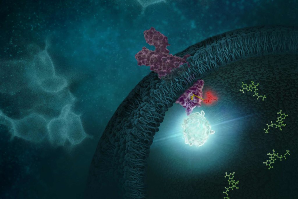
G protein-coupled receptors (GPCRs) comprise a large group of cell surface receptors, characterized by the unique structural property of crossing the cell membrane seven times. They respond to a diverse group of signaling molecules, such as peptides, neurotransmitters, cytokines, hormones and other small molecules (1). Upon activation, GPCRs interact with GTP-binding (G) proteins and arrestins to regulate a wide variety of signaling pathways. This broad range of functions makes GPCRs attractive targets for drug discovery. The importance of GPCR research was highlighted in 2012, with the Nobel Prize in chemistry being awarded to Robert Lefkowitz and Brian Kobilka “for studies of G-protein–coupled receptors”.
Based on structure and function, GPCRs are categorized into six classes, A–F. The class A GPCRs, or rhodopsin-like receptors, have been studied extensively due to their association with many types of diseases (2). Within the class A GPCRs is a group that share a highly conserved structural motif (3) and respond to chemokines—small “chemotactic cytokines” that stimulate cell migration, especially that of white blood cells (4). A subfamily of class A GPCRs respond to chemokines that have two cysteine residues near the N-terminus, known as CC chemokines. GPCRs activated by CC chemokines are called CC chemokine receptors or CCRs, and these interactions have been implicated in both pro- and anti-cancer pathways (5).
Understanding how a compound or drug affects cellular pathways often requires measuring kinetic changes over an extended period of time—from several hours to days. Live-cell kinetic cell-based assays that measure cell viability, cytotoxicity, apoptosis and other cellular pathways are great for collecting real-time data. You don’t necessarily need expensive equipment to run these types of assays. In the videos below, Dr. Sarah Mahan, a research scientist at Promega, demonstrates how you can easily get great 24-hour or multi-day kinetic data using a GloMax® Microplate Reader.
XWe use cookies and similar technologies to make our website work, run analytics, improve our website, and show you personalized content and advertising. Some of these cookies are essential for our website to work. For others, we won’t set them unless you accept them. To learn more about our approach to Privacy we invite you to Read More
By clicking “Accept All”, you consent to the use of ALL the cookies. However you may visit Cookie Settings to provide a controlled consent.
We use cookies and similar technologies to make our website work, run analytics, improve our website, and show you personalized content and advertising. Some of these cookies are essential for our website to work. For others, we won’t set them unless you accept them. To find out more about cookies and how to manage cookies, read our Cookie Policy.
If you are located in the EEA, the United Kingdom, or Switzerland, you can change your settings at any time by clicking Manage Cookie Consent in the footer of our website.
Necessary cookies are absolutely essential for the website to function properly. These cookies ensure basic functionalities and security features of the website, anonymously.
Cookie
Duration
Description
cookielawinfo-checbox-analytics
11 months
This cookie is set by GDPR Cookie Consent plugin. The cookie is used to store the user consent for the cookies in the category "Analytics".
cookielawinfo-checbox-functional
11 months
The cookie is set by GDPR cookie consent to record the user consent for the cookies in the category "Functional".
cookielawinfo-checbox-others
11 months
This cookie is set by GDPR Cookie Consent plugin. The cookie is used to store the user consent for the cookies in the category "Other.
cookielawinfo-checkbox-advertisement
1 year
The cookie is set by GDPR cookie consent to record the user consent for the cookies in the category "Advertisement".
cookielawinfo-checkbox-necessary
11 months
This cookie is set by GDPR Cookie Consent plugin. The cookies is used to store the user consent for the cookies in the category "Necessary".
cookielawinfo-checkbox-performance
11 months
This cookie is set by GDPR Cookie Consent plugin. The cookie is used to store the user consent for the cookies in the category "Performance".
gdpr_status
6 months 2 days
This cookie is set by the provider Media.net. This cookie is used to check the status whether the user has accepted the cookie consent box. It also helps in not showing the cookie consent box upon re-entry to the website.
lang
This cookie is used to store the language preferences of a user to serve up content in that stored language the next time user visit the website.
viewed_cookie_policy
11 months
The cookie is set by the GDPR Cookie Consent plugin and is used to store whether or not user has consented to the use of cookies. It does not store any personal data.
Analytical cookies are used to understand how visitors interact with the website. These cookies help provide information on metrics the number of visitors, bounce rate, traffic source, etc.
Cookie
Duration
Description
SC_ANALYTICS_GLOBAL_COOKIE
10 years
This cookie is associated with Sitecore content and personalization. This cookie is used to identify the repeat visit from a single user. Sitecore will send a persistent session cookie to the web client.
vuid
2 years
This domain of this cookie is owned by Vimeo. This cookie is used by vimeo to collect tracking information. It sets a unique ID to embed videos to the website.
WMF-Last-Access
1 month 18 hours 24 minutes
This cookie is used to calculate unique devices accessing the website.
_ga
2 years
This cookie is installed by Google Analytics. The cookie is used to calculate visitor, session, campaign data and keep track of site usage for the site's analytics report. The cookies store information anonymously and assign a randomly generated number to identify unique visitors.
_gid
1 day
This cookie is installed by Google Analytics. The cookie is used to store information of how visitors use a website and helps in creating an analytics report of how the website is doing. The data collected including the number visitors, the source where they have come from, and the pages visted in an anonymous form.
Advertisement cookies are used to provide visitors with relevant ads and marketing campaigns. These cookies track visitors across websites and collect information to provide customized ads.
Cookie
Duration
Description
IDE
1 year 24 days
Used by Google DoubleClick and stores information about how the user uses the website and any other advertisement before visiting the website. This is used to present users with ads that are relevant to them according to the user profile.
test_cookie
15 minutes
This cookie is set by doubleclick.net. The purpose of the cookie is to determine if the user's browser supports cookies.
VISITOR_INFO1_LIVE
5 months 27 days
This cookie is set by Youtube. Used to track the information of the embedded YouTube videos on a website.
Performance cookies are used to understand and analyze the key performance indexes of the website which helps in delivering a better user experience for the visitors.
Cookie
Duration
Description
YSC
session
This cookies is set by Youtube and is used to track the views of embedded videos.
_gat_UA-62336821-1
1 minute
This is a pattern type cookie set by Google Analytics, where the pattern element on the name contains the unique identity number of the account or website it relates to. It appears to be a variation of the _gat cookie which is used to limit the amount of data recorded by Google on high traffic volume websites.