The Case for Canine Immunotherapy
Cancer does not respect species boundaries. Each year more than four million dogs are diagnosed with cancer (1), making it the leading disease related cause of death in the canine population. Osteosarcoma, lymphoma, mast cell tumors and mammary carcinomas are among the most prevalent (1). In many cases, these tumors in dogs bear striking biological and molecular similarities to their human counterparts.
This convergence is the foundation of the Comparative Oncology (2) framework and One Health Initiatives. Companion pets, like dogs and cats, share our environments, our lifestyles and increasingly our therapeutic challenges. When research advances in veterinary oncology, it can open windows into human disease as well.

What Veterinary Checkpoint Immunotherapy Brings to Comparative Oncology
Immune checkpoint blockade, particularly targeting the programmed cell death protein 1 (PD-1) and its ligand, PD-L1, has transformed human oncology. By releasing the inhibitory break that tumor cells apply to T cells through the PD-1/PD-L1 axis, checkpoint inhibitors have produced durable remissions in melanoma, non-small cell lung cancer and dozens of other malignancies (2,3).
The biological rationale for translating this approach to veterinary oncology is compelling. Canine tumors express PD-L1 and exploit the same immune evasion pathway observed in humans. Yet, despite significant scientific interest, no PD-L1 targeted antibody has received regulatory approval for use in canine cancer (4), in part because of the scarcity of standardized, biologically relevant tools for evaluating candidate antibodies.
Filling that gap requires assay infrastructure purpose-built for canine biology, not human tools adapted for veterinary use. Our Canine PD-1/PD-L1 Blockade Bioassay is exactly such a tool. The Promega Canine PD-1/PD-L1 Blockade Bioassay is a functional, mechanism-of-action-based assay that quantifies the potency of biologics targeting the canine PD-1/PD-L1 immune checkpoint axis, while providing stability-indicating performance suitable for characterizing antibody candidates directed against canine PD-1 dependent cancers.
The Canine PD-1/PD-L1 Blockade Bioassay is a mechanistically grounded, luminescent cell-based assay that is designed specifically to quantify the potency and stability of biologics that block the canine PD-1/PD-L1 interaction. Unlike binding assays or endpoint ELISAs, this bioassay reflects the actual mechanism of action (MoA) of checkpoint-blocking antibodies in a biologically relevant cellular context.
The assay uses two cell lines: canine PD-L1-expressing CHO-K1 target cells paired with canine PD-1-expressing Jurkat effector cells that include a response-element-driven luciferase reporter. The architecture of the assay directly links the PD-1/PD-L1 pathway blocked to a quantifiable signal. The result is a dose-response assay that is sensitive and reproducible, with the convenience of a luminescent readout.
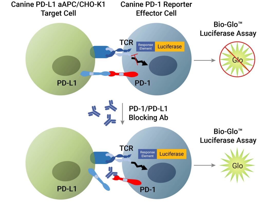
Principle of Canine PD-1/PD-L1 Blockade Bioassay. The Canine PD-1/PD-L1 Blockade Bioassay consists of two genetically engineered cell lines, the CHO-K1 target cells and the PD-1 reporter effector cell. When co-cultured, the PD-1/PD-L1 interaction inhibits TCR-mediated luminescence. When the PD-1/PD-L1 interaction is disrupted, TCR activation induces luminescence that can be detected by addition of Bio-Glo™ Reagent and quantitation with a luminometer.
Driving Discovery in Veterinary Oncology
The scientific value of a tool is ultimately demonstrated by the research it enables. Two papers published in 2025 illustrate how the Canine PD-1/PD-L1 Blockade Bioassay is enabling discovery in veterinary oncology. Both research groups (Song and colleagues at Kangwon National University and Di Palma Subran and colleagues the University of Bern) were looking to develop and functionally characterize canine PD-L1 targeted antibodies to address the absence of any approved anti-Canine PD-L1 immunotherapies. Both research groups developed novel antibodies targeting canine PD-L1 (cPD-L1), and they used the Canine PD-1/PD-L1 Blockade Bioassay to validate antibody activity. Each group also assessed functional immune activity in vitro using canine peripheral blood mononuclear cells (PBMCs). However, the two studies differed substantially in antibody format, engineering strategy, functional scope, and in vivo approach. Song et al. provide proof-of-concept that phage display against canine PD-L1 can yield conventional IgG antibodies with in vivo antitumor efficacy in a xenograft model, with the added value of cross-reactivity to human PD-L1. Di Palma Subran et al. demonstrate that nanobody-based engineering can achieve substantially superior potency through multivalent formatting and Fc-mediated effector function and introduce canine-specific Fc isotypes as a tool for studying immune mechanisms relevant to veterinary immunotherapy.
Comparative Oncology and One Health
The implications of this work extend beyond the clinic. Companion animals, especially dogs, serve as a uniquely powerful cancer model: they share human environments, develop cancers spontaneously, exhibit immune systems that parallel human immune systems and respond to therapies through mechanisms that often mirror human biology. These factors make canine research data generated through standardized tools and methods informative for human biology.
Learn more about the Canine PD-1/PD-L1 Blockade Bioassay and the expanding portfolio of our Veterinary and One Health research tools.
Literature Cited
- Chibuk, J. et al. (2021) Horizons in Veterinary Precision Oncology: Fundamentals of Cancer Genomics and Applications of Liquid Biopsy for the Detection, Characterization, and Management of Cancer in Dogs. Front. Vet. Sci. 8, 22 March. Accessed: 14 May 2026 https://doi.org/10.3389/fvets.2021.664718
- Vincze, O. (2025) Advancing Cancer Research Via Comparative Oncology. Nat. Rev. Cancer 25, 740–8. Accessed 18 May 2026.
- Lin, X. et al. Regulatory Mechanism of PD-1/PD-L1 in Cancers. Molecular Cancer 23, 108. Accessed 15 May 2026.
- Haslam, A. et al. (2024) The Landscape of Checkpoint Inhibitors in Oncology. Eur. J. Cancer 209, Sept. Accessed 15 May 2026. https://doi.org/10.1016/j.ejca.2024.114240
- Giuliano, A. et al. (2024) Checkpoint Inhibitors in Dogs: Are We There Yet? Cancers 16 (11) Accessed 15 May 2026 https://doi.org/10.3390/cancers16112003
- Song, M.Y. et al. (2025) Discovery and Functional Characterization of Canine PD-L1-Targeted Antibodies for Evaluating Antitumor Efficacy in a Canine Osteosarcoma Xenograft Model. Scientific Reports 15(7574). Accessed 15 May 2026.
- Di Plama-Subran, M. et al. (2025) Nanobody-based canine PD-L1-targeting immune checkpoint inhibitors for cancer therapy in dogs. Molecular Therapy: Oncology. 33(3):201036. Accessed 15 May 2026
This article was generated with AI assistance, and edited by Michele Arduengo, PhD.