This guest blog post is written by Aisosa Omere, Product Marketing Intern at Promega.
Metabolic diseases fundamentally arise from disrupted cellular communication. In type 2 diabetes, cellular responsiveness to insulin is impaired. Within cancer, tumors alter their metabolic pathways to gain a proliferative advantage. In both conditions, dysfunction extends beyond individual molecules or pathways and involves a complex, interconnected network of metabolites, enzymes, and signaling molecules that dynamically respond to environmental changes. Traditional approaches to studying these networks often required a compromise: stopping experiments, lysing cells, and analyzing the resulting components. Although effective, this method is inherently limited. It captures a snapshot of what was present, rather than how the biology was actually behaving.
That compromise is becoming less necessary. The evolution of bioluminescent tools is changing what is possible. Some allow researchers to watch protein behavior and drug engagement directly in living cells in real time. Others offer faster, more sensitive detection of metabolites at physiologically relevant concentrations, and are compatible enough to run multiple assays from the same experiment, making coordinated, multi-pathway profiling practical in a standard lab setting.
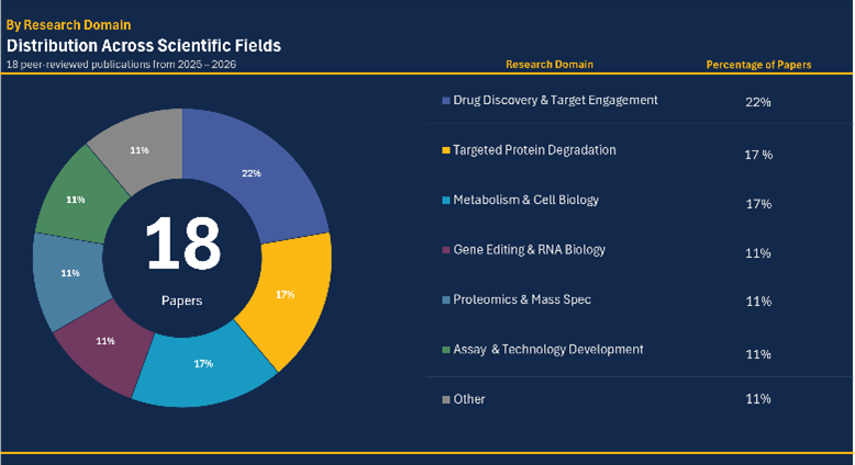
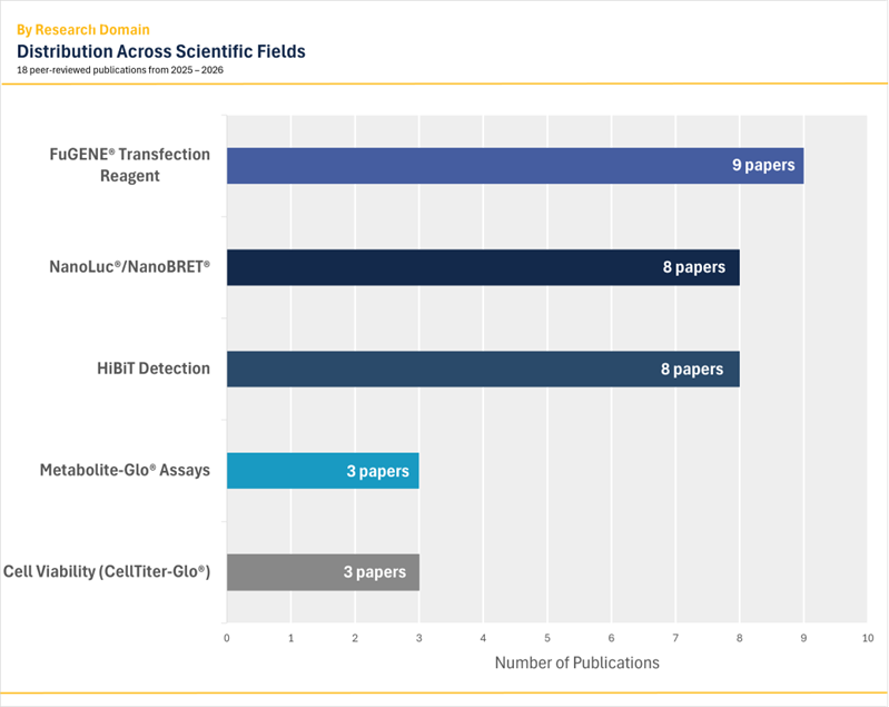
An analysis of eighteen peer-reviewed publications from 2025 and 2026 shows just how quickly these approaches are taking hold across metabolic disease research. What follows explores the tools making this possible and why this shift represents one of the most consequential methodological changes in metabolic disease research in recent years.
Section I: Why Metabolism Resists Simple Measurement
The metabolic state shifts rapidly in response to nutrients, drugs, and cellular stress, meaning a snapshot taken after cell lysis may not reflect what was happening moments before. Metabolites do not exist in isolation. They are part of pathways that are constantly feeding and regulating one another. Traditional single-endpoint assays miss this complexity on two fronts: they answer only the question of “how much metabolite is present?” and they do so one pathway at a time, making it difficult to capture the coordinated shifts that define metabolic disease.
Addressing these limitations has required different tools for different questions. For tracking protein behavior, trafficking, and drug engagement in their native cellular environment, bioluminescent live-cell approaches, such as NanoBRET and HiBiT-based assays, allow researchers to observe biology as it happens, without lysing the cells and halting the experiment. For metabolite detection, a new generation of Glo-based bioluminescent assays offers a faster, more sensitive alternative to older methods like radioactive tracers and colorimetric kits and crucially, their shared detection format makes running multiple assays from the same sample practical in a way it simply was not before.
An analysis of eighteen peer-reviewed publications from 2025–2026 found these approaches appearing across seven distinct research domains, a signal that the field has moved well beyond early adoption.
Section II: The Tools Closing the Gap
Several of the most frequently used tools in metabolic disease research belong to Promega’s Glo-based metabolite detection family. Among them are the Glucose Uptake-Glo™ Assay, Lactate-Glo™ Assay, Malate-Glo™ Assay, and Glutamine/Glutamate-Glo™ assays. Each measures a distinct metabolite from lysed cell samples or cells in culture using a bioluminescent readout—sensitive enough to detect physiologically relevant concentrations without the need for radioactive tracers or specialized instrumentation. The assays rely on enzymatic coupling that translates the presence of a specific metabolite into a proportional bioluminescent signal. Because many of these assays measure metabolites from the cell medium, researchers can measure multiple metabolites from a single aliquoted sample, enabling true multi-pathway analysis in a way that older, incompatible platforms could not.
Where the Glo-based assays excel at metabolite quantification from cell samples, NanoBRET and HiBiT-based technologies address a different but equally important question: what is happening inside living cells in real time. NanoBRET measures interactions between proteins and small molecules, including drug-target engagement, directly within intact cells without disrupting the experiment. HiBiT, a small peptide tag that produces a bioluminescent signal when paired with its complementary protein fragment, allows researchers to track protein levels, localization, and trafficking as they occur. Together, these live-cell approaches capture the dynamic cellular behavior that lysis-based methods, by definition, cannot—making them a natural complement to the metabolite detection tools described above.
Section III: Tracking Insulin Sensitivity in Living Muscle Cells
A recent study by Sivasengh et al. in Biology Open used a custom HiBiT-tagged GLUT4 construct alongside the Glucose Uptake-Glo™ assay to study how the circadian clock protein Per3 regulates insulin sensitivity in skeletal muscle cells. GLUT4 translocation to the cell surface is a key step in insulin-stimulated glucose uptake, and watching it happen dynamically in living cells, rather than inferring it after lysis, reveals the timing and magnitude of the response in a way that endpoint assays cannot. This matters because insulin resistance, the defining characteristic of type 2 diabetes, is fundamentally a problem of impaired GLUT4 trafficking. By pairing live-cell HiBiT-based tracking of transporter movement with bioluminescent detection of downstream glucose uptake from lysed samples, the researchers captured both the trafficking event and its functional consequence, a level of detail that older approaches would have required entirely separate experimental setups to achieve.
Section IV: The Multi-Analyte Advantage
Cappabianca et al. conducted a study published in Molecular Therapy Oncology that utilized Lactate-Glo™, Malate-Glo™, and Glutamine/Glutamate-Glo™ assays to examine how HDAC inhibition alters metabolic state in CAR T cells. Because each assay shares the same bioluminescent readout, the collected supernatant was divided into separate aliquots, one per assay, enabling multi-analyte profiling of lactate, malate, glutamine, and glutamate from a single experimental condition. Cancer metabolism involves coordinated alterations across multiple pathways simultaneously, and measuring those shifts from the same cell sample, under identical experimental conditions, provides a level of consistency and comparability that separate, sequential experiments cannot reliably replicate. The findings highlight that metabolic reprogramming in disease cannot be fully understood by measuring a single metabolite in isolation. By employing a panel of compatible bioluminescent assays, the researchers captured a multidimensional metabolic signature that previously would have required multiple, often incompatible platforms to piece together. This study exemplifies the broader shift in metabolic disease research from single-analyte measurements to integrated, multi-pathway profiling.
Section V: Metabolic Enzymes as Drug Targets
A study conducted by Gough et al. in SLAS Discovery developed a NanoBRET-based target engagement assay for Pyruvate Dehydrogenase Kinase (PDK), a mitochondrial enzyme central to glucose oxidation and implicated in both diabetes and cancer. PDK sits at the gateway between glycolysis and the TCA cycle, a critical junction in metabolic disease. Being able to measure drug engagement at that enzyme directly in living cells, rather than in a biochemical assay, is a meaningful step forward for metabolic drug discovery. This paper demonstrates that live-cell tools extend well beyond metabolite detection. They are enabling researchers to validate drug candidates against the metabolic machinery itself, within the native cellular environment, before committing to costly in vivo studies. In doing so, they are effectively bridging basic metabolic research and translational drug discovery.
Conclusion
Understanding metabolic disease requires measuring metabolism the way it operates: dynamically, in context, and across multiple pathways. The work highlighted here represents a broader shift toward that systems-level view — one enabled by tools that can observe protein behavior and drug engagement in living cells and quantify coordinated metabolic changes across multiple pathways from the same experiment. In the future, as these approaches are applied to increasingly complex models like organoids, patient-derived cells, and long-term time-course experiments, the picture of metabolic disease they reveal will only become more complete. This is not just a technical improvement. It is a fundamentally unique way of asking what goes wrong in disease and how we might fix it.
To learn more about Promega’s metabolic activity assays, visit: https://www.promega.com/products/energy-metabolism/
References
- Sivasengh, R.; Scott, A.; Gabriel, B. M. Live Cell GLUT4 Translocation Assay Reveals Per3 as a Novel Regulator of Circadian Insulin Sensitivity in Skeletal Muscle Cells. Biology Open, 2025, 14 (7). https://doi.org/10.1242/bio.061941.
- Gough, M. D.; Robers, M. B.; Corona, C. R.; Mehta, R. K.; Nyati, M. K.; Toogood, P. L. Development of a Cell-Based Target Engagement Assay for Pyruvate Dehydrogenase Kinase. SLAS Discovery, 2025, 32, 100227. https://doi.org/10.1016/j.slasd.2025.100227.
- Cappabianca, D.; Toulany, M.; Zima, S.; Shea, A.; Vidugiriene, J.; Lauer, A.; Sylvester, K.; Gnanasekar, V.; Foster, S.; Turaga, R.; Saha, K.; Ayuso, J.; Sodji, Q. H. Class I HDAC Inhibition Enhances the Stem-like Memory Properties of CRISPR-Engineered CAR T Cells in Neuroblastoma. Molecular Therapy Oncology, 2026, 34 (2), 201200. https://doi.org/10.1016/j.omton.2026.201200.