Model organisms are essential tools in the pursuit of understanding biological processes, elucidating the mechanisms of diseases, and developing potential treatments and therapies. Use of these organisms in scientific research has paved the way for groundbreaking discoveries across various fields of biology. In particular, non-mammalian models can be valuable due to characteristics such as rapid life cycles, low cost, and amenability to use with advanced genetic tools, including bioluminescent reporters such as NanoLuc® Luciferase.
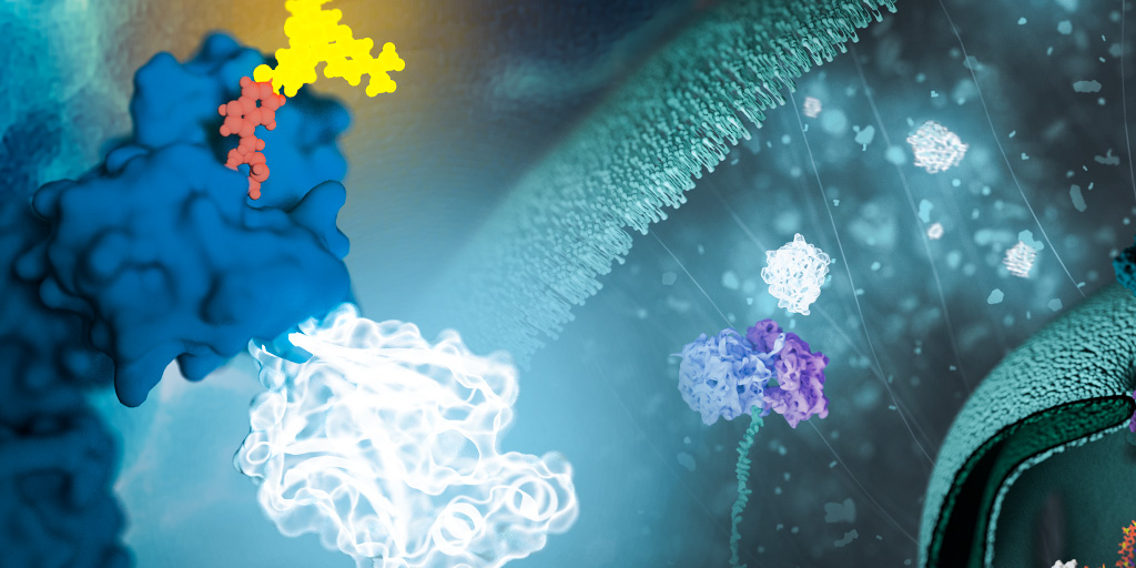
NanoLuc® is a small (19.1 kDa) luciferase enzyme originating from deep sea shrimp that is 100x brighter than firefly or Renilla luciferase. It utilizes a furimazine substrate to produce its bright glow-type luminescence. In the decade following its development, the NanoLuc® toolbox has expanded to include NanoBiT® complementation, NanoBRET™ energy transfer methods, and new reagents such as the Nano-Glo® Fluorofurimazine In Vivo Substrate (FFz) which was designed for in vivo detection of NanoLuc® Luciferase, NanoLuc® fusion proteins or reconstituted NanoBiT® Luciferase. In addition to the aqueous-soluble reagents increased substrate bioavailability in vivo, with fluorofurimazine, NanoLuc® and firefly luciferase can be used together in dual-luciferase molecular imaging studies.
Here we spotlight some recent research that demonstrates how the expanded NanoLuc® toolbox can be adapted to use in non-mammalian models, shedding new light on fundamental biological processes and advancing our understanding of complex mechanisms in these diverse organisms.
Planarians: Optimizing mRNA Delivery Methods
Planarians are studied primarily to advance understanding of regeneration and stem cell dynamics. Autofluoresence, the natural emission of light from biological structures, remains a large complication for planarian research. To manage this complication, a study from the University of Stanford was able to observe a signal from NanoLuc® mRNAs in vitro and in vivo planarian cells. The experimental conditions of the study showed luminescence intensities 3-4 magnitudes above typical background signals.
Additionally, this study used the optimized flurofurimazine substrate which allowed researchers to monitor kinetics of transfection and in vivo expression through time-lapse luminescence imaging. This method allows animals to recover after imaging, making it an attractive tool for screening DNA-based transgenic methods. Results of the study also show promise for testing additional reporters and being able to transfect other organisms. Additionally, this study demonstrated the use of NanoLuc® mRNA transfections to study post-translational modifications.
Drosophila: Detecting Fusion Proteins and Protein Interactions
Drosophila was one of the first non-mammalian model systems. For over 100 years researchers have used Drosophila to further our understanding of genetics and molecular biology. Today, Drosophila remains one of the most widely used model organisms. A recent study from Tsinghua University utilized Drosophila to study stop codon readthrough, a mechanism where translation continues past a stop codon and terminates at the next in-frame stop codon. This mechanism is used by RNA viruses and the synthesis of eukaryotic proteins. The headcase gene in Drosophila encodes two proteins, one of which is essential for tracheal development and is produced through translational readthrough. This study sought to measure readthrough throughout the life cycle of Drosophila using the NanoLuc® luciferase in
gain-of-function reporter lines. Additionally, researchers performed the Nano-Glo In-Gel Detection Assay to validate that the NanoLuc® protein was the product of translation past the stop codon.
Recent discoveries in Drosophila have also revealed a potential novel target for manipulating insect steroid hormone signaling in a membrane transporter called the Ecdysone Importer (EcI). A study from the University of California-Riverside used NanoBiT® Technology, a complementation reporter system based on NanLuc, and HEK293-EcI cells to develop a real time monitoring system and a stable cell line to detect insect steroid hormone entry into cultured cells. In this study, researchers fused the two subunits to two nuclear receptors that mediate insect steroid actions: ecdysone receptor (EcR) and retinoid X receptor (RXR). Results show that the EcR-RXR NanoBiT® assay can be effective to investigate the insect steroid importing function of EcI. Additionally, the NanoBiT® ecdysteroid sensor can be utilized in Drosophila S2 cells and cells from other insect species. This knowledge will aid in the effort to develop novel pharmacological tools by manipulating insect steroid hormone signaling.
Zebrafish: In Vivo Tracking and Imaging
Although the mouse is a powerful animal often used in cancer research, zebrafish are better suited for studies using high-throughput small molecule inhibitor screening, providing an in vivo alternative to inhibitor screening that is typically done biochemically or in cell culture. Researchers use in vivo tracking and imaging fluorescently labeled cancer cells to study early tumor growth and proliferation in zebrafish. Some of the chemical compounds used in studies as cancer growth inhibitors have been known to interfere with fluorescence-based measurements by amplifying background autofluorescence. Because of this, a recent study from the Institute of Molecular Genetics of the Czech Academy of Sciences stepped away from conventional fluorescent-based readouts and developed a pre-clinical screening model for drug discovery utilizing the NanoLuc® luciferase.

Researchers established the bioluminescent platform by labeling a zebrafish melanoma cancer cell line with EGFP and NanoLuc®. Following successful application of the double-reporter in vitro, researchers transplanted cells into zebrafish embryos. This study was able to validate bioluminescence in a zebrafish model of cancer cell growth and develop an assay measuring in vivo bioluminescence-based tumor growth at different time periods.
Check out this blog to learn more about the use of NanoLuc® in zebrafish.
C. elegans: Improvement in Sensitivity Over GFP
C. elegans is a free-living nematode that has become a key model organism in studying biology and neuroscience. In C. elegans, green fluorescent protein (GFP), is a commonly used reporter gene to observe cell morphology and in vivo gene expression. The use and sensitivity of GFP in whole-animal settings is limited by autofluorescence. A study from the University of California, San Diego developed an optimized method for detecting gene expression using the NanoLuc® luciferase instead of GFP. The study monitored rare transformation events in obligate intracellular pathogens in the intestine of C. elegans, as well as inducible promoter activity.
Researchers found the benefit of using NanoLuc® to be that intestinally expressed NanoLuc® was detected several million-fold more over background levels in a plate-reader setting. Comparatively, researchers detected intestinally expressed GFP sixfold over background. Additionally, NanoLuc® does not require ATP, and while monitoring gene expression, is not affected by any changes to ATP levels.
Conclusion
NanoLuc® has become a revolutionary tool in model organism research by allowing scientists to non-invasively monitor biological interactions and perform compound screening. By fusing NanoLuc® with target proteins, researchers can now track their behavior and interactions with exceptional precision. This technology has profound implications across scientific disciplines and is essential for illuminating new insights from model organism studies.
Related Posts
Kerstin Hurd
Latest posts by Kerstin Hurd (see all)
- Revolutionizing Food Security: How Biotechnology Contributes to Sustainability and Safety - June 26, 2024
- Reviewing the Importance of circRNA - November 14, 2023
- Glowing Testimonies: A Review of NanoLuc® Use in Model Organisms - October 6, 2023