It’s FINALLY time to announce the winners of the 2019 Promega iGEM Grant! We received over 150 applications this year, so picking the top 10 was very tough. As always, we’re impressed by the amazing work iGEM teams are doing in the lab and in their communities. The 10 winners listed below will receive $2,000 in free Promega products.
Good luck to all teams competing in iGEM this year, and congratulations to our winners! Don’t forget that Promega has free technical support for all teams competing in iGEM. Our scientists are excited to help out. You can also check out our iGEM Sponsor page, which has tools and resources to help make your project a success. Continue reading “Announcing the 2019 Promega iGEM Grant Winners”
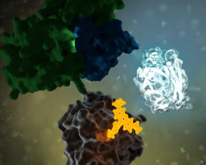
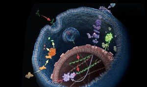
No protein is an island. Within a cell, protein-protein interactions (PPIs) are involved in highly regulated and specific pathways that control gene expression and cell signaling. The disruption of PPIs can lead to a variety of disease states, including cancer.
Two general approaches are commonly used to study PPIs. Real-time assays measure PPI activity in live cells using fluorescent or luminescent tags. A second approach includes methods that measure a specific PPI “after the fact”; popular examples include a reporter system, such as the classic yeast two-hybrid system.
The stage is set. You’ve spent days setting up this experiment. Your bench is spotless. All the materials you need to finally collect data are laid neatly before you. You fetch your cells from the incubator, add your detection reagents, and carefully slide the assay plate into the luminometer. It whirs and buzzes, and data begin to appear on the computer screen. But wait!
These data are garbage!
Don’t let this dramatic person be you. Here are 8 tips from us on things to watch out for before you start your next luminescent assay. Make sure you’ll be getting good data before wasting precious sample!
For a few years beginning late in 2013, warmer ocean conditions in the eastern Pacific prompted the appearance of unexpected species and toxic algal blooms that devastated others. When temperatures cooled in 2017, the marine ecosystems seemed to be returning to normal. Except for the pyrosomes. Although these previously rare organisms did start to wash up on beaches during the periods of warming, they began to appear by the millions from Oregon to Alaska that spring.
Photo by Steven Grace.
Some combination of ideal conditions led pyrosomes to multiply, dominate the ocean surface and wash up on beaches along the US and Canadian Pacific Coasts. Pyrosomes typically exist offshore, far below the surface in warm, tropical waters all over the world. Their sudden proliferation in other areas is likely due to the warm, Pacific ocean “blob,” although atypical sea currents and changes in pyrosome diet have been offered as other possible explanations.
While the appearance of pyrosomes impeded the efforts of fisherman by clogging nets and filling hooks, greater ecological effects have yet to be observed. As we celebrate World Oceans Month, pyrosomes offer a mesmerizing example of the astounding biological diversity our oceans have to offer and, perhaps, a cautionary tale of the impact climate change can have on those marine lifeforms.
The pyrosome species common in the NE Pacific, Pyrosoma atlanticum, goes by a few other colorful names. Each name reveals something captivating about these creatures. Commonly called “sea pickles” due their size, shape and bumpy texture (like a transparent cucumber), these are not single organisms, but colonies formed by hundreds or thousands of individual multicellular animals call zooids.
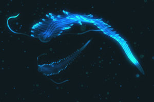
Fascinating bioluminescent organisms floating on dark waters of the ocean. Polychaete tomopteris.
Today’s blog comes to you from the Promega North America Branch Office.
In nature, the ability to “glow” is actually quite common. Bioluminescence, the chemical reaction involving the molecule luciferin, is a useful adaptation for many lifeforms. Fireflies, mushrooms and creatures of the ocean deep use their internal lightshows to cope with a variety of situations. Used for hunting, communicating, ridding cells of oxygen, and simply surviving in the darkness of the ocean depths, bioluminescence is one of nature’s more flashy, and advantageous traits.
In new research published in April in the journal Scientific Reports, MBARI researchers Séverine Martini and Steve Haddock found that three-quarters of all sea animals make their own light. The study reviewed 17 years of video from Monterey Bay, Calif in oceans that descended to 2.5 miles, to determine the commonality of bioluminescence in the deep waters.
Martini and Haddock’s observations concluded that 76 percent off all observed animals produced some light, including 97 to 99.7 cnidarians (jellyfish), half of fish, and most polychaetes (worms), cephalopods (squid), and crustaceans (shrimp).
Most of us are familiar with the fabled anglerfish, the menacing deep-sea creature known for attracting ignorant prey with a glowing lure attached to their head. As you descend below 200 meters, where light no longer penetrates, you will be surprised at the unexpected color display of the oceans’ sea life. Bioluminescence is not simply an exotic phenomenon, but an important ecological trait that the oceans’ sea creatures have wholeheartedly adopted to cope with complete darkness.
Fluorescence resonance energy transfer (FRET) probes or sensors are commonly used to measure cellular events. The probes typically have a matched pair of fluorescent proteins joined by a ligand-binding or responsive protein domain. Changes in the responsive domain are reflected in conformational changes that either bring the two fluorescent proteins together or drive them apart. The sensors are measured by hitting the most blue-shifted fluorescent protein with its excitation wavelength (donor). The resulting emission is transferred to the most red-shifted fluorescent protein in the pair, and the result is ultimately emission from the red-shifted protein (acceptor).
As pointed out by Aper, S.J.A. et al. below, FRET sensors face challenges of photobleaching, autofluorescence, and, in the case of exciting cyan-excitable donors, phototoxicity. Another challenge to using FRET sensors comes when employing optogenetic regulators to initiate the event you wish to monitor. Optogenetic regulators respond to specific wavelengths and initiate signaling. The challenge comes when the FRET donor excitation overlaps with the optogenetic initiation wavelengths. Researchers have sought to alleviate many of these challenges by exchanging the fluorescent donor for a bioluminescent donor, making bioluminescence resonance energy transfer (BRET) probes. In the three papers described below, the authors chose NanoLuc® Luciferase as the BRET donor due to its extremely bright signal.
When I was a post-doc at UT Southwestern, I was fortunate to interact with two Nobel prize winners, Johann Deisenhofer and Fred Gilman. Johann once helped me move a -80°C freezer into his lab when we lost power in my building. I once replaced my boss at small faculty mixer with a guest speaker and had a drink with Fred Gilman and several other faculty members from around the university. Among the faculty, one professor had a cell phone on his belt, an odd sight in 1995. Fred Gilman asked him what it was and why he had it. It was so his lab could notify him of good results anytime of the day. Fred laughed and told him to get rid of it – if it’s good data, it will survive until morning.
I was reminded of this story when I read a recent paper by Bodle, C.R. et al (1) about the development of a NanoBiT® Complementation Assay (2) to measure interactions of Regulators of G Protein Signaling (RGS) with Gα proteins in cells. (Fred Gilman was the first to isolate a G protein and that led to him being a co-recipient of the Nobel Prize in 1994). The authors created over a dozen NanoBiT Gα:RGS domain pairs and found they could classify different RGS proteins by the speed of the interaction in a cellular context. The interactions were readily reversible with known inhibitors and were suitable for high-throughput screening due to Z’ factors exceeding 0.5. The study paves the way for future work to identify broad spectrum RGS domain:Gα inhibitors and even RGS domain-specific inhibitors. This is the second paper applying NanoBiT Technology to GPCR studies (3).
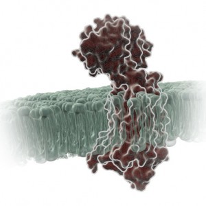
A Little Background… A primary function of GPCRs is transmission of extracellular signals across the plasma membrane via coupling with intracellular heterotrimeric G proteins. Upon receptor stimulation, the Gα subunit dissociates from the βγ subunit, initiating the cascade of downstream second messenger pathways that alter transcription (4). The Gα subunits are actually slow GTPases that propagate signals when GTP is bound but shutdown and reassociate with the βγ subunit when GTP is cleaved to GDP. This activation process is known as the GTPase cycle. G proteins are extremely slow GTPases.
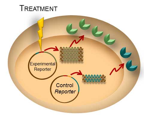
Transient transfection is often used to perform reporter assays. We have advocated using a dual-reporter system for decades to normalize the data obtained and gain a clearer understanding of your results. The experimental reporter should vary with treatment and the control reporter should vary little with treatment. The control reporter thus serves as a marker to help you understand the relative activity of your experimental reporter. The bioluminescent Dual-Luciferase® method allows for sequential detection of the second reporter in a single sample providing a simple two-step normalization method. Here are seven ways in which dual-reporter assays help you avoid misinterpreting results.
Simply comparing the ratio of the experimental to the control reporter can resolve differences in:
Number of Cells/Well: When manually pipeting cells into a 96-well plate, there is always a chance of having variable numbers of cells in each well. This variation is cell number will affect the experimental and control reporters equally, so the ratio of experimental:control reporter activity will eliminate false interpretation of the experimental data–whether it affects an entire row or column on the plate or individual wells.
Transfection Efficiency: The variations in transfection efficiency will equally affect both the experimental and control reporters so the ratio of activity in dual-reporter assays will normalize the data.
Cell Viability: Often, reporter assays look at the dose response curve of a particular compound with regard to gene expression. Ideally, if a compound causes a change in the experimental reporter the control reporter will demonstrate little effect. However, if the compound is toxic, both the experimental and control will be altered and the ratio will tell you whether the compound truly affects reporter activity or just kills the cells.
Lysis Efficiency: When lysing a plate of cells, you could encounter situations where rows or columns lyse differently, especially if you are using manual disruption or get interrupted mid-plate. The difference is lysis will affect the experimental and control equally so the ratio will remove the variation.
Temperature: Ideally, a plate should be equilibrated to ambient room temperature before proceeding to the reporter assay. Plates can cool at different rates or researchers anxious to record data may read the data early. Temperature variations will affect both reporters so the ratio will limit the affect on the data.
Measurement Time: Repetition of data is a hallmark of good science. You are often called upon to repeat experiments sometimes days or weeks apart. Let’s say you repeat your experiment one week after the initial experiment. The first time you measured the response, you waited 10 minutes after reagent addition to read, this week you waited 30 minutes. This will affect both reporters equally and therefore the ratio will allow you to more easily compare the data from this week and last week.
Bonus Benefit from Dual-Luciferase®, Dual-Glo® and the NanoGlo® Dual Luciferase Reporter Systems: NoLysate Splitting: Promega dual-reporter assays are designed for same-well multiplexing so there is no chance of variations creeping into your data due to unequal splitting of the cellular lysate to measure two separate reporter activities.
Since the introduction of the first bioluminescent dual-luciferase assay in 1995, this approach has been used in countless studies to advance our scientific understanding of cellular gene regulation.
A paper published on October 2 in the Journal of Virology describes an exciting development in the world of influenza research—the construction of a luciferase reporter virus that does not affect virulence and can be used to track development and spread of infection in mice.
Insertion of luciferase reporter genes into viruses has been accomplished before for several viruses, but has not been successful for influenza. Construction of influenza reporter viruses is complicated because the viral genome is small and all the viral genes are critical for infection. Therefore, replacement of an existing gene with a reporter gene or insertion of additional reporter sequences without affecting the virus’s ability to replicate and cause infection has proven difficult. To be successful, a reporter gene needs to be small enough to insert into the viral genome without eliminating any other vital functionality.
Search the PubMed database for “dual-luciferase” and citations abound. The Dual-Luciferase® Reporter Assay is a powerful tool that allows researchers to ask a multitude of questions about gene control and expression in a system that itself could be normalized and internally controlled. For more than 15 years, firefly and Renilla luciferases have formed the basis of a range of powerful assays and research tools for scientists who are asking questions about the deep and complex genetic and cellular story associated with cancer. Here we talk a bit of about bioluminescent chemistries, some of the newest bioluminescent tools available, and how some of these tools can be used to probe the deeper questions of cell biology, including cancer biology.
XWe use cookies and similar technologies to make our website work, run analytics, improve our website, and show you personalized content and advertising. Some of these cookies are essential for our website to work. For others, we won’t set them unless you accept them. To learn more about our approach to Privacy we invite you to Read More
By clicking “Accept All”, you consent to the use of ALL the cookies. However you may visit Cookie Settings to provide a controlled consent.
We use cookies and similar technologies to make our website work, run analytics, improve our website, and show you personalized content and advertising. Some of these cookies are essential for our website to work. For others, we won’t set them unless you accept them. To find out more about cookies and how to manage cookies, read our Cookie Policy.
If you are located in the EEA, the United Kingdom, or Switzerland, you can change your settings at any time by clicking Manage Cookie Consent in the footer of our website.
Necessary cookies are absolutely essential for the website to function properly. These cookies ensure basic functionalities and security features of the website, anonymously.
Cookie
Duration
Description
cookielawinfo-checbox-analytics
11 months
This cookie is set by GDPR Cookie Consent plugin. The cookie is used to store the user consent for the cookies in the category "Analytics".
cookielawinfo-checbox-functional
11 months
The cookie is set by GDPR cookie consent to record the user consent for the cookies in the category "Functional".
cookielawinfo-checbox-others
11 months
This cookie is set by GDPR Cookie Consent plugin. The cookie is used to store the user consent for the cookies in the category "Other.
cookielawinfo-checkbox-advertisement
1 year
The cookie is set by GDPR cookie consent to record the user consent for the cookies in the category "Advertisement".
cookielawinfo-checkbox-necessary
11 months
This cookie is set by GDPR Cookie Consent plugin. The cookies is used to store the user consent for the cookies in the category "Necessary".
cookielawinfo-checkbox-performance
11 months
This cookie is set by GDPR Cookie Consent plugin. The cookie is used to store the user consent for the cookies in the category "Performance".
gdpr_status
6 months 2 days
This cookie is set by the provider Media.net. This cookie is used to check the status whether the user has accepted the cookie consent box. It also helps in not showing the cookie consent box upon re-entry to the website.
lang
This cookie is used to store the language preferences of a user to serve up content in that stored language the next time user visit the website.
viewed_cookie_policy
11 months
The cookie is set by the GDPR Cookie Consent plugin and is used to store whether or not user has consented to the use of cookies. It does not store any personal data.
Analytical cookies are used to understand how visitors interact with the website. These cookies help provide information on metrics the number of visitors, bounce rate, traffic source, etc.
Cookie
Duration
Description
SC_ANALYTICS_GLOBAL_COOKIE
10 years
This cookie is associated with Sitecore content and personalization. This cookie is used to identify the repeat visit from a single user. Sitecore will send a persistent session cookie to the web client.
vuid
2 years
This domain of this cookie is owned by Vimeo. This cookie is used by vimeo to collect tracking information. It sets a unique ID to embed videos to the website.
WMF-Last-Access
1 month 18 hours 24 minutes
This cookie is used to calculate unique devices accessing the website.
_ga
2 years
This cookie is installed by Google Analytics. The cookie is used to calculate visitor, session, campaign data and keep track of site usage for the site's analytics report. The cookies store information anonymously and assign a randomly generated number to identify unique visitors.
_gid
1 day
This cookie is installed by Google Analytics. The cookie is used to store information of how visitors use a website and helps in creating an analytics report of how the website is doing. The data collected including the number visitors, the source where they have come from, and the pages visted in an anonymous form.
Advertisement cookies are used to provide visitors with relevant ads and marketing campaigns. These cookies track visitors across websites and collect information to provide customized ads.
Cookie
Duration
Description
IDE
1 year 24 days
Used by Google DoubleClick and stores information about how the user uses the website and any other advertisement before visiting the website. This is used to present users with ads that are relevant to them according to the user profile.
test_cookie
15 minutes
This cookie is set by doubleclick.net. The purpose of the cookie is to determine if the user's browser supports cookies.
VISITOR_INFO1_LIVE
5 months 27 days
This cookie is set by Youtube. Used to track the information of the embedded YouTube videos on a website.
Performance cookies are used to understand and analyze the key performance indexes of the website which helps in delivering a better user experience for the visitors.
Cookie
Duration
Description
YSC
session
This cookies is set by Youtube and is used to track the views of embedded videos.
_gat_UA-62336821-1
1 minute
This is a pattern type cookie set by Google Analytics, where the pattern element on the name contains the unique identity number of the account or website it relates to. It appears to be a variation of the _gat cookie which is used to limit the amount of data recorded by Google on high traffic volume websites.