Real-time, up-to-the-minute access to information provides new opportunities for scientists to monitor cellular events in ever more meaningful ways. Real-time cytotoxicity and cell viability assay reagents now allow constant monitoring of cell health status without the need to lyse or remove aliquots from plates for measurement. With a real-time approach, data can be collected from cell cultures or microtissues at multiple time points after addition of a drug compound or other event, and the response to treatment continually observed.
The CellTox™ Green assay is a real-time assay that monitors cytotoxicity using a fluorescent DNA binding dye, which binds DNA released from cells upon loss of membrane integrity. The dye cannot enter intact, live cells and so fluorescence only occurs upon cell death, correlating with cytotoxicity. Here’s a quick overview showing how the assay works:
More Data Using Fewer Samples and Reagents
The ability to continually monitor cytotoxicity in this way makes it easy to conduct more than one type of analysis on a single sample. Assays can be combined to determine not only the timing of cytotoxicity, but to also understand related events happening in the same cell population. As long as the readouts can be distinguished from one another multiple assays can be performed in the same well, providing more informative data while using less cells, plates and reagents.
Combining assays in this way can reveal critical information regarding mechanism of cell death. For example, assay combinations can be used to determine whether cells are dying from apoptosis or necrosis, or to distinguish nonproliferation from cell death. Combining CellTox Green with an endpoint luminescent caspase assay or a real-time apoptosis assay allows you to determine whether observed cytotoxic effects are due to apoptosis. Cytotoxic and anti-proliferative effects can be distinguished by combining the cytotoxicity assay with a luminescent or fluorescent cell viability assay.
Continue reading “A Better Way to Understand How and Why Cells Die” Several weeks ago, I came across an article on ScienceNews.org about how Wikipedia is becoming a scientific resource, whether we like it or not. Scientists are reading Wikipedia, the article said, and it’s affecting how they write. The article cited


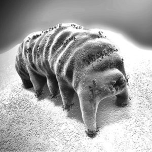 Everyone has their favorite microscopic creature—you all do have a favorite, right? Mine is unquestionably the tardigrade. Tardigrades, also called water bears or moss piglets, are microscopic invertebrates that are composed of five segments: one head segment and four body segments, each with a pair of legs. They are 0.1–1.2mm in length, making them easy to see under low magnification, and have a brain and well-developed nervous system. Tardigrades are found in just about every environment on earth. Termed “extremophiles”, they have adapted to survive in even extremely harsh environments. Your neighborhood pond? The Himalayas? Antarctica? Deep sea? Tardigrades live in all those places.