Welcome to Your Biotechnology Field Trip at the BTC Institute!

BTCI provides our students an opportunity that they could never get in the classroom.
—Jim Geoffrey, Biology Teacher, Kaukauna High School

Kaukauna High School students arrive at the BTC for a biotechnology fieldtrip.

Your bus has arrived and parked in the circular driveway at the front of the BioPharmaceutical Technology Center on the Promega Corporation campus in Fitchburg, WI. Your BTC Institute hosts – and instructors – for your field trip are Barbara Bielec (K-12 Program Director) and Ryan Olson (Biotechnology Instructor). They’ll greet you in the Atrium and direct you to a conference room where you can leave coats and backpacks, and then to the lab you’ll be working in during your visit.

Here’s a taste of what happened next for students from Random Lake High School and Wonewoc High School on December 3rd, and from Kaukauna High School on December 4th.

Continue reading “Welcome to Your Biotechnology Field Trip at the BTC Institute!”

Do you want to build a snowman? Developing and optimizing a qPCR assay to detect ice-nucleating activity

Snowflakes---MA-400x600

Over the last few months we have published several blogs about qPCR—from basic pointers on avoiding contamination in these sensitive reactions to a collection of tips for successful qPCR. Today we look in depth at a paper that describes the design and and optimization of a qPCR assay, and in keeping with the season of winter in the Northern hemisphere, it is only fitting that the assay tests for the abundance and identity of ice-nucleating bacteria.

Ice-nucleating bacteria are gram-negative bacteria that occur in the environment and are able to “catalyze” the formation ice crystals at warmer temperatures because of the expression of specific, ice-nucleating proteins on their outer membrane. Ice-nucleating bacteria are found in abundance on crop plants, especially grains, and are estimated to cause one-billion dollars in crop damage from frost in the United States alone.

In addition to their abundance on crop plants, ice-nucleating bacteria are also found on natural vegetation and have been isolated from soil, snow, hail, cloud water, in the air above crops under dry conditions and during rain fall. They have even been isolated from soil, seedlings and snow in remote locations in Antarctica. For the bacteria, ice nucleation may be a method to promote dissemination through rain and snow.

Although ice-nucleating bacteria have been isolated from clouds, ice and rain, little is known about their true contribution to precipitation or other events such as glaciation. Are such bacteria the only source of warm-temperature (above temperatures at which ice crystals form without a catalyst) ice nucleation? Can they trigger precipitation directly? What are the factors that trigger their release from vegetation into the atmosphere? Can we determine their abundance and variety in the environment?

Continue reading “Do you want to build a snowman? Developing and optimizing a qPCR assay to detect ice-nucleating activity”

Real-Time (quantitative) qPCR for Quantitating Library Prep before NGS

Real-Time (or quantitative, qPCR) monitors PCR amplification as it happens and allows you to measure starting material in your reaction.

This the last in a series of four blogs about Quantitation for NGS is written by guest blogger Adam Blatter, Product Specialist in Integrated Solutions at Promega.

When it comes to nucleic acid quantitation, real-time or quantitative (qPCR) is considered the gold standard because of its unmatched performance in senstivity, specificity and accuracy. qPCR relies on thermal cycling, consisting of repeated cycles of heating an cooling for DNA melting and enzyamtic replication. Detection instrumentation is capable of measuring the accumulation of DNA product after each round of amplification in real time.

Because PCR amplifies specific regions of DNA, the method is highly sensitive, specific to DNA, and it can determine whether a sample is truly able to be amplified. Degraded DNA or free nucleotides, which might otherwise skew your quantiation, will not contribute to the signal, and your measurement will be more accurate.

However, while qPCR does provide technical advantages, the method requires special instrumentation, specialized reagents and is a more time-consuming process. In addition, you will probably need to optimize your qPCR assay for each of your targets to achieve your desired results.

Because of the added complexity and cost, qPCR is a technique suited for post-library quantitation when you need to know the exact amount of amplifiable, adapter-ligated DNA.  PCR is the only method capable of specifically targeting these library constructs over other DNA that may be present. This specificity is important because accurate normalization is especially critical for producing even coverage in multiplex experiments where equimolar amounts of several libraries are added to a pooled sample. This normalization process is essential  if your are screening for rare variants that might be lost in background and go undetected if underrepresented in a mixed pool.


Read Part 1: When Every Step Counts: Quantitation for NGS

Read Part 2: Nucleic Acid Quantitation by UV Absorbance: Not for NGS

Read Part 3: Fluorescence Dye-Based Quantitation: Sensitive and Specific for NGS Applications

ISO 18385: The Creation of a “Forensic Grade” Standard

Today’s blog is from guest blogger Charles Stollberg. Charlie is a Promega production scientist in the Genetic Identity group and is focused on manufacturing inventory material and production process improvements. He’s been with the company for about 4 years. He graduated from UW-Whitewater in 2007 with a bachelor’s degree in cell biology. Prior to Promega, he worked in a small genetics lab studying lethal recessive traits in cattle.

ForensicGradeLogoForensic DNA laboratories rely on reagent and plastics manufacturers to supply high-quality products with minimal interference from contaminating DNA. With the increasing sensitivity of short tandem repeat (STR) amplification systems, levels of DNA that were previously undetected may now generate partial profiles. To address the concern of laboratories worldwide regarding the potential of low-level DNA contamination in consumables, ISO 18385 was developed to provide requirements for minimizing the risk of human DNA contamination events during the manufacturing process. Many of you may not have heard of ISO 18385, so I’d like to give you an introduction to how the standard came to be.

Continue reading “ISO 18385: The Creation of a “Forensic Grade” Standard”

Fluorescence Dye-Based Quantitation: Sensitive and Specific for NGS Applications

This is the third post in a series of blogs on quantitation for NGS applications written by guest blogger Adam Blatter, Product Specialist in Integrated Solutions at Promega.

Fluorescent dye-based quantitation uses specially designed DNA binding compounds that intercalate only with double stranded DNA molecules. When excited by a specific wavelength of light, only dye in the DNA-bound state will fluoresce. These aspects of the technique contribute to low background signal, and therefore the ability to accurately and specifically detect very low quantities of DNA in solution, even the nanogram quantities used in NGS applications.

For commercial NGS systems, such as the Nextera Rapid Capture Enrichment Protocol by Illumina, this specificity and sensitivity of quantitation are critical. The Nextera protocol is optimized for 50ng of total genomic DNA. A higher mass input of genomic DNA can result in incomplete tagmentation, and larger insert sizes, which can adversely affect enrichment. A lower mass input of genomic DNA or low-quality DNA can generate smaller than expected inserts, which can be lost during subsequent cleanup steps, giving lower diversity of inserts.

Continue reading “Fluorescence Dye-Based Quantitation: Sensitive and Specific for NGS Applications”

NanoBiT™ Assay: Transformational Technology for Studying Protein Interactions Named a Top 10 Innovation of 2015

21416943-wb-cr-uw-nanobit-pm-page-hero
For three out of the last four years, we have been honored to have one of our key technologies named a Top 10 Innovation by The Scientist. This year the innovative NanoBiT™ Assay (NanoLuc® Binary Technology) received the recognition. NanoBiT™ is a structural complementation reporter based on NanoLuc® Luciferase, a small, bright luciferase derived from the deep sea shrimp Oplophorus gracilirostris.

Using plasmids that encode the NanoBiT complementation reporter, you can make fusion proteins to “report” on protein interactions that you are studying. One of the target proteins is fused to the 18kDa subunit; the other to the 11 amino acid subunit. The NanoBiT™ subunits are stable, exhibiting low self-affinity, but produce an ultra-bright signal upon association. So, if your target proteins interact, the two subunits are brought close enough to each other to associate and produce a luminescent signal. The strong signal and low background associated with a luminescent system, and the small size of the complementation reporter, all help the NanoBiT™ assay overcome the limitations associated with traditional methods for studying protein interactions.

The small size reduces the chances of steric interference with protein interactions. The ultra bright signal, means that even interactions among proteins present in very low amounts can be detected and quantified–without over-expressing large quantities of non-native fusion proteins and potentially disrupting the normal cellular environment. And the NanoBiT™ assay can be performed in real time, in live cells.

The NanoBiT™ assay is already being deployed in laboratories to help advance understanding of fundamental cell biology. You can see how one researcher is already taking full advantage of this innovative technology in the video embedded below:

Visit the Promega web site to see more examples more examples how the NanoBiT™ assay can break through the traditional limitations for studying protein interactions in cells.

You can read the Top 10 article in The Scientist here.

Circulating Biomarkers and Their Applications in Gastric Cancer

cancer cellCancer. It has been the nemesis of medical science for decades. We declare war on it, we wax philosophical about finding a cure for it. We talk about it as if it were a single enemy, but it isn’t—Cancer is not a disease, it is hundreds of diseases. These diseases manifest in every region of the human body. Many cancers, if diagnosed early, can be treated successfully. Unfortunately, there are many forms of cancers that have no external signs and very few symptoms at the early stages. Such is the enemy we face: we know what it is, we know we need to kill it early and in many cases we know how to do it, what we don’t know is how to catch it early.

Gastric cancer kills approximately 745,000 people a year worldwide, making it the third most common cause of cancer-related deaths (1). It has such a high mortality rate because usually it is not detected until the disease has progressed to the later stages (IIIA–IV; 2). When detected this late, the 5-year survival rate ranges from 7–27%, with the median survival being less than 12 months (2). In contrast, when diagnosed early (i.e., cancer that is limited to the submucosal layer) it is curable with an endoscopic mucosal dissection or a minimally invasive surgery. The difference between these two outcomes is time. The earlier the cancer is detected, the better the prognosis.

Currently, upper endoscopy is the primary screening technique for detecting precancerous lesions as well as gastric cancer in the early stages. This technique has a number of downsides: it is invasive, it can have serious side effects (although these are uncommon), and it is expensive and highly dependent on the skill of the endoscopist.  For these reasons, endoscopy screening is likely to suffer from poor participation rates. In addition, endoscopy is not a practical approach in low-income countries. There is clearly a need for a less invasive, sensitive screening test that will detect gastric cancer at an early stage. Continue reading “Circulating Biomarkers and Their Applications in Gastric Cancer”

A Better GTPase Assay for Drug Development

Cover of October Issue of Assay and Drug Development Technologies featuring GTPase-Glo™ Assay.

The path to drug development is strewn with obstacles: Identifying targets; configuring assays to help identify targets or drugs; uncovering the right compound to affect the selected target without off-target effects and screening multiple compounds to eliminate or identify potential drugs. Without the right tools, compounds or target, identifying potential disease therapies becomes nearly impossible.

When it comes to a drug target for cancer, the Ras protein family is at the top of the list because the proteins are expressed ubiquitiously and found mutated in many types of cancer. Because Ras proteins are involved in transducing signals from the surface of cells, many of the resulting mutations produce an activated Ras, inducing uncontrolled expression of the genes that Ras controls. Ras proteins are small GTPases (20–25kDa) that comprise a larger superfamily of proteins divided into five subfamilies: Ras, Rho, Rab, Arf, and Ran. These proteins control diverse cellular activities, including cellular differentiation, proliferation, cell division, nuclear import and export, and vesicle transport. GTPases are guanosine-nucleotide-binding proteins with affinity for GDP or GTP and are able to hydrolyze GTP. When bound to GTP, GTPases are active (turned on) and interact with downstream proteins in the signaling cascade. When GTPases are bound to GDP, the proteins are inactivated (turned off) and no longer transduce signals.

Continue reading “A Better GTPase Assay for Drug Development”

Nucleic Acid Quantitation by UV Absorbance: Not for NGS

schematic diagram of UV-Vis Absorbance Method
For UV-Vis Spectrophotometry, light is split into its component wavelengths and directed through a solution. Molecules in the solution absorb specific wavelengths of light.

This is the second in a series of four blogs about Quantitation for NGS is written by guest blogger Adam Blatter, Product Specialist in Integrated Solutions at Promega.

Perhaps the most ubiquitous quantitation method is UV-spectrophotometry (also called absorbance spectroscopy). This technique takes advantage of the Beer-Lambert Law: an observation that many compounds absorb UV-Visible light at unique wavelengths, and that for a fixed path length the absorbance of a solution is directly proportional to the concentration of the absorbing species. DNA, for example has a peak absorbance at 260nm (A260nm).

This method is user friendly, quick and easy. But, it has significant limitations, especially when quantitating samples for NGS applications.

Continue reading “Nucleic Acid Quantitation by UV Absorbance: Not for NGS”

When Every Step Counts: Quantitation for NGS

13170MA-800x277This series of blogs about Quantitation for NGS is written by guest blogger Adam Blatter, Product Specialist in Integrated Solutions at Promega.

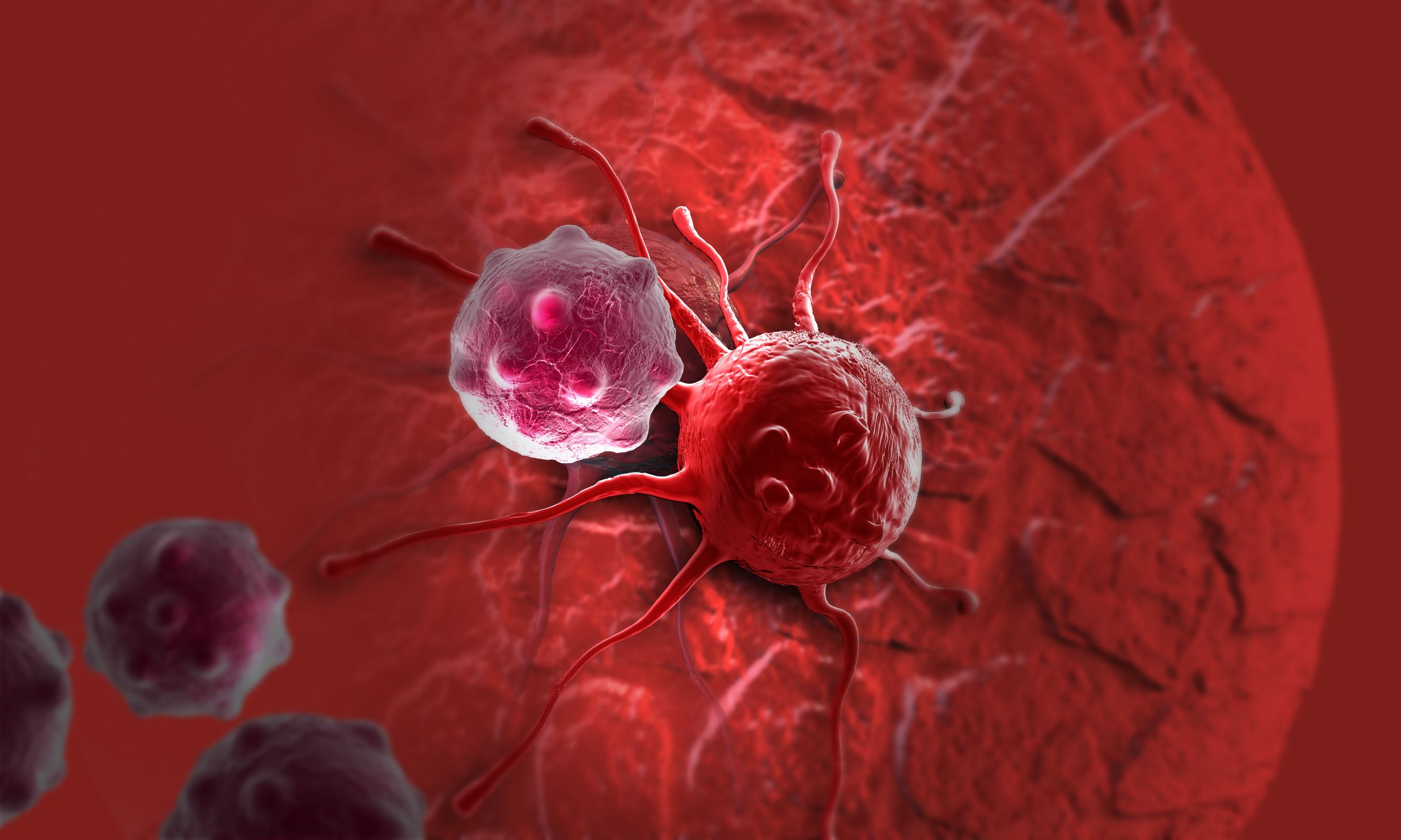
As sequencing technology races toward ever cheaper, faster and more accurate ways to read entire genomes, we find ourselves able to study biological systems at a level never before possible. From basic science to translational research, massively parallel sequencing (also known as next-generation sequencing or NGS) has opened up new avenues of inquiry in genomics, oncology and ecology.

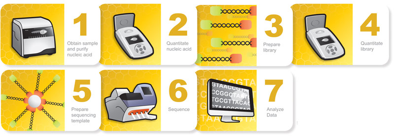
Many commercial sequencing platforms have been established (e.g., Illumina, IonTorrent, 454, PacBio), and new technologies are developed every day to enable new and unique applications. However, all of these platforms and technologies work off the same general principle: nucleic acid must be extracted from a sample, arranged into platform-specific library constructs, and loaded into the sequencer. Regardless of the sample type or the platform used, every step throughout this workflow is critical for successful results. An often overlooked part of the NGS workflow is sample quantitation. Here we are presenting the first in a series of four short blogs about the critical step of quantitation in NGS workflows.

Sample input is critical to NGS in terms of both quality and quantity. Knowing how much DNA you have, often in nanogram quantities, can make the difference between success and failure. There are several key points in the NGS workflow where sample quantitation is important before you can proceed:

  • After initial nucleic acid extraction from the sample matrix and before proceeding with library preparation
  • Post-library preparation when pooling barcoded libraries for multiplexing
  • Final pooled library quantitation immediately before loading for sequencing

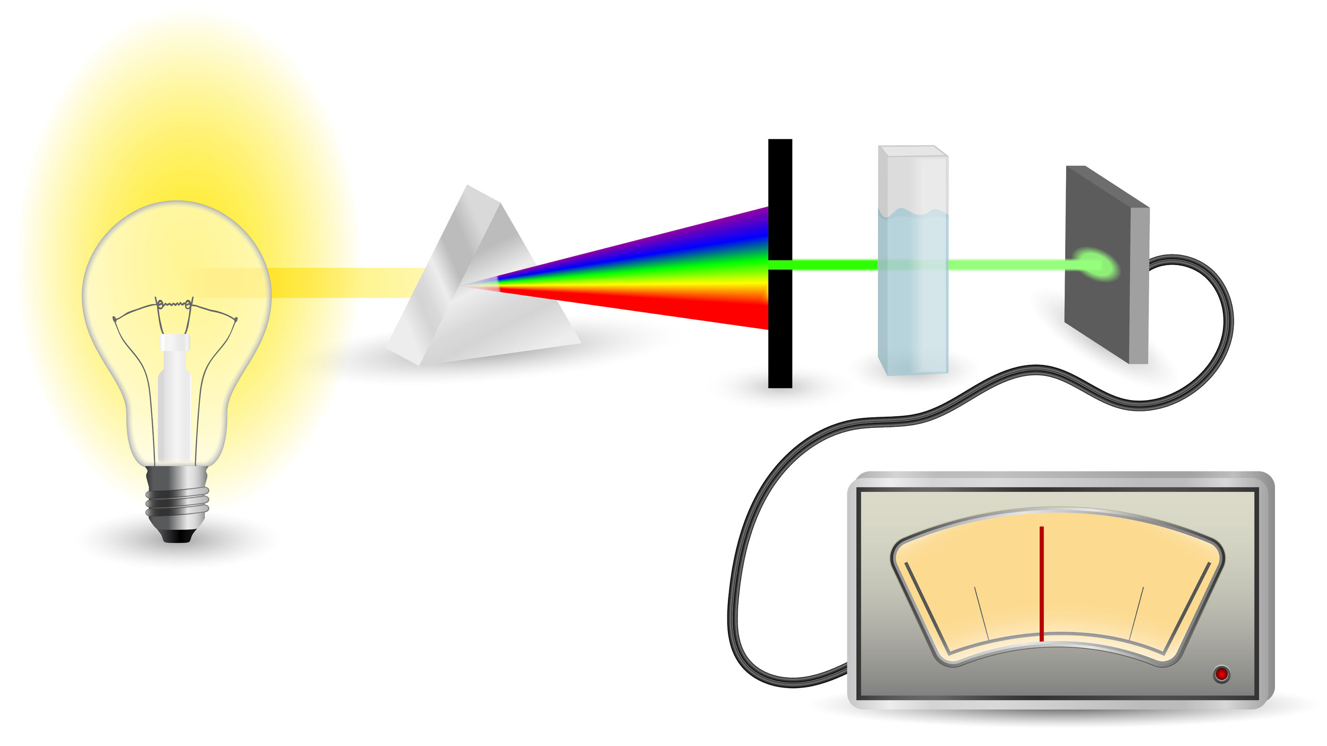
There are several common methods for quantitating nucleic acids: UV-spectroscopy, Fluorescence spectoscopy, real-time quantitative PCR (qPCR). Because of inherent differences in sensitivity, specificity, time and cost, each of these techniques pose certain advantages and disadvantages with respect to the specific sample you are quantitating. Our next three blogs will discuss each of these methods against the backdrop of quantitating samples for NGS applications.


Read Part 2: Nucleic Acid Quantitation by UV Absorbance: Not for NGS

Read Part 3: Fluorescence Dye-Based Quantitation: Sensitive and Specific for NGS Applications

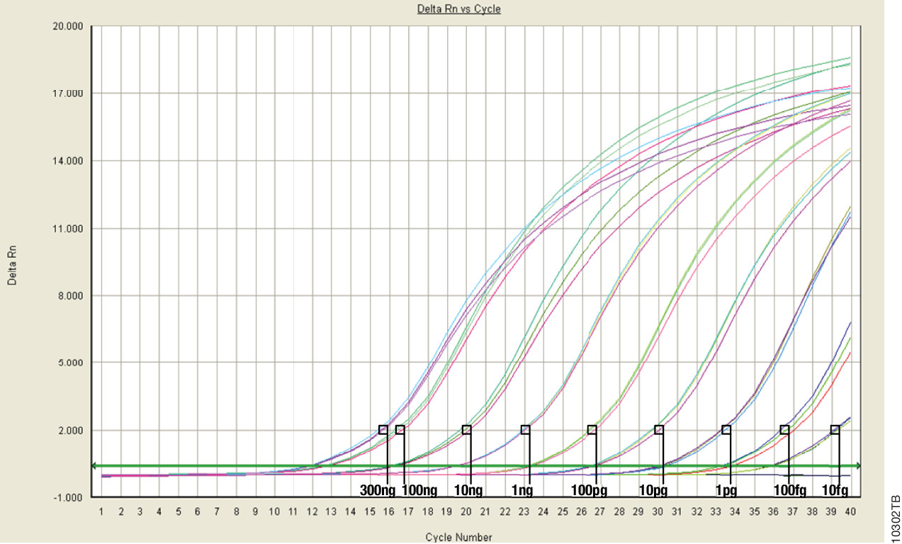
Read Part 4: Real-Time (Quantitative) qPCR for Quantitating Library Prep before NGS