Will Warmer Weather Wake the Sleeping Giant (Viruses)?

Artist's conception of Mimivirus structure, the first of the giant viruses identified.
Artist’s conception of Mimivirus structure, the first of the giant viruses identified.

Following the discovery of Mimivirus (1) the first virus with a particles large enough to be visible under the light microscope, two additional “giant” viruses infecting Acanthamoeba have been discovered Pandoravirus (2) and Pithovirus sibericum (3), the latter from a 30,000 year old Siberian permafrost. A fourth type was recently isolated from the same sample of permafrost by Legendre et al, and named Mollivirus sibericum (4).

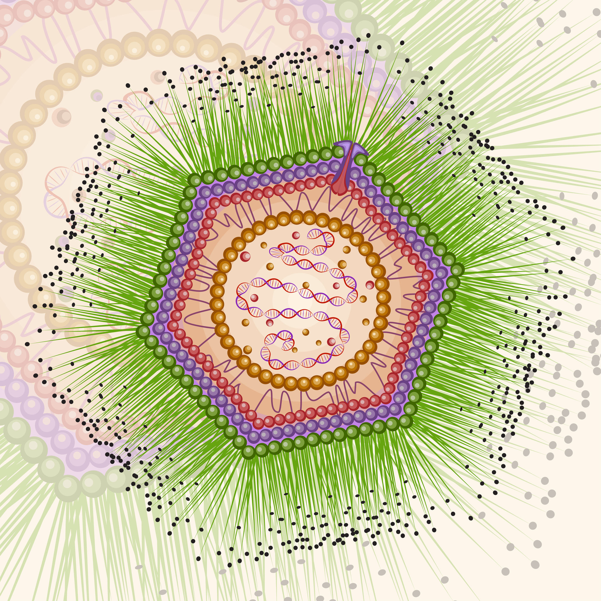
Mollivirus sibericum has an approximately spherical virion (0.6 µm diameter) with a 651kb GC-rich genome that encodes 523 proteins. To further characterize the virus the researchers performed transcromic- and proteomic-based time course experiments.

For the particle proteome and infectious cycle analysis, proteins were extracted and then run a 4–12% polyacrylamide gel, and trypsin digests were performed in-gel before nano LC-MS/MS analysis of the resulting peptides. Proteomic studies of the particle showed that it lacked an embarked transcription apparatus, but revealed an unusual presence of many ribosomal and ribosome-related proteins.

When the researchers explored the proteome during the course of an entire infectious cycle, the relative proportions of Mollivirus-, mitochondrion-, and Acanthamoeba encoded proteins were found to vary consistently with an infectious pattern that preserved the cellular host integrity as long as possible and with the release of newly formed virus particles through exocytosis.

In an interesting footnote, the authors of this study point out the fact that two different viruses retain their infectivity in prehistorical permafrost layers should be a concern in the context of global warming and the potential to expose humans to primeval viruses.

References

1. La Scola, B. et al.   (2003) A giant virus in amoebae. Science  299, 2033.
2. Philippe, N. et al. (2013) Pandoraviruses. Amoeba virus with genomes up to 2.5Mb reaching that of parasitic eukaryotes. Science 341,281–6.
3. Legendre, M. et al. (2014) Thirty thousand year old distant relative of giant icosahedral DNA viruses with a pandoravirus morphology. Proc.Natl. Acad. Sci. 111, 4274–9.
4. Legendre, M. et al. (2015)  In depth study of Mollivirus sibercum, a new 30,000 year old giant virus infecting Acanthamoeba.  Proc. Natl. Acad. Sci. 112, E5327–35 (online).

How Fruit Flies (and maybe Pigeons?) Navigate; A New Report

A rock dove, similar in plumage to a pigeon.

Several years ago an intriguing story of successful navigation in complex situation, by pigeons, the birds most often compared to rats, caught my eye.

Our backyard once had a coop full of pigeons, so I’m not a total stranger to their navigation abilities (nor am I a pigeon expert). My favorites were the tumbling pigeons.

But it didn’t take much time researching that article from 2012, to learn that one of the more hotly debated how-do-they-do-it topics is animal navigation, in particular, the ability of pigeons to navigate back to home/point A when released at point B.

So when it appeared online today, in Nature Materials, the story “A Magnetic Protein Biocompass” caught my eye.

Continue reading “How Fruit Flies (and maybe Pigeons?) Navigate; A New Report”

There and Back Again, Part 1

In 2014, Promega created a special incentive to reward field science consultants who help the scientific community take advantage of the our on-site stocking program. The winners had to meet ambitious criteria to receive 2 round-trip tickets to anywhere in the world, a week of paid vacation and spending money. Our four winners from 2014 will share photos and stories about their journeys in a semi-regular Friday feature on the Promega Connections Blog.

Today’s travelogue comes to us from Mica Zaragoza, a senior client rep, who used his award to travel to Australia and New Zealand.

When initially introduced to the ambitious Helix award, I was amazed at the prospect of selecting anywhere in the world to travel, while blogging about my the adventures. Both humbled and amazed to receive this opportunity, my wife and I embarked on a journey across the Pacific.

Hyde Park in Sydney, Australia.

Sydney, Australia

Departing our home in Chicago, my wife Crystal and I started our journey with a 5-hour trip to San Francisco for a layover before the 14 hour journey to Sydney. After jumping into the future (Thurs became Saturday), our first visit was to Central Sydney’s Hyde Park.

Taking jet lag into consideration, we decided to double-down by freshening up and dropping luggage to kick off our day at 7:30am. My first Australian purchase? Coffee! Continue reading “There and Back Again, Part 1”

The Promise of miRNAs as Therapeutic Agents in Treating Disease

When researchers first identified a new family of seemingly non-functional “junk” RNA molecules, it’s unlikely they could have predicted the power and promise of these nucleic acids. The small, non-coding, single-stranded RNAs – typically 21-25 base pairs in length – were first discovered over 20 years ago in C. elegans, yet they were quickly found to be ubiquitous in species from worms to flies to plants to mammals. The role of these novel RNAs in the regulation of developmental pathways in worms, coupled with their prevalence, inspired researchers to better understand their significance.

We now know that miRNAs (for microRNAs) serve as post-transcriptional repressors of gene expression by targeting degradation of mRNA or interfering with mRNA translation. While small, each can have a big effect; a single miRNA can regulate dozens to hundreds of distinct target genes. They’ve been implicated in a variety of critical cellular processes such as differentiation, development, metabolism, signal transduction, apoptosis and proliferation.

Tissue-specific expression patterns revealed that specific miRNAs are enriched in mammalian tissues including adult brain, lung, spleen, liver, kidney and heart.  More compelling was the identification of abnormal miRNA expression in tumorigenic cell lines. It’s no wonder that this growing family quickly became ripe for exploration in disease development.

Basic research on miRNA is making its way into the clinic.
Research on miRNA is making its way into the clinic.

Within only a few years, a rapidly expanding body of research supported the theory that miRNA expression may indeed play a role in the development of human diseases including cardiovascular disease, cancer, diabetes, cystic fibrosis, and liver disease. Investigations into the expression of miRNAs in cardiovascular disease, in particular, have demonstrated not only their value as disease markers, but also how their dysregulation is linked to disease processes.

More recently a new possibility is being explored: can miRNA be manipulated to interfere with disease progression? Continue reading “The Promise of miRNAs as Therapeutic Agents in Treating Disease”

Support Young Scientists: Mentors Needed for Dane County Biotechnology Youth Apprentices

I moved back to Madison from the east coast last September and I have to say it’s been really great being back in the Midwest… the Youth Apprenticeship Program opened doors to opportunities for me that may never have existed if I had not participated in the program. It established the foundation of my entire resume throughout college, which was crucial to the genetic counseling application process. — Kristin Gunderson, Genetic Counselor, Carbone Cancer Center (Kristin worked in the lab of Dr. Deane Mosher, UW School of Medicine Public Health, under the mentorship of Dr. Bianca Tomasini-Johannson and is a 2006 high school graduate.)

Yang Chen, 2015 graduate, at her worksite in the lab of Dr. Xuehua Zhong, UW-Madison Department of Genetics; mentor: Dean Sanders. Yang is currently a freshman at UW-Madison, majoring in microbiology.

OK, we are not going to be shy about it: We need any assistance our readers may be able to provide to help us find additional mentors for high school juniors and seniors who are enrolled in the Dane County Youth Apprenticeship Program in Biotechnology.

The good news is that there are 32 students who have elected to participate in the program, given their strong interests in the life sciences and in particular, biotechnology. They represent 14 public high schools in the area. They (1) complete all necessary classes for graduation; (2) attend a hour-hour intensive laboratory course at the BTC Institute from 4:30–8:30pm on Wednesdays; and, (3) work in laboratory settings throughout the community.  (For details, please visit: http://www.btci.org/k12/yap/yap.html) Continue reading “Support Young Scientists: Mentors Needed for Dane County Biotechnology Youth Apprentices”

Interview Day Checklist

Ribbon to remember

I’m a list person. You may know people like me—we are the ones who start compiling a list of items to pack for vacation a month in advance; we wouldn’t be caught in a grocery store without a carefully curated grocery list (often organized by department), and we have been known to write down previously completed items on our to-do list just to experience the satisfaction of crossing them off. The internet is full of lists and I love comparing other people’s checklists against my own to make sure I have what I need.

Some call my list-making zeal a curse, some call it a gift. Whatever you call it, I’d like to share with you my suggestions of items to bring to your next onsite interview (in list form, of course). Whether you are as passionate about lists as I am or not, I think it can help. Packing for an onsite interview in advance can help you feel calm, confident and prepared; which is exactly what an interviewer wants to see. When getting ready for an interview, be sure to pack:

Continue reading “Interview Day Checklist”

Yersinia pestis Reveals More Secrets From the Grave

Yersinia pestis. By A.Myasnikov for Wiki (Self made work) [CC0], via Wikimedia Commons

Fridays are generally reserved for fun posts to share prior to the weekend. As we all know, fun is relative and to me, the latest news about how long Yersinia pestis has been entwined with human history is intriguing. I enjoy writing about the latest historical finding of Y. pestis even if I do earn a black reputation among my blogging colleagues (pun intended). Therefore, as soon as I saw the Cell article about Y. pestis found in Bronze age human teeth, I knew my blog topic was at hand.

Y. pestis has long been suspected in several plagues that occurred in the last two millennia. Publications in 2011 and 2013 used DNA extracted from teeth of human remains dated to the 14th century Black Death and 6th century Plague of Justinian to confirm Y. pestis was the causative agent in those devastating plagues. These results beg the question: How long has Y. pestis been infecting humans? The phylogenic trees generated from recent studies suggested Y. pestis has been with humans for as little as 2,600 years and as long as and 28,000 years. Equipped with these DNA-based tools, Rasmussen et al. asked if they could find evidence of Y. pestis in older human remains.

Continue reading Yersinia pestis Reveals More Secrets From the Grave”

IdeZ Protease: A New Tool for the Characterization of Antibodies

12335MB

Therapeutic monoclonal antibodies are large, complex molecules that undergo numerous post translational modifications (PTMs).  In-depth characterization of antibody PTMs remains a significant hurdle because their large size (~150 kDa) makes mass spectrometry analysis extremely challenging.

IdeS protease specifically cleaves IgGs into Fab and Fc fragments. This enzyme is highly specific and cleaves human IgG specifically at one site in the lower hinge region.  Because of the exquisite specificity of the enzyme, it produces highly homogeneous Fc and Fab fragments which are then readily analyzed using techniques such as mass spectrometry or HPLC.

Are you looking for proteases to use in your research?
Explore our portfolio of proteases today.

One of the drawbacks of IdeS is that it exhibits poor activity against mouse IgGs. IdeZ Protease is an immunoglobulin-degrading enzyme from Streptococcus equi subspecies zooepidemicus. It is an engineered recombinant protease overexpressed in E. coli. Like IdeS Protease, IdeZ Protease specifically cleaves IgG molecules below the hinge region to yield F(ab′)2 and Fc fragments.  Reduction of the digestion products produces three fragments of ~25kDa that are readily analyzed by LC-MS.

One of the key advantages of the IdeZ Protease is that it has significantly improved activity against mouse IgG2a and IgG3 subclasses compared to IdeS Protease. IdeZ Protease does not cleave mouse IgG1 or IgG2b.

Key technical parameters when digesting mouse IgGs utilizing IdeZ are the following:

• Add 1 unit of IdeZ Protease per 1µg of IgG to be digested.
• IdeZ Protease is most active in buffers at or near neutral pH. The recommended digestion buffer is 50mM sodium phosphate, 150mM NaCl (pH 6.6).
• Mouse IgG2a and IgG3 typically require 2–4 hours at 37°C  for complete digestion.
• IdeZ Protease has a histidine tag for easy removal if so desired.

Interrogating Protein Interactions: An Infographic for NanoBRET™ Assay Design

Yesterday my fellow blogger, Kari, posted a review of the ACS Chemical Biology paper describing a new BRET platform for analyzing protein-protein interactions. If you are interested in studying induction and inhibition of protein interactions in real time, take a look at the infographic below to learn how to develop a NanoBRET™ Assay to monitor your protein of interest.

21421668-IG-CR-NanoBRET-Infographic-Final

About the Development of an Improved BRET Assay: NanoBRET

"Protein BRD4 PDB 2oss" by Emw - Own work. Licensed under CC BY-SA 3.0 via Wikimedia Commons - https://commons.wikimedia.org/wiki/File:Protein_BRD4_PDB_2oss.png#/media/File:Protein_BRD4_PDB_2oss.png
“Protein BRD4 PDB 2oss” by Emw – Own work. Licensed under CC BY-SA 3.0 via Wikimedia Commons – https://commons.wikimedia.org/wiki/File:Protein_BRD4_PDB_2oss.png#/media/File:Protein_BRD4_PDB_2oss.png

One of the more exciting reporter molecules technologies available came online in the past year, with the launch of the Promega NanoBRET™ technology. While it’s easy for me, a science writer at Promega, to brag, seriously, this is a very cool protein interactions tool.

A few of the challenges facing protein-protein interactions researchers include:

  • The ability to quantitatively characterize protein-protein interactions
  • Ability to examine protein-protein interactions in situ, in the context of the living cell

A goal of the NanoBRET™ developers was to improve the sensitivity and dynamic range of traditional BRET technology, in order to address these challenges.

In May 2015 these researchers published an article outlining their efforts to create NanoBRET technology in ACS Chemical Biology, in an article entitled, “NanoBRET—A Novel BRET Platform for the Analysis of Protein-Protein Interactions”. Here is a brief look at their work.

Continue reading “About the Development of an Improved BRET Assay: NanoBRET”