Making the Switch from FRET to BRET: Applications of NanoLuc® Luciferase with Fluorescent Protein Acceptors for Sensing Cellular Events

A Bioluminescent Alternative

Fluorescence resonance energy transfer (FRET) probes or sensors are commonly used to measure cellular events. The probes typically have a matched pair of fluorescent proteins joined by a ligand-binding or responsive protein domain. Changes in the responsive domain are reflected in conformational changes that either bring the two fluorescent proteins together or drive them apart. The sensors are measured by hitting the most blue-shifted fluorescent protein with its excitation wavelength (donor). The resulting emission is transferred to the most red-shifted fluorescent protein in the pair, and the result is ultimately emission from the red-shifted protein (acceptor).

As pointed out by Aper, S.J.A. et al. below, FRET sensors face challenges of photobleaching, autofluorescence, and, in the case of exciting cyan-excitable donors, phototoxicity. Another challenge to using FRET sensors comes when employing optogenetic regulators to initiate the event you wish to monitor. Optogenetic regulators respond to specific wavelengths and initiate signaling. The challenge comes when the FRET donor excitation overlaps with the optogenetic initiation wavelengths. Researchers have sought to alleviate many of these challenges by exchanging the fluorescent donor for a bioluminescent donor, making bioluminescence resonance energy transfer (BRET) probes. In the three papers described below, the authors chose NanoLuc® Luciferase as the BRET donor due to its extremely bright signal.

Continue reading “Making the Switch from FRET to BRET: Applications of NanoLuc® Luciferase with Fluorescent Protein Acceptors for Sensing Cellular Events”

Probing RGS:Gα Protein Interactions with NanoBiT Assays

gpcr_in_membrane_on_white2

When I was a post-doc at UT Southwestern, I was fortunate to interact with two Nobel prize winners, Johann Deisenhofer and Fred Gilman.  Johann once helped me move a -80°C freezer into his lab when we lost power in my building. I once replaced my boss at small faculty mixer with a guest speaker and had a drink with Fred Gilman and several other faculty members from around the university. Among the faculty, one professor had a cell phone on his belt, an odd sight in 1995. Fred Gilman asked him what it was and why he had it. It was so his lab could notify him of good results anytime of the day. Fred laughed and told him to get rid of it – if it’s good data, it will survive until morning.

I was reminded of this story when I read a recent paper by Bodle, C.R. et al (1) about the development of a NanoBiT® Complementation Assay (2) to measure interactions of Regulators of G Protein Signaling (RGS) with Gα proteins in cells. (Fred Gilman was the first to isolate a G protein and that led to him being a co-recipient of the Nobel Prize in 1994). The authors created over a dozen NanoBiT Gα:RGS domain pairs and found they could classify different RGS proteins by the speed of the interaction in a cellular context. The interactions were readily reversible with known inhibitors and were suitable for high-throughput screening due to Z’ factors exceeding 0.5. The study paves the way for future work to identify broad spectrum RGS domain:Gα inhibitors and even RGS domain-specific inhibitors. This is the second paper applying NanoBiT Technology to GPCR studies (3).

A Little Background…
A primary function of GPCRs is transmission of extracellular signals across the plasma membrane via coupling with intracellular heterotrimeric G proteins. Upon receptor stimulation, the Gα subunit dissociates from the βγ subunit, initiating the cascade of downstream second messenger pathways that alter transcription (4). The Gα subunits are actually slow GTPases that propagate signals when GTP is bound but shutdown and reassociate with the βγ subunit when GTP is cleaved to GDP. This activation process is known as the GTPase cycle. G proteins are extremely slow GTPases.

Continue reading “Probing RGS:Gα Protein Interactions with NanoBiT Assays”

Restoring Memory in Alzheimer’s Mice with Microbubbles and Ultrasound

Neurons with amyloid plaques.

Imagine driving in your car and suddenly not recognizing where you were going and having no idea how to find your way home. What if you looked across the breakfast table at your spouse and no longer recognized them?  Or maybe you have to brace yourself every time you visit your parent, waiting for the day when they won’t know who you are. This is the reality for the estimated 50 million (worldwide) Alzheimer’s disease sufferers and their families.

In a world with an aging population, Alzheimer’s is a growing problem. Recent estimates suggest that 11% of people over the age of 65 have Alzheimer’s disease. For people 85 and older, that number increases to 32% (1).

Alzheimer’s disease is a devastating degenerative brain disease. It is the most common cause of dementia and is characterized by a decline in cognitive skills such as memory, language skills, communication and problem-solving abilities. These symptoms make it difficult for people with Alzheimer’s to perform everyday activities. It also is difficult to diagnose, even more, difficult to treat, and, as of now, impossible to cure.

Continue reading “Restoring Memory in Alzheimer’s Mice with Microbubbles and Ultrasound”

Gut Microbes and Hypertension: Demonstrating a Causal Link

Most of us are aware that the human body is covered by and full of microorganisms. And we understand that most of these microorganisms are helpful, both in terms of competition with and protection against invading microorganisms, and in the gut, as agents of digestion.

Bacillus subtilis, an example of Firmicutes, and not a good gut microbe. By Y tambe (original uploader) - Own work, CC BY-SA 3.0, https://commons.wikimedia.org/w/index.php?curid=49528
Bacillus subtilis, an example of Firmicutes, and not a good gut microbe. By Y tambe (original uploader) – Own work, CC BY-SA 3.0, https://commons.wikimedia.org/w/index.php?curid=49528

In the past decade, however, research has brought compelling details implicating gut microbes in obesity, cancer, insulin resistance and such central nervous system disorders as depression, austism spectrum disorder and multiple sclerosis (Adnan, S. et al.). Yet the mechanisms and details of these associations have not been fully demonstrated.

Gut microbes have been proven to be connected to thickening of heart vasculature, known as atherosclerosis. Researchers have demonstrated that bacteria metabolize choline and L-carnitine from food to trimethylamine, which crosses the gut barrier into circulation and reaches the liver. In the liver, trimethylamine is metabolized to the atherogenic molecule triethylamine-N-oxide (Gregory, J.C. et al., Brown and Hazen). These studies are among the few that provide a direct connection between gut microbes and a pathological condition.

Continue reading “Gut Microbes and Hypertension: Demonstrating a Causal Link”

NanoBiT Assay Applied to Study Role of SOD1 in ALS

NanoBiT Protein Complementation
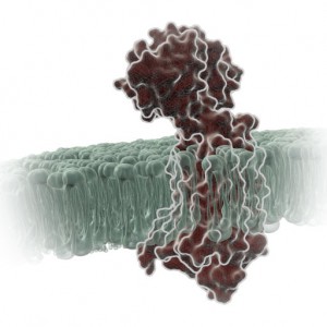
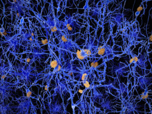
Back in 2015 the Ice Bucket Challenge brought Amyotrophic Lateral Sclerosis (ALS) to public attention, initiating worldwide pleas for more funding of research toward a cure for this fatal disease, which is characterized by progressive degeneration of motor neurons. In spite of many efforts over the last few decades, the precise cause of ALS is still unknown.

The complexity of the problem of ALS pathogenesis is highlighted in the review “Decoding ALS: from genes to mechanism”  published in Nature in November 2016. The review highlights a long list of genetic factors implicated in ALS, grouping them into genes affecting protein quality control, RNA stability/function, and the cytoskeletal structure of neuronal cells.

Mutations in the antioxidant enzyme superoxide dismutase (SOD1) were the first to be associated with ALS. According to the review, more than 170 SOD1 mutations causing ALS have since been identified. Many of these mutations are thought to result in misfolding of SOD1, contributing to toxicity when the misfolded protein accumulates within the cell.

A paper by Oh-hashi et al., published in Cell Biochemistry and Function in October 2016 used the NanoBiT protein complementation assay to investigate the effect of two common ALS-associated SOD1 mutations on dimerization of the SOD1 protein. Continue reading “NanoBiT Assay Applied to Study Role of SOD1 in ALS”

The Role of the NanoLuc® Reporter in Investigating Ligand-Receptor Interactions

Luminescent reporter assays are powerful research tools for a variety of applications. Last March we presented a webinar on this topic, Understanding Luminescent Reporter Assay Design, which proved to enlighten many who registered. The webinar addressed the importance of careful experimental design when using a luminescent reporter such as Promega’s Firefly or NanoLuc® Luciferase.

Reporters provide a highly sensitive, quantifiable metric for cellular events such as gene expression, protein function and signal transduction. Luminescent reporters have become even more valuable for live, real-time measurement of various processes in living cells. This is backed by the fact that a growing number of scientific publications reference the use of the NanoLuc® Luciferase reporter and demonstrate its effectiveness as a reporter assay. Continue reading “The Role of the NanoLuc® Reporter in Investigating Ligand-Receptor Interactions”

Bioassay for Cannabinoid Receptor Agonists Designed with NanoBiT™ Techology

Cannabinoids. What are they? Sometimes, Wikipedia can give a nice definition:

Tetrahydrocannabinol (THC), a partial agonist of the CB1 and CB2 cannabinoid receptors. Wikipedia Commons

A cannabinoid is one of a class of diverse chemical compounds that acts on cannabinoid receptors in cells that alter neurotransmitter release in the brain. Ligands for these receptor proteins include the endocannabinoids (produced naturally in the body by animals), the phytocannabinoids (found in Cannabis and some other plants), and synthetic cannabinoids (manufactured artificially).

Synthetic cannabinoids (SCs) were originally created for the scientific investigation of two cannabinoid receptors, CB1 and CB2, but have made their way to the streets as “safe” and “legal” alternatives to marijuana.

The problem is that these SCs engage the cannabinoid receptors more completely and with higher affinity than anything derived from marijuana. As a result, SCs can produce serious side effects that often require medical attention. In fact, you are 30 times more likely to seek emergency medical attention following the use of an SC than with natural cannabinoid sources like marijuana.

Continue reading “Bioassay for Cannabinoid Receptor Agonists Designed with NanoBiT™ Techology”

Improved Method for the Rapid Analysis of Monoclonal Antibodies Using IdeS

ides_ab

Therapeutic monoclonal antibodies (MAbs) are inherently heterogeneous due to a wide range of both enzymatic and chemical modifications, such as oxidation, deamidation and glycosylation which may occur during expression, purification or storage. For identification and functional evaluation of these modifications, stability studies
are typically performed by employing stress conditions such as exposure to chemical oxidizers, elevated pH and temperature.

To characterize MAbs, a variety of analytical techniques are chosen, such as size exclusion chromatography and ion exchange chromatography. However, due to the large size of the intact MAbs, these methods lack structural resolution. Often, the chromatographic peaks resolved by SEC and IEC methods are collected and further analyzed by peptide mapping to obtain more detailed information. Peptide mapping, in which antibodies are cleaved into small peptides through protease digestion followed by LC–MS/MS analysis, is generally the method of choice for detection and quantitation of site-specific modifications. However sample preparation and lengthy chromatographic separation make peptide mapping impractical for the analysis of large numbers of samples. In contrast to peptide mapping analysis, the middle-down approach offers the advantage of high-throughput and specificity for antibody characterization.

Limited proteolysis of IgG molecules by the IdeS enzyme has been introduced for antibody characterization due to its high cleavage specificity and simple digestion procedure.

Continue reading “Improved Method for the Rapid Analysis of Monoclonal Antibodies Using IdeS”

Improving the Success of Your Transfection

12150558-plasmid_with_cell_membrane3

Not every lab has a tried and true transfection protocol that can be used by all lab members. Few researchers will use the same cell type and same construct to generate data. Many times, a scientist may need to transfect different constructs or even different molecules (e.g., short-interfering RNA [siRNA]) into the same cell line, or test a single construct in different cultured cell lines. One construct could be easily transfected into several different cell lines or a transfection protocol may work for several different constructs. However, some cells like primary cells can be difficult to transfect and some nucleic acids will need to be optimized for successful transfection. Here are some tips that may help you improve your transfection success.

Transfect healthy, actively dividing cells at a consistent cell density. Cells should be at a low passage number and 50–80% confluent when transfected. Using the same cell density reduces variability for replicates. Keep cells Mycoplasma-free to ensure optimal growth.

Transfect using high-quality DNA. Transfection-quality DNA is free from protein, RNA and chemical contamination with an A260/A280 ratio of 1.7–1.9. Prepare purified DNA in sterile water or TE buffer at a final concentration of 0.2–1mg/ml.

Continue reading “Improving the Success of Your Transfection”

Shining Light on a Superbug: Clostridium difficile

Antibiotic-resistant bacteria and their potential to cause epidemics with no viable treatment options have been in the news a lot. These “superbugs,” which have acquired genes giving them resistance to common and so-called “last resort” antibiotics, are a huge concern as effective treatment options dwindle. Less attention has been given to an infection that is not just impervious to antibiotics, but is actually enabled by them.

33553646_l

Clostridium difficile Infection (CDI) is one of the most common healthcare-associated infections and a significant global healthcare problem. Clostridium difficile (C. diff), a Gram-positive anaerobic bacterium, is the source of the infection. C. diff spores are very resilient to environmental stressors, such as pH, temperature and even antibiotics, and can be found pretty much everywhere around us, including on most of the food we eat. Ingesting the spores does not usually lead to infection inside the body without also being exposed to antibiotics.

Individuals taking antibiotics are 7-10 times more likely to acquire a CDI. Antibiotics disrupt the normal flora of the intestine, allowing C. diff to compete for resources and flourish. Once exposed to the anaerobic conditions of the human gut, these spores germinate into active cells that embed into the tissue lining the colon. The bacteria are then able to produce the toxins that can cause disease and result in severe damage, or even death.

Continue reading “Shining Light on a Superbug: Clostridium difficile”