We have all been hearing a lot about RT-PCR, rRT-PCR and RT-qPCR lately, and for good reason. Real-Time Reverse Transcriptase Polymerase Chain Reaction (rRT-PCR) is the technique used in by the Center for Disease Control (CDC) to test for COVID-19. Real-time RT-PCR, or quantitative RT-PCR (RT-qPCR)*, is a specialized PCR technique that visualizes the amplification of the target sequence as it happens (in real-time) and allows you to measure the amount of starting target material in your reaction. For more about RT-PCR for COVID-19 testing, read this blog.
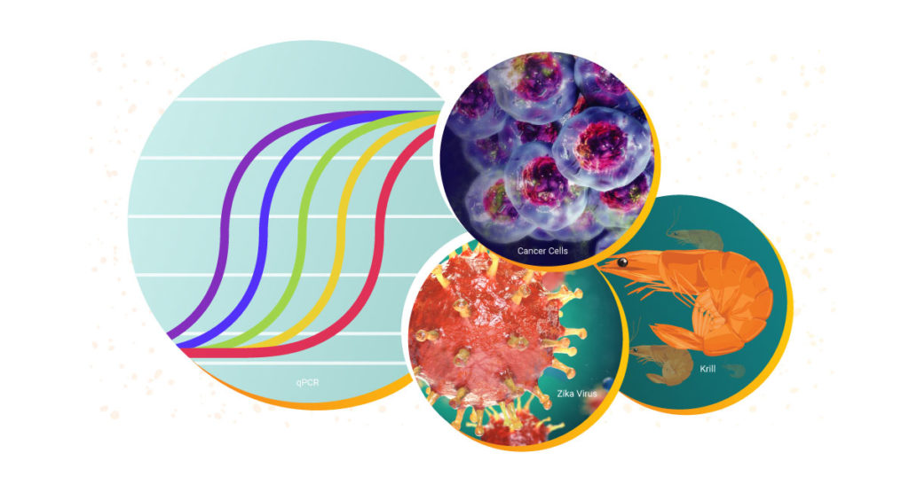
Both qPCR and RT-qPCR are powerful tools for scientists to have at their disposal. These fundamental techniques are used to study biological processes in a wide range of areas. Over the decades, Promega has supported researchers with RT-qPCR and qPCR reagents and systems to study everything from diseases like COVID-19 and cancer to viruses in elephants and the circadian rhythm of krill.
The three winners of the 2019 Real-Time PCR Grants have been hard at work in the six months since receiving their grants. Each winner was eligible to receive up to $10,000 in free PCR reagents as well as the opportunity to collaborate with our knowledgeable technical service and training teams.
Abbeah Navasca is a plant pathology researcher with the Tagum Agricultural Development Company, Inc. (TADECO*, Philippines). She is developing treatments for viral infections that affect one of Philippines’ largest and most valuable agricultural exports: bananas. As a result of the qPCR grant, she and two of her colleagues were able to participate in sample preparation and analysis workshops with Promega Technical Services experts in Singapore. During her visit, the team worked through strategies for plant sample preparation and amplified those samples with the GoTaq® 1-Step RT-qPCR System. We had a chance to ask her more before she headed back to her lab.
Remdesivir (RDV or GS-5734) was used in the treatment of the first case of the SARS-CoV-2 (formerly 2019-nCoV ) in the United States (1). RDV is not an approved drug in any country but has been requested by a number of agencies worldwide to help combat the SARS-CoV-2 virus (2). RDV is an adenine nucleotide monophosphate analog demonstrated to inhibit Ebola virus replication (3). RDV is bioactivated to the triphosphate form within cells and acts as an alternative substrate for the replication-necessary RNA dependent RNA polymerase (RdRp). Incorporation of the analog results in early termination of the primer extension product resulting in the inhibition.
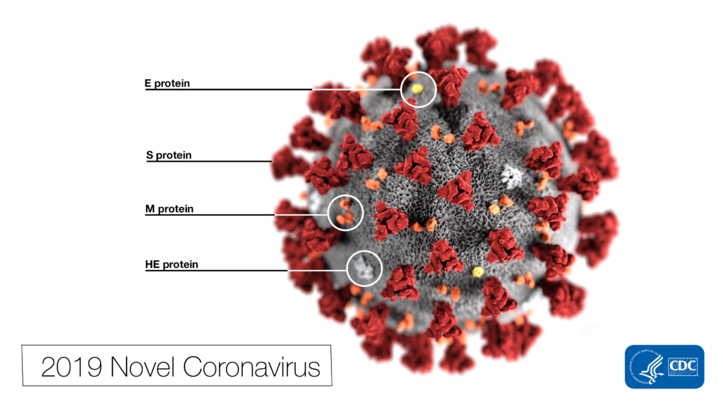
This illustration, created at the Centers for Disease Control and Prevention (CDC), reveals ultrastructural morphology exhibited by coronaviruses. Photo Credit: Alissa Eckert, MS; Dan Higgins, MAM CDC
Why all the interest in RDV as a treatment for SARS-CoV-2 ? Much of the interest in RDV is due to a series of studies performed by collaborating groups at the University of North Carolina Chapel Hill (Ralph S. Baric’s lab) and Vanderbilit University Medical Center (Mark R. Denison’s lab) in collaboration with Gilead Sciences.
The three 2019 Real-Time PCR Grant Winners have been hard at work in the six months since winning their grants. Each winner was eligible to receive up to $10,000 in free PCR reagents as well as the opportunity to collaborate with our knowledgeable technical service and training teams.
One of the 2019 winners, Alberto Biscontin (University of Padova, Italy), performs research in the fields of Neurogenetics and Chronobiology. He is looking to shed greater light on the circadian rhythms of the Antarctic krill. Alberto published his most recent analysis in Nature and GoTaq® qPCR Master Mix helped him validate expression of genes for his study.
His qPCR data showed support for internal mechanisms that not only support daily living but also clarified the overwintering process of the krill. Now that Alberto has sized up some zooplankton, we asked him to share a little more about himself and his research:
Q: How long have you been a researcher? A: I have been a researcher since 2012.
Q: How did you decide to research Antarctic krill? A: In 2013, I had the opportunity to join the international Antarctic research program PolarTime. [It] brought together eight research groups with different scientific expertise to study seasonal and daily rhythms in the Antarctic krill Euphausia superba.
Q: When you are not busy at the bench, what do you like to do? A: Traveling. I love strolling through open-air markets.
Q: Are there any tips or tricks you have learned that make your job easier? A: You can easily switch from a classic RT-PCR protocol to a cheaper and faster One-step protocol using the same primers and temperatures.
Q: What comes next? A: I would like to characterize the clock machinery of other polar organisms to understand whether high latitude clocks have developed similar strategies to cope with [the] polar environment. Moreover, a better understanding of marine circadian clocks could help to shed light on the evolution of the animal circadian machinery.
You can find Alberto’s most recent publication in Nature Scientific Reports. The 2020 Real-Time PCR Grant will be coming soon. Be sure to follow us on social media for the most up-to-date information regarding the 2020 Grant, including application deadlines and winner notifications!
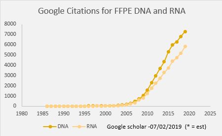
Peer-reviewed publications containing data dervived from analysis of nucleic acids isolated from FFPE samples have increased dramatically since 2006.
Formalin Fixed Paraffin Embedded samples (FFPE) have been a mainstay of the pathology lab for over 100 years. Initially FFPE blocks were sectioned, stained with simple dyes and used for studying morphology, but now a variety of biomolecules can be analyzed in these samples. Over the past 10 years we have discovered that there is a treasure trove of genomics data waiting to be unearthed in FFPE tissue. While viral RNAs and miRNA were some of the first molecules found to be present and accessible for analysis starting in the 1990s, improvements to DNA and RNA extraction methods have demonstrated that PCR, qPCR, SNP genotyping, Exome and WGS are possible. This has resulted scientific publications of DNA and RNA data generated from FFPE samples starting in 2006, and today we see immense amounts of data generated from FFPE—with nearly 2000 citations in 2018 reporting sequencing of FFPE samples.
Depending on the type of project, prospective or
retrospective, the genomics scientist has an opportunity to affect the
probability of success by better understanding the fixation process. The
challenge with FFPE is the host of variables that have the potential to
negatively affect downstream assays.
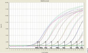
qPCR monitors amplification in real and allows you to measure starting material.
For those of us well versed in traditional, end-point PCR, wrapping our minds and methods around real-time or quantitative (qPCR) can be challenging. Here at Promega Connections, we are beginning a series of blogs designed to explain how qPCR works, things to consider when setting up and performing qPCR experiments, and what to look for in your results.
First, to get our bearings, let’s contrast traditional end-point PCR with qPCR.
End-Point PCR
qPCR
Visualizes by agarose gel the amplified product AFTER it is produced (the end-point)
Visualizes amplification as it happens (in real time) via a detection instrument
Does not precisely measure the starting DNA or RNA
Measures how many copies of DNA or RNA you started with (quantitative = qPCR)
Less expensive; no special instruments required
More expensive; requires special instrumentation
Basic molecular biology technique
Requires slightly more technical prowess
Quantitative PCR (qPCR) can be used to answer the same experimental questions as traditional end-point PCR: Detecting polymorphisms in DNA, amplifying low-abundance sequences for cloning or analysis, pathogen detection and others. However, the ability to observe amplification in real-time and detect the number of copies in the starting material can quantitate gene expression, measure DNA damage, and quantitate viral load in a sample and other applications.
Anytime that you are performing a reaction where something is copied over and over in an exponential fashion, contaminants are just as likely to be copied as the desired input. Quantitative PCR is subject to the same contamination concerns as end-point PCR, but those concerns are magnified because the technique is so sensitive. Avoiding contamination is paramount for generating qPCR results that you can trust.
Use aerosol-resistant pipette tips, and have designated pipettors and tips for pre- and post-amplification steps.
Wear gloves and change them frequently.
Have designated areas for pre- and post-amplification work.
Use reaction “master mixes” to minimize variability. A master mix is a ready-to-use mixture of your reaction components (excluding primers and sample) that you create for multiple reactions. Because you are pipetting larger volumes to make the reaction master mix, and all of your reactions are getting their components from the same master mix, you are reducing variability from reaction to reaction.
Dispense your primers into aliquots to minimize freeze-thaw cycles and the opportunity to introduce contaminants into a primer stock.
These are very basic tips that are common to both end-point and qPCR, but if you get these right, you are off to a good start no matter what your experimental goals are.
If you are looking for more information regarding qPCR, watch this supplementary video below.
Are you looking for more in-depth information about qPCR? Check out our qPCR and RT-qPCR Guide!
Scientists have had a love-hate relationship with PCR amplification for decades. Real-time or quantitative PCR (qPCR) can be an amazingly powerful tool, but just like traditional PCR, it can be quite frustrating. There are several parameters that can influence the success of your PCR assay. We’ve highlighted ten things to consider when trying to improve your qPCR results.
Over the last few months we have published several blogs about qPCR—from basic pointers on avoiding contamination in these sensitive reactions to a collection of tips for successful qPCR. Today we look in depth at a paper that describes the design and and optimization of a qPCR assay, and in keeping with the season of winter in the Northern hemisphere, it is only fitting that the assay tests for the abundance and identity of ice-nucleating bacteria.
Ice-nucleating bacteria are gram-negative bacteria that occur in the environment and are able to “catalyze” the formation ice crystals at warmer temperatures because of the expression of specific, ice-nucleating proteins on their outer membrane. Ice-nucleating bacteria are found in abundance on crop plants, especially grains, and are estimated to cause one-billion dollars in crop damage from frost in the United States alone.
In addition to their abundance on crop plants, ice-nucleating bacteria are also found on natural vegetation and have been isolated from soil, snow, hail, cloud water, in the air above crops under dry conditions and during rain fall. They have even been isolated from soil, seedlings and snow in remote locations in Antarctica. For the bacteria, ice nucleation may be a method to promote dissemination through rain and snow.
Although ice-nucleating bacteria have been isolated from clouds, ice and rain, little is known about their true contribution to precipitation or other events such as glaciation. Are such bacteria the only source of warm-temperature (above temperatures at which ice crystals form without a catalyst) ice nucleation? Can they trigger precipitation directly? What are the factors that trigger their release from vegetation into the atmosphere? Can we determine their abundance and variety in the environment?
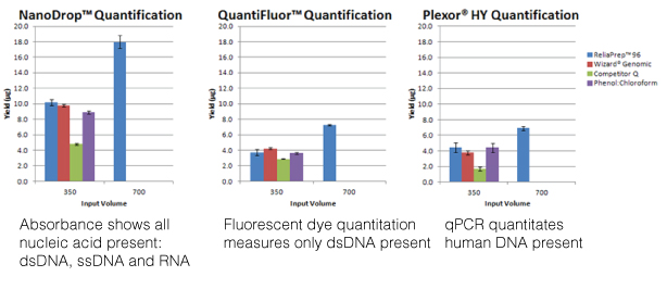
For most molecular biology applications, knowing the amount of nucleic acid present in your purified sample is important. However, one quantitation method might serve better than another, depending on your situation, or you may need to weigh the benefits of a second method to assess the information from the first. Our webinar “To NanoDrop® or Not to NanoDrop®: Choosing the Most Appropriate Method for Nucleic Acid Quantitation” given by Doug Wieczorek, one of our Applications Scientists, discussed three methods for quantitating nucleic acid and outlined their strengths and weaknesses.
Yesterday I listened in on the Webinar “Getting the Most Out of Your DNA Analysis from Purification to Downstream Assays”, presented by Eric Vincent–a Product Manager in the Promega Genomics group.
This is the webinar for you if you have ever wondered about the relative advantages and disadvantages of the many methods available for DNA purification, quantitation and analysis, or if you are comparing options for low- to high-throughput DNA purification. Eric presents a clear analyses of each of the steps in a basic DNA workflow: Purification, Quantitation, Quality Determination, and Downstream Analysis, providing key considerations and detailing the potential limitations of the methods commonly used at each step.
The DNA purification method chosen has an affect on the quality and integrity of the DNA isolated, and can therefore affect performance in downstream assays. Accuracy of quantitation also affects success, and the various downstream assays themselves (such as end-point PCR, qPCR, and sequencing) each have different sensitivities to factors such as DNA yield, quality, and integrity, and the presence of inhibitors. Continue reading “DNA Purification, Quantitation and Analysis Explained”
XWe use cookies and similar technologies to make our website work, run analytics, improve our website, and show you personalized content and advertising. Some of these cookies are essential for our website to work. For others, we won’t set them unless you accept them. To learn more about our approach to Privacy we invite you to Read More
By clicking “Accept All”, you consent to the use of ALL the cookies. However you may visit Cookie Settings to provide a controlled consent.
We use cookies and similar technologies to make our website work, run analytics, improve our website, and show you personalized content and advertising. Some of these cookies are essential for our website to work. For others, we won’t set them unless you accept them. To find out more about cookies and how to manage cookies, read our Cookie Policy.
If you are located in the EEA, the United Kingdom, or Switzerland, you can change your settings at any time by clicking Manage Cookie Consent in the footer of our website.
Necessary cookies are absolutely essential for the website to function properly. These cookies ensure basic functionalities and security features of the website, anonymously.
Cookie
Duration
Description
cookielawinfo-checbox-analytics
11 months
This cookie is set by GDPR Cookie Consent plugin. The cookie is used to store the user consent for the cookies in the category "Analytics".
cookielawinfo-checbox-functional
11 months
The cookie is set by GDPR cookie consent to record the user consent for the cookies in the category "Functional".
cookielawinfo-checbox-others
11 months
This cookie is set by GDPR Cookie Consent plugin. The cookie is used to store the user consent for the cookies in the category "Other.
cookielawinfo-checkbox-advertisement
1 year
The cookie is set by GDPR cookie consent to record the user consent for the cookies in the category "Advertisement".
cookielawinfo-checkbox-necessary
11 months
This cookie is set by GDPR Cookie Consent plugin. The cookies is used to store the user consent for the cookies in the category "Necessary".
cookielawinfo-checkbox-performance
11 months
This cookie is set by GDPR Cookie Consent plugin. The cookie is used to store the user consent for the cookies in the category "Performance".
gdpr_status
6 months 2 days
This cookie is set by the provider Media.net. This cookie is used to check the status whether the user has accepted the cookie consent box. It also helps in not showing the cookie consent box upon re-entry to the website.
lang
This cookie is used to store the language preferences of a user to serve up content in that stored language the next time user visit the website.
viewed_cookie_policy
11 months
The cookie is set by the GDPR Cookie Consent plugin and is used to store whether or not user has consented to the use of cookies. It does not store any personal data.
Analytical cookies are used to understand how visitors interact with the website. These cookies help provide information on metrics the number of visitors, bounce rate, traffic source, etc.
Cookie
Duration
Description
SC_ANALYTICS_GLOBAL_COOKIE
10 years
This cookie is associated with Sitecore content and personalization. This cookie is used to identify the repeat visit from a single user. Sitecore will send a persistent session cookie to the web client.
vuid
2 years
This domain of this cookie is owned by Vimeo. This cookie is used by vimeo to collect tracking information. It sets a unique ID to embed videos to the website.
WMF-Last-Access
1 month 18 hours 24 minutes
This cookie is used to calculate unique devices accessing the website.
_ga
2 years
This cookie is installed by Google Analytics. The cookie is used to calculate visitor, session, campaign data and keep track of site usage for the site's analytics report. The cookies store information anonymously and assign a randomly generated number to identify unique visitors.
_gid
1 day
This cookie is installed by Google Analytics. The cookie is used to store information of how visitors use a website and helps in creating an analytics report of how the website is doing. The data collected including the number visitors, the source where they have come from, and the pages visted in an anonymous form.
Advertisement cookies are used to provide visitors with relevant ads and marketing campaigns. These cookies track visitors across websites and collect information to provide customized ads.
Cookie
Duration
Description
IDE
1 year 24 days
Used by Google DoubleClick and stores information about how the user uses the website and any other advertisement before visiting the website. This is used to present users with ads that are relevant to them according to the user profile.
test_cookie
15 minutes
This cookie is set by doubleclick.net. The purpose of the cookie is to determine if the user's browser supports cookies.
VISITOR_INFO1_LIVE
5 months 27 days
This cookie is set by Youtube. Used to track the information of the embedded YouTube videos on a website.
Performance cookies are used to understand and analyze the key performance indexes of the website which helps in delivering a better user experience for the visitors.
Cookie
Duration
Description
YSC
session
This cookies is set by Youtube and is used to track the views of embedded videos.
_gat_UA-62336821-1
1 minute
This is a pattern type cookie set by Google Analytics, where the pattern element on the name contains the unique identity number of the account or website it relates to. It appears to be a variation of the _gat cookie which is used to limit the amount of data recorded by Google on high traffic volume websites.