Paving New Ways for Drug Discovery & Development: Targeted Protein Degradation

The Dana-Farber Targeted Protein Degradation Webinar Series discusses new discoveries and modalities in protein degradation.

In this webinar, Senior Research Scientist, Dr. Danette Daniels, focuses primarily on proteolysis-targeting chimeras, or PROTACs. A variety of topics are covered including the design, potency, and efficacy of PROTACs in targeted protein degradation. Watch the video below to learn more about how PROTACs are shifting perspectives through fascinating research and discoveries in targeted protein degradation.

Learn more about targeted protein degradation and PROTACS here.

From Live Cells to Lysates: Adapting NanoBiT to a Biochemical Assay Format

The ability to target protein interactions with low solubility or weak binding affinities can present a significant challenge when it comes to drug screening. The beauty of these types of challenges we often face in the lab is that finding solutions to these problems doesn’t necessarily require brand new tools. Sometimes we already have the right tools in our arsenal and, with just a little creativity and collaboration, they can be adapted to address the challenge at hand.

In the following video, Dr. Mohamed (Soly) Ismail, a Postdoctoral Fellow at the Downward Lab of the Francis Crick Institute, presents the perfect example of this with his novel approach to the NanoBiT® Protein:Protein Interaction Assay. Through a collaboration with Promega R&D Scientists, Dr. Ismail has translated the assay into a cell-free, biochemical format, termed the NanoBiT Biochemical Assay (NBBA).

Watch the subtitled version of the video here >

Continue reading “From Live Cells to Lysates: Adapting NanoBiT to a Biochemical Assay Format”

Looking Back: Cell-Free Expression Systems Helped to Characterize Proteins Involved in Hypoxia Response

Structur of a HIF-1a-pVHL-ElonginB-ElonginC complex
Structure of a HIF-1a-pVHL-ElonginB-ElonginC complex

William G. Kaelin Jr., Sir Peter J. Ratcliffe and Gregg L. Semenza were awarded the 2019 Nobel Prize in Physiology or Medicine for their discoveries of how cells sense and adapt to oxygen availability.

Kaelin and Ratcliffe’s labs focused their efforts on the transcription factor HIF (hypoxia-inducible factor). This transcription factor is critical in the cellular adaptation of to changes in oxygen availability.

When oxygen levels are elevated cells contain very little HIF. Ubiquitin is added to the HIF protein via the VHL complex and it is degraded in the proteasome.  When oxygen levels are low (hypoxia) the amount of HIF increases.

In 2001 both groups published articles characterizing the interaction between VHL and HIF, and these articles were referenced by the Nobel Prize Organization in their press release about this year’s award. (1,2). Both studies demonstrated that under the normal oxygen conditions hydroxylation of proline residue P564 enabled VHL to recognize and bind to HIF.

The use of cell free expression (i.e., TNT Coupled Transcription/Translation System) by both labs was key in the characterization of the VHL:HIF interaction The labs utilized HIF and VHL 35-S labeled proteins generated via the TNT system under both normal or in a hypoxic work station to:

  • Determine the affect of ferrous chloride and cobaltous chloride on the interaction
  • Map the specific region of HIF required for the interaction to occur (556-574)
  • Determine the effect of HIF point mutations on the interaction
  • Use synthetic peptides to block the interaction
  • Conclude that a factor in mammalian cells was necessary for the interaction to occur.

Literature Cited

  1. Ivan, M et al. (2001) HIF Targeted for VHL-Mediated Destruction by Proline Hydroxylation: Implications for O2 Sensing. Science 292: 464–67.
  2. Jaakkola, P. et al. (2001) Targeting of HIF-α to the von Hippel-Lindau Ubiquitylation of Complex by O2– Regulated Prolyl Hydroxylation. Science 202, 468–72 .

Related Posts

When Proteins Get Together: Shedding (Blue) Light on Cellular LOV

NanoBRETNo protein is an island. Within a cell, protein-protein interactions (PPIs) are involved in highly regulated and specific pathways that control gene expression and cell signaling. The disruption of PPIs can lead to a variety of disease states, including cancer.

Two general approaches are commonly used to study PPIs. Real-time assays measure PPI activity in live cells using fluorescent or luminescent tags. A second approach includes methods that measure a specific PPI “after the fact”; popular examples include a reporter system, such as the classic yeast two-hybrid system.

Continue reading “When Proteins Get Together: Shedding (Blue) Light on Cellular LOV”

A BiT or BRET, Which is Better?

Now that Promega is expanding its offerings of options for examining live-cell protein interactions or quantitation at endogenous protein expression levels, we in Technical Services are getting the question about which option is better. The answer is, as with many assays… it depends! First let’s talk about what are the NanoBiT and NanoBRET technologies, and then we will provide some similarities and differences to help you choose the assay that best suits your individual needs.

Continue reading “A BiT or BRET, Which is Better?”

Probing RGS:Gα Protein Interactions with NanoBiT Assays

gpcr_in_membrane_on_white2

When I was a post-doc at UT Southwestern, I was fortunate to interact with two Nobel prize winners, Johann Deisenhofer and Fred Gilman.  Johann once helped me move a -80°C freezer into his lab when we lost power in my building. I once replaced my boss at small faculty mixer with a guest speaker and had a drink with Fred Gilman and several other faculty members from around the university. Among the faculty, one professor had a cell phone on his belt, an odd sight in 1995. Fred Gilman asked him what it was and why he had it. It was so his lab could notify him of good results anytime of the day. Fred laughed and told him to get rid of it – if it’s good data, it will survive until morning.

I was reminded of this story when I read a recent paper by Bodle, C.R. et al (1) about the development of a NanoBiT® Complementation Assay (2) to measure interactions of Regulators of G Protein Signaling (RGS) with Gα proteins in cells. (Fred Gilman was the first to isolate a G protein and that led to him being a co-recipient of the Nobel Prize in 1994). The authors created over a dozen NanoBiT Gα:RGS domain pairs and found they could classify different RGS proteins by the speed of the interaction in a cellular context. The interactions were readily reversible with known inhibitors and were suitable for high-throughput screening due to Z’ factors exceeding 0.5. The study paves the way for future work to identify broad spectrum RGS domain:Gα inhibitors and even RGS domain-specific inhibitors. This is the second paper applying NanoBiT Technology to GPCR studies (3).

A Little Background…
A primary function of GPCRs is transmission of extracellular signals across the plasma membrane via coupling with intracellular heterotrimeric G proteins. Upon receptor stimulation, the Gα subunit dissociates from the βγ subunit, initiating the cascade of downstream second messenger pathways that alter transcription (4). The Gα subunits are actually slow GTPases that propagate signals when GTP is bound but shutdown and reassociate with the βγ subunit when GTP is cleaved to GDP. This activation process is known as the GTPase cycle. G proteins are extremely slow GTPases.

Continue reading “Probing RGS:Gα Protein Interactions with NanoBiT Assays”

Studying Mitochondrial Fission with NanoBiT Complementation Assay

3D depiction of NanoBiT Protein Complementation

Updated February 15, 2021

If you’re interrogating two proteins to understand the conditions under which they interact, a complementation system enables you to tag each protein. Interaction of the tagged proteins facilitates the complementation of the subunits, resulting in a signal. Here we discuss the NanoBiT complementation assay and describe its use to study mitochondrial fission.

Continue reading “Studying Mitochondrial Fission with NanoBiT Complementation Assay”

NanoBiT™ Assay: Transformational Technology for Studying Protein Interactions Named a Top 10 Innovation of 2015

21416943-wb-cr-uw-nanobit-pm-page-hero
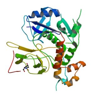
For three out of the last four years, we have been honored to have one of our key technologies named a Top 10 Innovation by The Scientist. This year the innovative NanoBiT™ Assay (NanoLuc® Binary Technology) received the recognition. NanoBiT™ is a structural complementation reporter based on NanoLuc® Luciferase, a small, bright luciferase derived from the deep sea shrimp Oplophorus gracilirostris.

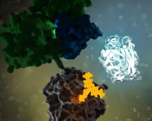
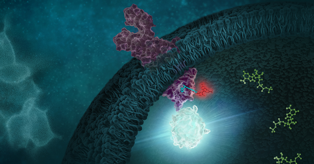
Using plasmids that encode the NanoBiT complementation reporter, you can make fusion proteins to “report” on protein interactions that you are studying. One of the target proteins is fused to the 18kDa subunit; the other to the 11 amino acid subunit. The NanoBiT™ subunits are stable, exhibiting low self-affinity, but produce an ultra-bright signal upon association. So, if your target proteins interact, the two subunits are brought close enough to each other to associate and produce a luminescent signal. The strong signal and low background associated with a luminescent system, and the small size of the complementation reporter, all help the NanoBiT™ assay overcome the limitations associated with traditional methods for studying protein interactions.

The small size reduces the chances of steric interference with protein interactions. The ultra bright signal, means that even interactions among proteins present in very low amounts can be detected and quantified–without over-expressing large quantities of non-native fusion proteins and potentially disrupting the normal cellular environment. And the NanoBiT™ assay can be performed in real time, in live cells.

The NanoBiT™ assay is already being deployed in laboratories to help advance understanding of fundamental cell biology. You can see how one researcher is already taking full advantage of this innovative technology in the video embedded below:

Visit the Promega web site to see more examples more examples how the NanoBiT™ assay can break through the traditional limitations for studying protein interactions in cells.

You can read the Top 10 article in The Scientist here.

About the Development of an Improved BRET Assay: NanoBRET

"Protein BRD4 PDB 2oss" by Emw - Own work. Licensed under CC BY-SA 3.0 via Wikimedia Commons - https://commons.wikimedia.org/wiki/File:Protein_BRD4_PDB_2oss.png#/media/File:Protein_BRD4_PDB_2oss.png
“Protein BRD4 PDB 2oss” by Emw – Own work. Licensed under CC BY-SA 3.0 via Wikimedia Commons – https://commons.wikimedia.org/wiki/File:Protein_BRD4_PDB_2oss.png#/media/File:Protein_BRD4_PDB_2oss.png

One of the more exciting reporter molecules technologies available came online in the past year, with the launch of the Promega NanoBRET™ technology. While it’s easy for me, a science writer at Promega, to brag, seriously, this is a very cool protein interactions tool.

A few of the challenges facing protein-protein interactions researchers include:

  • The ability to quantitatively characterize protein-protein interactions
  • Ability to examine protein-protein interactions in situ, in the context of the living cell

A goal of the NanoBRET™ developers was to improve the sensitivity and dynamic range of traditional BRET technology, in order to address these challenges.

In May 2015 these researchers published an article outlining their efforts to create NanoBRET technology in ACS Chemical Biology, in an article entitled, “NanoBRET—A Novel BRET Platform for the Analysis of Protein-Protein Interactions”. Here is a brief look at their work.

Continue reading “About the Development of an Improved BRET Assay: NanoBRET”

If We Could But Peek Inside the Cell …Quantifying, Characterizing and Visualizing Protein:Protein  Interactions

14231183 WB MS Protein Interactions Hero Image 600x214

Robert Hooke first coined the term “cell” after observing  plant cell walls through a light microscope—little empty chambers, fixed in time and space. However,  cells are anything but fixed.

Cells are dynamic: continually responding to a shifting context of time, environment, and signals from within and without. Interactions between the macromolecules within cells, including proteins, are ever changing—with complexes forming, breaking up, and reforming in new ways. These interactions provide a temporal and special framework for the work of the cell, controlling gene expression, protein production, growth, cell division and cell death.

Visualizing and measuring protein:protein interactions at the level of the cell without perturbing them is the goal of every cell biologist.

A recent article by Thomas Machleidt et al. published in ACS Chemical Biology, describes a new technology that brings us closer to being able to realize that goal.

Continue reading “If We Could But Peek Inside the Cell …Quantifying, Characterizing and Visualizing Protein:Protein  Interactions”