Investigation of Remdesivir as a Possible Treatment for SARS-CoV-2 (2019-nCoV)

Remdesivir (RDV or GS-5734) was used in the treatment of the first case of the SARS-CoV-2 (formerly 2019-nCoV ) in the United States (1). RDV is not an approved drug in any country but has been requested by a number of agencies worldwide to help combat the SARS-CoV-2 virus (2). RDV is an adenine nucleotide monophosphate analog demonstrated to inhibit Ebola virus replication (3). RDV is bioactivated to the triphosphate form within cells and acts as an alternative substrate for the replication-necessary RNA dependent RNA polymerase (RdRp). Incorporation of the analog results in early termination of the primer extension product resulting in the inhibition.
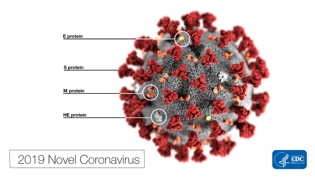
 Note the spikes that adorn the outer surface of the virus, which impart the look of a corona surrounding the virion, when viewed electron microscopically. In this view, the protein particles E, S, M, and HE, also located on the outer surface of the particle, have all been labeled as well. A novel coronavirus virus was identified as the cause of an outbreak of respiratory illness first detected in Wuhan, China in 2019.
This illustration, created at the Centers for Disease Control and Prevention (CDC), reveals ultrastructural morphology exhibited by coronaviruses. Photo Credit: Alissa Eckert, MS; Dan Higgins, MAM CDC

Why all the interest in RDV as a treatment for SARS-CoV-2 ? Much of the interest in RDV is due to a series of studies performed by collaborating groups at the University of North Carolina Chapel Hill (Ralph S. Baric’s lab) and Vanderbilit University Medical Center (Mark R. Denison’s lab) in collaboration with Gilead Sciences. 

Continue reading “Investigation of Remdesivir as a Possible Treatment for SARS-CoV-2 (2019-nCoV)”

Targeted Protein Degradation: A Bright Future for Drug Discovery

targeted protein degradation and protacs

Our cells have evolved multiple mechanisms for “taking out the trash”—breaking down and disposing of cellular components that are defective, damaged or no longer required. Within a cell, these processes are balanced by the synthesis of new components, so that DNA, RNA and proteins are constantly undergoing turnover.

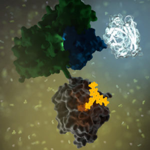
Proteins are degraded by two major components of the cellular machinery. The discovery of the lysosome in the mid-1950s provided considerable insight into the first of these degradation mechanisms for extracellular and cytosolic proteins. Over the next several decades, details of a second protein degradation mechanism emerged: the ubiquitin-proteasome system (UPS). Ubiquitin is a small, highly conserved polypeptide that is used to selectively tag proteins for degradation within the cell. Multiple ubiquitin tags are generally attached to a single targeted protein. This ill-fated, ubiquitinated protein is then recognized by the proteasome, a large protein complex with proteolytic activity. Ubiquitination is a multistep process, involving several specialized enzymes. The final step in the process is mediated by a family of ubiquitin ligases, known as E3.

Continue reading “Targeted Protein Degradation: A Bright Future for Drug Discovery”

Popular Papers from Promega Authors

Promega is a chemistry and instrument supplier to scientists in diverse industries and research labs around the world. True. But we are more than just a supply company; we are scientists dedicated to supporting the work of other scientists. We want the science behind the technologies we develop to be both vetted and valued by the scientific community at large, which is one reason our scientists take the time to prepare and submit manuscripts to peer-reviewed journals. Here we call out some of our published research papers that were highly read in 2019. In the journal ACS Chemical Biology alone, five Promega-authored papers were among the top 10 most read papers in 2019. Here’s a quick review of the highlights from these ACS papers.

Continue reading “Popular Papers from Promega Authors”

Designing a Reporter Construct for Analyzing Gene Regulation

Bioluminescent reporter assays are an excellent choice for analyzing gene regulation because they provide higher sensitivity, wider dynamic range and better signal-to-background ratios compared to colorimetric or fluorescent assays. In a typical genetic reporter assay, cells are transfected with a vector that contains the sequence of interest cloned upstream of a reporter gene, and the reporter activity is used to determine how the target sequence influences gene expression under experimental conditions. A second control reporter encoded on the same or a different plasmid is an essential internal control. The secondary reporter is used to normalize the data and compensate for variability caused by differences in cell number, lysis efficiency, cell viability, transfection efficiency, temperature, and measurement time. 

Basic Introduction to the Strategy of Reporter Gene Assays

For genetic reporter assays, using a secondary control vector with a weak promoter like PGK or TK to ensures that the control does not interfere with activation of your primary reporter vector. Transfection of high amounts of the control plasmid or putting the control reporter under control of a strong promoter like CMV or SV40 often leads to transcriptional squelching or other interference with the experimental promoter (i.e., trans effects). Reporter assays can also be used to quantitatively evaluate microRNA activity by inserting miRNA target sites downstream or 3´ of the reporter gene. For example, the pmirGLO Dual-Luciferase miRNA Target Expression Vector is based on dual-luciferase technology, with firefly luciferase as the primary reporter to monitor mRNA regulation and Renilla luciferase as a control reporter for normalization.

Here in Technical Services we often talk with researchers who are just starting their project and looking for advice on designing their genetic reporter vector. They have questions like:

  • How much of the upstream promoter region should be included in the vector?
  • How many copies of a response element will be needed to provide a good response?
  • Does the location of the element or surrounding sequence alter gene regulation?
Continue reading “Designing a Reporter Construct for Analyzing Gene Regulation”

NanoLuc: Tiny Tag with a Big Impact

Synthetic biology—genetically engineering an organism to do or make something useful—is the central goal of the iGEM competition each year. After teams conquer the challenge of cloning their gene, the next hurdle is demonstrating that the engineered gene is expressing the desired protein (and possibly quantifying the level of expression), which they may do using a reporter gene.

Reporters can also play a more significant role in iGEM projects when teams design their organism with reporter genes to detect and signal the presence of specific molecules, like environmental toxins or biomarkers. Three of the iGEM teams Promega sponsored this year opted to incorporate some version of NanoLuc® Luciferase into their projects.

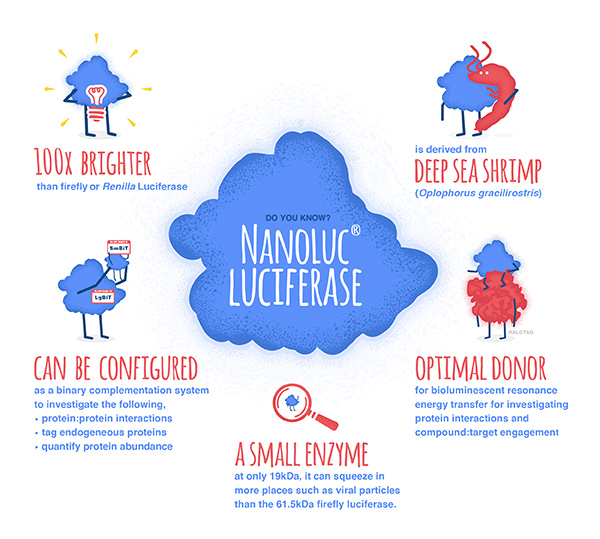
NanoLuc® luciferase is a small monomeric enzyme (19.1kDa, 171 amino acids) based on the luciferase from the deep sea shrimp Oplophorus gracilirostris. This engineered enzyme uses a novel substrate, furimazine, to produce high-intensity, glow-type luminescence in an ATP-independent reaction. Unlike other molecules for tagging and detecting proteins, NanoLuc® luciferase is less likely to interfere with enzyme activity and affect protein production due to its small size.

NanoLuc® Luciferase has also been engineered into a structural complementation reporter system, NanoBiT® Luciferase, that contains a Large subunit (LgBiT) and two small subunit options: low affinity SmBiT and high affinity HiBiT. Together, these NanoLuc® technologies provide a bioluminescent toolbox that was used by the iGEM teams to address a diverse set of biological challenges.

Here is an overview of each team’s project and how they incorporated NanoLuc® technology.

Continue reading “NanoLuc: Tiny Tag with a Big Impact”

Choosing a Tag for Your Protein

You have identified and cloned your protein of interest, but you want to explore its function. A protein fusion tag might help with your investigation. However, choosing a tag for your protein depends on what experiments you are planning. Do you want to purify the protein? Would you like to identify interacting proteins by performing pull-down assays? Are you interested in examining the endogenous biology of the protein? Here we cover the advantages and disadvantages of some protein tags to help you select the one that best suits your needs.

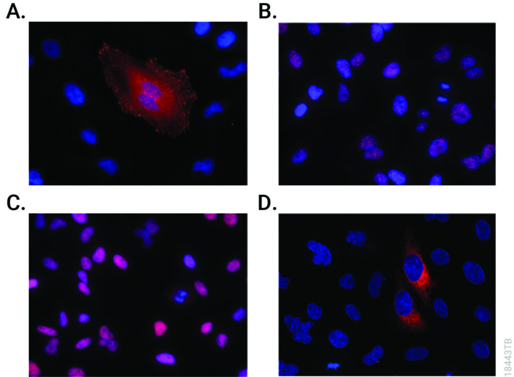
Immunofluorescent detection of HiBiT-tagged proteins in CRISPR-edited cell pools and clones using the Anti-HiBiT Monoclonal Antibody.
CRISPR-Cas9 editing knocked-in HiBiT at the endogenous locus of proteins with varying subcellular localization. Fixed CRISPR-modified clones or pools of cells were imaged by immunofluorescent staining using the Anti-HiBiT Monoclonal Antibody (red) and Hoechst dye (blue). Panel A. VCL-HiBiT pool. Panel B. SMARCA4-HiBiT clone. Panel C. HDAC2-HiBiT clone. Panel D. HSP90B1-HiBiT pool.

Affinity Tags

The most commonly used protein tags fall under the category of affinity tags. This means that the tag binds to another molecule or metal ion, making it easy to purify or pull down your protein of interest. In all cases, the tag will be fused to your protein of interest at either the amino (N) or carboxy (C) terminus by cloning into an expression vector. This protein fusion can then be expressed in cells or cell-free systems, depending on the promoter the vector contains.

Continue reading “Choosing a Tag for Your Protein”

I Have My Luciferase Vector, Now What?

Choosing and Optimizing Transfection Methods

Here in Technical Services we often talk with researchers at the beginning of their project about how to carefully design and get started with their experiments. It is exciting when you have selected the Luciferase Reporter Vector(s) that will best suit your needs; you are going to make luminescent cells! But, how do you pick the best way to get the vector into your cells to express the reporter? What transfection reagent/method will work best for your cell type and experiment? Do you want to do transient (short-term) transfections, or are you going to establish a stable cell line?

Continue reading “I Have My Luciferase Vector, Now What?”

CRISPR/Cas9, NanoBRET and GPCRs: A Bright Future for Drug Discovery

GPCRs

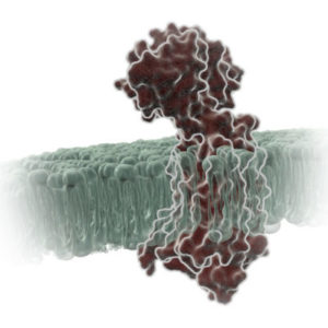
G protein-coupled receptors (GPCRs) are a large family of receptors that traverse the cell membrane seven times. Functionally, GPCRs are extremely diverse, yet they contain highly conserved structural regions. GPCRs respond to a variety of signals, from small molecules to peptides and large proteins. Many GPCRs are involved in disease pathways and, not surprisingly, they present attractive targets for both small-molecule and biologic drugs.

In response to a signal, GPCRs undergo a conformational change, triggering an interaction with a G protein—a specialized protein that binds GDP in its inactive state or GTP when activated. Typically, the GPCR exchanges the G protein-bound GDP molecule for a GTP molecule, causing the activated G protein to dissociate into two subunits that remain anchored to the cell membrane. These subunits relay the signal to various other proteins that interact with or produce second-messenger molecules. Activation of a single G protein can result, ultimately, in the generation of thousands of second messengers.

Given the complexity of GPCR signaling pathways and their importance to human health, a considerable amount of research has been devoted to GPCR interactions, both with specific ligands and G proteins.

Continue reading “CRISPR/Cas9, NanoBRET and GPCRs: A Bright Future for Drug Discovery”

Lighting Up GPCR Research with Bioluminescent Tagging

G Protein-Coupled Receptors (GPCRs) are a very large, diverse family of transmembrane receptors in eukaryotes. These receptors detect molecules outside the cell and activate internal signaling pathways by coupling with G proteins. Once a GPCR is activated, β-arrestins translocate to the cell membrane and bind to the occupied receptor, uncoupling it from G proteins and promoting its internalization.

Reporter tags are useful for studying the dynamics of GPCRs and associated proteins, but large tags can disrupt the receptors’ native functioning, and often overexpression of the tagged protein is required to obtain sufficient signal. Here is one example of how researchers have used the small, bright NanoLuc® luciferase to overcome these common challenges and answer questions about GPCRs.

Continue reading “Lighting Up GPCR Research with Bioluminescent Tagging”

Why You Don’t Need to Select a Wavelength for a Luciferase Assay

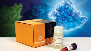
Promega kit depicted; test involves wavelength for a luciferase assay.

It’s a question I’m asked probably once a week. “What wavelength do I select on my luminometer when performing a luciferase assay?” The question is a good and not altogether unexpected one, especially for those new to bioluminescent assays. The answer is that in most cases, you don’t and in fact shouldn’t select a wavelength (the exception to this rule is if you’re measuring light emitted in two simultaneous luciferase reactions). To understand why requires a bit of an explanation of absorbance, fluorescence, and luminescence assays, and the differences among them.

Absorbance, fluorescence, and luminescence assays are all means to quantify something of interest, be that a genetic reporter, cell viability, cytotoxicity, apoptosis, or other markers. In principle, they are all similar. For example, a genetic reporter assay is an indicator of gene expression. The promoter of a gene of interest can be cloned upstream of a reporter such as β-galactosidase, GFP, or firefly luciferase. The amount of each of these reporters that is transcribed into mRNA and translated into protein by the cell is indicative of the endogenous expression of the gene of interest.

Continue reading “Why You Don’t Need to Select a Wavelength for a Luciferase Assay”