I Have My Luciferase Vector, Now What?

Choosing and Optimizing Transfection Methods

Here in Technical Services we often talk with researchers at the beginning of their project about how to carefully design and get started with their experiments. It is exciting when you have selected the Luciferase Reporter Vector(s) that will best suit your needs; you are going to make luminescent cells! But, how do you pick the best way to get the vector into your cells to express the reporter? What transfection reagent/method will work best for your cell type and experiment? Do you want to do transient (short-term) transfections, or are you going to establish a stable cell line?

Continue reading “I Have My Luciferase Vector, Now What?”

CRISPR/Cas9, NanoBRET and GPCRs: A Bright Future for Drug Discovery

GPCRs

G protein-coupled receptors (GPCRs) are a large family of receptors that traverse the cell membrane seven times. Functionally, GPCRs are extremely diverse, yet they contain highly conserved structural regions. GPCRs respond to a variety of signals, from small molecules to peptides and large proteins. Many GPCRs are involved in disease pathways and, not surprisingly, they present attractive targets for both small-molecule and biologic drugs.

In response to a signal, GPCRs undergo a conformational change, triggering an interaction with a G protein—a specialized protein that binds GDP in its inactive state or GTP when activated. Typically, the GPCR exchanges the G protein-bound GDP molecule for a GTP molecule, causing the activated G protein to dissociate into two subunits that remain anchored to the cell membrane. These subunits relay the signal to various other proteins that interact with or produce second-messenger molecules. Activation of a single G protein can result, ultimately, in the generation of thousands of second messengers.

Given the complexity of GPCR signaling pathways and their importance to human health, a considerable amount of research has been devoted to GPCR interactions, both with specific ligands and G proteins.

Continue reading “CRISPR/Cas9, NanoBRET and GPCRs: A Bright Future for Drug Discovery”

Lighting Up GPCR Research with Bioluminescent Tagging

G Protein-Coupled Receptors (GPCRs) are a very large, diverse family of transmembrane receptors in eukaryotes. These receptors detect molecules outside the cell and activate internal signaling pathways by coupling with G proteins. Once a GPCR is activated, β-arrestins translocate to the cell membrane and bind to the occupied receptor, uncoupling it from G proteins and promoting its internalization.

Reporter tags are useful for studying the dynamics of GPCRs and associated proteins, but large tags can disrupt the receptors’ native functioning, and often overexpression of the tagged protein is required to obtain sufficient signal. Here is one example of how researchers have used the small, bright NanoLuc® luciferase to overcome these common challenges and answer questions about GPCRs.

Continue reading “Lighting Up GPCR Research with Bioluminescent Tagging”

Why You Don’t Need to Select a Wavelength for a Luciferase Assay

Promega kit depicted; test involves wavelength for a luciferase assay.

It’s a question I’m asked probably once a week. “What wavelength do I select on my luminometer when performing a luciferase assay?” The question is a good and not altogether unexpected one, especially for those new to bioluminescent assays. The answer is that in most cases, you don’t and in fact shouldn’t select a wavelength (the exception to this rule is if you’re measuring light emitted in two simultaneous luciferase reactions). To understand why requires a bit of an explanation of absorbance, fluorescence, and luminescence assays, and the differences among them.

Absorbance, fluorescence, and luminescence assays are all means to quantify something of interest, be that a genetic reporter, cell viability, cytotoxicity, apoptosis, or other markers. In principle, they are all similar. For example, a genetic reporter assay is an indicator of gene expression. The promoter of a gene of interest can be cloned upstream of a reporter such as β-galactosidase, GFP, or firefly luciferase. The amount of each of these reporters that is transcribed into mRNA and translated into protein by the cell is indicative of the endogenous expression of the gene of interest.

Continue reading “Why You Don’t Need to Select a Wavelength for a Luciferase Assay”

The Simplex Things In Life: Utilizing Artificial Intelligence Models to Better Understand Autism

Autism Spectrum Disorder, or ASD, is nothing if not unique.

The way ASD manifests itself in people is unique; although it most often presents as some form of variable impairment in social interaction and communication, each individual has behaviors and habits that are as unique to them as snowflakes are to one another.

ASD has also proven itself to be a uniquely challenging disorder to study. In the past decade, de novo (new) mutations have been identified as key contributors to causality of ASD. However, the majority of these identified de novo mutations are located in protein-coding genes, which comprise only 1–2% of the entire human genome.

Up to this point, a majority of previous research has focused on identifying mutations located in the 20,000 identified genes in the protein-coding region, which would seem like a promising approach. Genes are the genetic blueprints for creating proteins, which control and perform crucial tasks in our bodies, such as fighting off infections, communicating between your organs, tissues, and cells as chemical messengers, and regulating your blood sugar levels. It seems like basic math: Genes + Mutations = Mutated Proteins. Mutated Proteins = Disrupted Protein Function.

However, it has been observed that all the known genes that are ASD-associated can explain only a minor fraction of new autism cases, and it is estimated that known de novo mutations in the protein-coding region contribute to not more than 30% of cases for individuals who have no family history of autism (better known as simplex ASD). This provides evidence to suggest mutations contributing to autism must additionally occur elsewhere in the genome. Continue reading “The Simplex Things In Life: Utilizing Artificial Intelligence Models to Better Understand Autism”
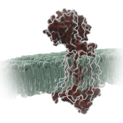
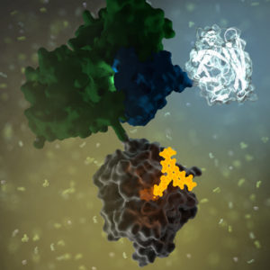
When Proteins Get Together: Shedding (Blue) Light on Cellular LOV

NanoBRET

No protein is an island. Within a cell, protein-protein interactions (PPIs) are involved in highly regulated and specific pathways that control gene expression and cell signaling. The disruption of PPIs can lead to a variety of disease states, including cancer.

Two general approaches are commonly used to study PPIs. Real-time assays measure PPI activity in live cells using fluorescent or luminescent tags. A second approach includes methods that measure a specific PPI “after the fact”; popular examples include a reporter system, such as the classic yeast two-hybrid system.

Continue reading “When Proteins Get Together: Shedding (Blue) Light on Cellular LOV”

A BiT or BRET, Which is Better?

Now that Promega is expanding its offerings of options for examining live-cell protein interactions or quantitation at endogenous protein expression levels, we in Technical Services are getting the question about which option is better. The answer is, as with many assays… it depends! First let’s talk about what are the NanoBiT and NanoBRET technologies, and then we will provide some similarities and differences to help you choose the assay that best suits your individual needs.

Continue reading “A BiT or BRET, Which is Better?”

The Role of the NanoLuc® Reporter in Investigating Ligand-Receptor Interactions

Luminescent reporter assays are powerful research tools for a variety of applications. Last March we presented a webinar on this topic, Understanding Luminescent Reporter Assay Design, which proved to enlighten many who registered. The webinar addressed the importance of careful experimental design when using a luminescent reporter such as Promega’s Firefly or NanoLuc® Luciferase.

Reporters provide a highly sensitive, quantifiable metric for cellular events such as gene expression, protein function and signal transduction. Luminescent reporters have become even more valuable for live, real-time measurement of various processes in living cells. This is backed by the fact that a growing number of scientific publications reference the use of the NanoLuc® Luciferase reporter and demonstrate its effectiveness as a reporter assay. Continue reading “The Role of the NanoLuc® Reporter in Investigating Ligand-Receptor Interactions”

Shining Light on a Superbug: Clostridium difficile

Antibiotic-resistant bacteria and their potential to cause epidemics with no viable treatment options have been in the news a lot. These “superbugs,” which have acquired genes giving them resistance to common and so-called “last resort” antibiotics, are a huge concern as effective treatment options dwindle. Less attention has been given to an infection that is not just impervious to antibiotics, but is actually enabled by them.

33553646_l

Clostridium difficile Infection (CDI) is one of the most common healthcare-associated infections and a significant global healthcare problem. Clostridium difficile (C. diff), a Gram-positive anaerobic bacterium, is the source of the infection. C. diff spores are very resilient to environmental stressors, such as pH, temperature and even antibiotics, and can be found pretty much everywhere around us, including on most of the food we eat. Ingesting the spores does not usually lead to infection inside the body without also being exposed to antibiotics.

Individuals taking antibiotics are 7-10 times more likely to acquire a CDI. Antibiotics disrupt the normal flora of the intestine, allowing C. diff to compete for resources and flourish. Once exposed to the anaerobic conditions of the human gut, these spores germinate into active cells that embed into the tissue lining the colon. The bacteria are then able to produce the toxins that can cause disease and result in severe damage, or even death.

Continue reading “Shining Light on a Superbug: Clostridium difficile”

For Protein Complementation Assays, Design is Everything

Most, if not all, processes within a cell involve protein-protein interactions, and researchers are always looking for better tools to investigate and monitor these interactions. One such tool is the protein complementation assay (PCA). PCAs use  a reporter, like a luciferase or fluorescent protein, separated into two parts (A and B) that form an active reporter (AB) when brought together. Each part of the split reporter is attached to one of a pair of proteins (X and Y) forming X-A and Y-B. If X and Y interact, A and B are brought together to form the active enzyme (AB), creating a luminescent or fluorescent signal that can be measured. The readout from the PCA assay can help identify conditions or factors that drive the interaction together or apart.

A key consideration when splitting a reporter is to find a site that will allow the two parts to reform into an active enzyme, but not be so strongly attracted to each other that they self-associate and cause a signal, even in the absence of interaction between the primary proteins X and Y. This blog will briefly describe how NanoLuc® Luciferase was separated into large and small fragments (LgBiT and SmBiT) that were individually optimized to create the NanoBiT® Assay and show how the design assists in monitoring protein-protein interactions.

Continue reading “For Protein Complementation Assays, Design is Everything”