How Do You Solve a Problem Like Malaria?

malaria_researcher
Photo courtesy of NIH/NIAID

Malaria affects nearly half of the world’s population, with almost 80% of cases in sub-Saharan Africa and India. While there have been many strides in education and prevention campaigns over the last 30 years, there were over 200 million cases documented in 2017 with over 400,000 deaths, and the majority were young children. Despite being preventable and treatable, malaria continues to thrive in areas that are high risk for transmission. Recently, clinicians started rolling out use of the first approved vaccine, though clinical trials showed it is only about 30% effective. Meanwhile, researchers must continue to focus on innovative efforts to improve diagnostics, treatment and prevention to reduce the burden in these areas.

Continue reading “How Do You Solve a Problem Like Malaria?”

Dual-Luciferase or Dual-Glo Luciferase Assay System? Which one should I choose for my reporter assays?

Confused woman

I’ve got a set of experiments planned that, if all goes well, will provide me with the answer I have been seeking for months. Plus, my supervisor is eagerly awaiting the results because she needs the data for a grant application, so I don’t want to mess it up. However, I am faced with a choice for my firefly and Renilla luciferase reporter assays: Do I use the Dual-Luciferase® Reporter Assay System or Dual-Glo® Luciferase Assay System? What’s the difference? How do I decide which to use? I’m so confused! Help!

Sound familiar? Not to worry! The choice is not difficult once you know how these assays work and how they differ.

Continue reading “Dual-Luciferase or Dual-Glo Luciferase Assay System? Which one should I choose for my reporter assays?”

Executing a NanoBRET™ Experiment: From Start to Data

This is a guest post from Katarzyna Dubiel, marketing intern in Cellular Analysis and Proteomics.

“The objective of my experiment was to test the NanoBRET™ assay as if I was a customer, independent of the research and development team which develops the assay.”

Designing and implementing a new assay can be a challenging process with many unexpected troubleshooting steps. We wanted to know what major snags a scientist new to the NanoBRET™ Assay would encounter. To determine this, we reached out to Laurence Delauriere, a senior applications scientist at Promega-France, who had never previously performed a NanoBRET™ assay. Laurence went step-by-step through the experimental process looking at the CRAF-BRAF interaction in multiple cell lines. In an interview, Laurence provided us with some tips and insights from her work implementing the new NanoBRET™ assay.

In a few words, can you explain NanoBRET?
“NanoBRET is used to monitor protein: protein interactions in live cells. It is a bioluminescence resonance energy transfer (BRET) based assay that uses NanoLuc® luciferase as the BRET energy donor and HaloTag® protein labeled with the HaloTag® NanoBRET™ 618 fluorescent ligand as the energy acceptor to measure the interaction of two binding partners.” Continue reading “Executing a NanoBRET™ Experiment: From Start to Data”

Shining Stars: Cool NanoLuc® Plasmid Constructs Available Through the Addgene Repository

Researchers having been sharing plasmids ever since there were plasmids to share. Back when I was in the lab, if you read a paper and saw an interesting construct you wished to use, you could either make it yourself or you could “clone by phone”.  One of my professors was excellent at phone cloning with labs around the world and had specific strategies and tactics for getting the plasmids he wanted. Addgene makes this so much easier to share your constructs from lab to lab. Promega supports the Addgene mission statement: Accelerate research and discovery by improving access to useful research materials and information.  Many of our technology platforms like HaloTag® Fusion Protein, codon-optimized Firefly luciferase genes (e.g., luc2), and NanoLuc® Luciferase are present in the repository. We encourage people to go to Addgene to get new innovative tools. Afterall, isn’t science better when we share?

I’d like to focus on some tools in the Addgene collection based on NanoLuc® Luciferase (NLuc).  The creation of NanoLuc® Luciferase and the optimal substrate furimazine is a good story (1).  From a deep sea shrimp to a compact powerhouse of bioluminescence, NLuc is 100-fold brighter than our more common luciferases like firefly (FLuc) and Renilla (RLuc) luciferase.  This is important not so much for how bright you can make a reaction but for how sensitive you can make a reaction.  NLuc requires 100-fold less protein to produce the same amount of light from a Fluc or RLuc reaction.  NLuc lets you work at physiological concentrations.  NLuc is bright enough to detect endogenous tagged genes generated through the CRISPR/Cas9 knock-in.  NLuc is very inviting for endogenous tagging as it is only 19kDa.  An example is the CRISPaint-NLuc construct (Plasmid #67178) for use in the system outlined in Schmid-Burgk, J.L. et al (2).

Two applications of NanoLuc® Technology have caught my attention through coupling the luciferase with fluorescent proteins to make better imaging reporters and biosensors. Continue reading “Shining Stars: Cool NanoLuc® Plasmid Constructs Available Through the Addgene Repository”

Making the Switch from FRET to BRET: Applications of NanoLuc® Luciferase with Fluorescent Protein Acceptors for Sensing Cellular Events

A Bioluminescent Alternative

Fluorescence resonance energy transfer (FRET) probes or sensors are commonly used to measure cellular events. The probes typically have a matched pair of fluorescent proteins joined by a ligand-binding or responsive protein domain. Changes in the responsive domain are reflected in conformational changes that either bring the two fluorescent proteins together or drive them apart. The sensors are measured by hitting the most blue-shifted fluorescent protein with its excitation wavelength (donor). The resulting emission is transferred to the most red-shifted fluorescent protein in the pair, and the result is ultimately emission from the red-shifted protein (acceptor).

As pointed out by Aper, S.J.A. et al. below, FRET sensors face challenges of photobleaching, autofluorescence, and, in the case of exciting cyan-excitable donors, phototoxicity. Another challenge to using FRET sensors comes when employing optogenetic regulators to initiate the event you wish to monitor. Optogenetic regulators respond to specific wavelengths and initiate signaling. The challenge comes when the FRET donor excitation overlaps with the optogenetic initiation wavelengths. Researchers have sought to alleviate many of these challenges by exchanging the fluorescent donor for a bioluminescent donor, making bioluminescence resonance energy transfer (BRET) probes. In the three papers described below, the authors chose NanoLuc® Luciferase as the BRET donor due to its extremely bright signal.

Continue reading “Making the Switch from FRET to BRET: Applications of NanoLuc® Luciferase with Fluorescent Protein Acceptors for Sensing Cellular Events”

Probing RGS:Gα Protein Interactions with NanoBiT Assays

gpcr_in_membrane_on_white2

When I was a post-doc at UT Southwestern, I was fortunate to interact with two Nobel prize winners, Johann Deisenhofer and Fred Gilman.  Johann once helped me move a -80°C freezer into his lab when we lost power in my building. I once replaced my boss at small faculty mixer with a guest speaker and had a drink with Fred Gilman and several other faculty members from around the university. Among the faculty, one professor had a cell phone on his belt, an odd sight in 1995. Fred Gilman asked him what it was and why he had it. It was so his lab could notify him of good results anytime of the day. Fred laughed and told him to get rid of it – if it’s good data, it will survive until morning.

I was reminded of this story when I read a recent paper by Bodle, C.R. et al (1) about the development of a NanoBiT® Complementation Assay (2) to measure interactions of Regulators of G Protein Signaling (RGS) with Gα proteins in cells. (Fred Gilman was the first to isolate a G protein and that led to him being a co-recipient of the Nobel Prize in 1994). The authors created over a dozen NanoBiT Gα:RGS domain pairs and found they could classify different RGS proteins by the speed of the interaction in a cellular context. The interactions were readily reversible with known inhibitors and were suitable for high-throughput screening due to Z’ factors exceeding 0.5. The study paves the way for future work to identify broad spectrum RGS domain:Gα inhibitors and even RGS domain-specific inhibitors. This is the second paper applying NanoBiT Technology to GPCR studies (3).

A Little Background…
A primary function of GPCRs is transmission of extracellular signals across the plasma membrane via coupling with intracellular heterotrimeric G proteins. Upon receptor stimulation, the Gα subunit dissociates from the βγ subunit, initiating the cascade of downstream second messenger pathways that alter transcription (4). The Gα subunits are actually slow GTPases that propagate signals when GTP is bound but shutdown and reassociate with the βγ subunit when GTP is cleaved to GDP. This activation process is known as the GTPase cycle. G proteins are extremely slow GTPases.

Continue reading “Probing RGS:Gα Protein Interactions with NanoBiT Assays”

NanoBiT Assay Applied to Study Role of SOD1 in ALS

NanoBiT Protein ComplementationBack in 2015 the Ice Bucket Challenge brought Amyotrophic Lateral Sclerosis (ALS) to public attention, initiating worldwide pleas for more funding of research toward a cure for this fatal disease, which is characterized by progressive degeneration of motor neurons. In spite of many efforts over the last few decades, the precise cause of ALS is still unknown.

The complexity of the problem of ALS pathogenesis is highlighted in the review “Decoding ALS: from genes to mechanism”  published in Nature in November 2016. The review highlights a long list of genetic factors implicated in ALS, grouping them into genes affecting protein quality control, RNA stability/function, and the cytoskeletal structure of neuronal cells.

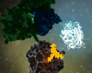
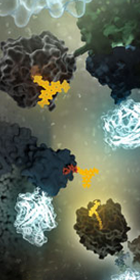
Mutations in the antioxidant enzyme superoxide dismutase (SOD1) were the first to be associated with ALS. According to the review, more than 170 SOD1 mutations causing ALS have since been identified. Many of these mutations are thought to result in misfolding of SOD1, contributing to toxicity when the misfolded protein accumulates within the cell.

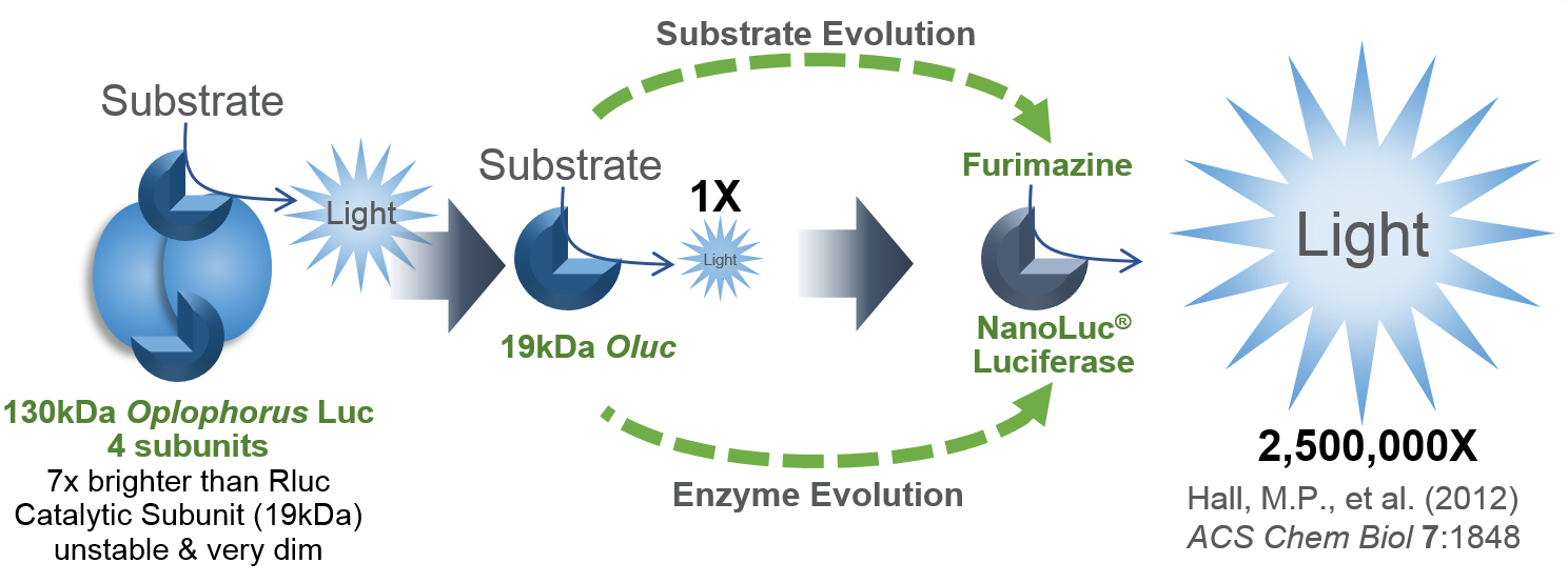
A paper by Oh-hashi et al., published in Cell Biochemistry and Function in October 2016 used the NanoBiT protein complementation assay to investigate the effect of two common ALS-associated SOD1 mutations on dimerization of the SOD1 protein. Continue reading “NanoBiT Assay Applied to Study Role of SOD1 in ALS”

The Role of the NanoLuc® Reporter in Investigating Ligand-Receptor Interactions

Luminescent reporter assays are powerful research tools for a variety of applications. Last March we presented a webinar on this topic, Understanding Luminescent Reporter Assay Design, which proved to enlighten many who registered. The webinar addressed the importance of careful experimental design when using a luminescent reporter such as Promega’s Firefly or NanoLuc® Luciferase.

Reporters provide a highly sensitive, quantifiable metric for cellular events such as gene expression, protein function and signal transduction. Luminescent reporters have become even more valuable for live, real-time measurement of various processes in living cells. This is backed by the fact that a growing number of scientific publications reference the use of the NanoLuc® Luciferase reporter and demonstrate its effectiveness as a reporter assay. Continue reading “The Role of the NanoLuc® Reporter in Investigating Ligand-Receptor Interactions”

Shining Light on a Superbug: Clostridium difficile

Antibiotic-resistant bacteria and their potential to cause epidemics with no viable treatment options have been in the news a lot. These “superbugs,” which have acquired genes giving them resistance to common and so-called “last resort” antibiotics, are a huge concern as effective treatment options dwindle. Less attention has been given to an infection that is not just impervious to antibiotics, but is actually enabled by them.

33553646_l

Clostridium difficile Infection (CDI) is one of the most common healthcare-associated infections and a significant global healthcare problem. Clostridium difficile (C. diff), a Gram-positive anaerobic bacterium, is the source of the infection. C. diff spores are very resilient to environmental stressors, such as pH, temperature and even antibiotics, and can be found pretty much everywhere around us, including on most of the food we eat. Ingesting the spores does not usually lead to infection inside the body without also being exposed to antibiotics.

Individuals taking antibiotics are 7-10 times more likely to acquire a CDI. Antibiotics disrupt the normal flora of the intestine, allowing C. diff to compete for resources and flourish. Once exposed to the anaerobic conditions of the human gut, these spores germinate into active cells that embed into the tissue lining the colon. The bacteria are then able to produce the toxins that can cause disease and result in severe damage, or even death.

Continue reading “Shining Light on a Superbug: Clostridium difficile”

A NanoBRET™ Biosensor for GPCR:G protein Interaction with the Kinetics and Temporal Resolution of Patch Clamping

Electrophysiologists are talented scientists/artists who see into the events of the cell with amazing detail.
Electrophysiology experiments provide a view into the cell with amazing detail. The paper reviewed here describes a molecular reporter biosensor (NanoBRET) that can offer the same kind of temporal and spatial resolution traditionally reserved for extremely labor-intensive experiments like patch clamp analysis.

I confess that I struggled through biophysics, and my Bertil Hille textbook Ion Channels of Excitable Membranes lies neglected somewhere in a box in my basement (I have not tossed it into the recycle bin—I can’t bear too, I spent too much time bonding with that book in graduate school).

My struggles in that graduate class and my attendance at the seminars of my grad school colleagues who were conducting electrophysiological studies left me with a sincere awe and appreciation of both the genius and the artistry required to produce nice electrophysiology data. The people who are good at these experiments are artists—they have the golden touch when it comes to generating that megaohm seal between a piece of cell membrane and a finely pulled glass pipette. And, they are brilliant scientists, they really understand the physics, the chemistry and the biology of the cells they study from a perspective that very few scientists ever develop.

Electrophysiology data, which often demonstrate the gating of a single channel protein in response to a single stimulus in real time–ions crossing a membrane through a single protein–are amazing for their ability, unlike virtually any other experimental data for the story they can tell about what is going on in a cell in real time under physiological conditions.

So when I read the paper recently published by Mashuo et al. in Science SignalingDistinct profiles of functional discrimination among G proteins determine the action of G protein-coupled receptors”, this sentence really caught my attention:

When constructs were ectopically expressed in HEK 293T/17 cells, we obtained very similar kinetics for the GPCR-driven responses between NanoBRET™ biosensors and the patch clamp recordings.

They continue:

Indeed, the activation rates that we observed were very similar to those of GPCR-stimulated GIRKs [G protein-coupled, inwardly rectifying K+ channel] in native cells, suggesting that the conditions of this assay closely match the in vivo setting. This finding further demonstrates the ability of the system to resolve the fast, physiological relevant kinetics of GPCR signaling.

A reporter biosensor that can resolve events similarly to patch clamping?!  Amazing. Continue reading “A NanoBRET™ Biosensor for GPCR:G protein Interaction with the Kinetics and Temporal Resolution of Patch Clamping”