NanoLuc® luciferase gave researchers a new way to see endogenous biology: how proteins behave in their native environments within cells and tissues. This small (~19kDa) enzyme, engineered from a luciferase found in the deep-sea shrimp Oplophorus gracilirostris, is up to 150X brighter than firefly or Renilla luciferase. For an overview of NanoLuc® luciferase applications, see: NanoLuc® Luciferase Powers More than Reporter Assays.
The small size of NanoLuc® luciferase, as well as the lack of a requirement for ATP to generate a bioluminescent signal, make it particularly attractive as a reporter for in vivo bioluminescent imaging, both in cells and live animals. Expression of a small reporter molecule as a fusion protein is less likely to interfere with the biological function of the target protein. NanoLuc® Binary Technology (NanoBiT®) takes this approach a step further by creating a complementation reporter system where one subunit is just 11 amino acids in length. This video explains how the high-affinity version of NanoBiT® complementation (HiBiT) works:
What happens when you engineer a small, super bright luciferase? A generation of new tools. We’ve highlighted many of the papers and new applications that NanoLuc® luciferase has enabled on this blog. Introduced first as a reporter enzyme to assess promoter activity, NanoLuc® has since become the foundation for bioluminescent tools that reveal endogenous protein dynamics, target engagement, protein degradation, immunodetection and more. Much of that biology was largely invisible before these tools existed. So where did NanoLuc® come from, and how does one enzyme power so many research capabilities? Read on for a primer on the technologies and applications built from this enzyme over the last decade.
The ability to target protein interactions with low solubility or weak binding affinities can present a significant challenge when it comes to drug screening. The beauty of these types of challenges we often face in the lab is that finding solutions to these problems doesn’t necessarily require brand new tools. Sometimes we already have the right tools in our arsenal and, with just a little creativity and collaboration, they can be adapted to address the challenge at hand.
In the following video, Dr. Mohamed (Soly) Ismail, a Postdoctoral Fellow at the Downward Lab of the Francis Crick Institute, presents the perfect example of this with his novel approach to the NanoBiT® Protein:Protein Interaction Assay. Through a collaboration with Promega R&D Scientists, Dr. Ismail has translated the assay into a cell-free, biochemical format, termed the NanoBiT Biochemical Assay (NBBA).
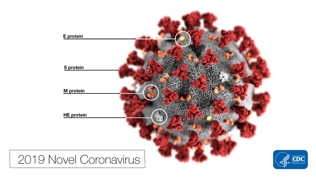
Remdesivir (RDV or GS-5734) was used in the treatment of the first case of the SARS-CoV-2 (formerly 2019-nCoV ) in the United States (1). RDV is not an approved drug in any country but has been requested by a number of agencies worldwide to help combat the SARS-CoV-2 virus (2). RDV is an adenine nucleotide monophosphate analog demonstrated to inhibit Ebola virus replication (3). RDV is bioactivated to the triphosphate form within cells and acts as an alternative substrate for the replication-necessary RNA dependent RNA polymerase (RdRp). Incorporation of the analog results in early termination of the primer extension product resulting in the inhibition.
This illustration, created at the Centers for Disease Control and Prevention (CDC), reveals ultrastructural morphology exhibited by coronaviruses. Photo Credit: Alissa Eckert, MS; Dan Higgins, MAM CDC
Why all the interest in RDV as a treatment for SARS-CoV-2 ? Much of the interest in RDV is due to a series of studies performed by collaborating groups at the University of North Carolina Chapel Hill (Ralph S. Baric’s lab) and Vanderbilit University Medical Center (Mark R. Denison’s lab) in collaboration with Gilead Sciences.
Bioluminescent reporter assays are an excellent choice for analyzing gene regulation because they provide higher sensitivity, wider dynamic range and better signal-to-background ratios compared to colorimetric or fluorescent assays. In a typical genetic reporter assay, cells are transfected with a vector that contains the sequence of interest cloned upstream of a reporter gene, and the reporter activity is used to determine how the target sequence influences gene expression under experimental conditions. A second control reporter encoded on the same or a different plasmid is an essential internal control. The secondary reporter is used to normalize the data and compensate for variability caused by differences in cell number, lysis efficiency, cell viability, transfection efficiency, temperature, and measurement time.
Basic Introduction to the Strategy of Reporter Gene Assays
For genetic reporter assays, using a secondary control vector with a weak promoter like PGK or TK to ensures that the control does not interfere with activation of your primary reporter vector. Transfection of high amounts of the control plasmid or putting the control reporter under control of a strong promoter like CMV or SV40 often leads to transcriptional squelching or other interference with the experimental promoter (i.e., trans effects). Reporter assays can also be used to quantitatively evaluate microRNA activity by inserting miRNA target sites downstream or 3´ of the reporter gene. For example, the pmirGLO Dual-Luciferase miRNA Target Expression Vector is based on dual-luciferase technology, with firefly luciferase as the primary reporter to monitor mRNA regulation and Renilla luciferase as a control reporter for normalization.
Here in Technical Services we often talk with researchers who are just starting their project and looking for advice on designing their genetic reporter vector. They have questions like:
How much of the upstream promoter region should be included in the vector?
How many copies of a response element will be needed to provide a good response?
Does the location of the element or surrounding sequence alter gene regulation?
You have identified and cloned your protein of interest, but you want to explore its function. A protein fusion tag might help with your investigation. However, choosing a tag for your protein depends on what experiments you are planning. Do you want to purify the protein? Would you like to identify interacting proteins by performing pull-down assays? Are you interested in examining the endogenous biology of the protein? Here we cover the advantages and disadvantages of some protein tags to help you select the one that best suits your needs.
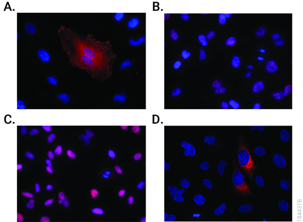
CRISPR-Cas9 editing knocked-in HiBiT at the endogenous locus of proteins with varying subcellular localization. Fixed CRISPR-modified clones or pools of cells were imaged by immunofluorescent staining using the Anti-HiBiT Monoclonal Antibody (red) and Hoechst dye (blue). Panel A. VCL-HiBiT pool. Panel B. SMARCA4-HiBiT clone. Panel C. HDAC2-HiBiT clone. Panel D. HSP90B1-HiBiT pool.
Affinity Tags
The most commonly used protein tags fall under the category of affinity tags. This means that the tag binds to another molecule or metal ion, making it easy to purify or pull down your protein of interest. In all cases, the tag will be fused to your protein of interest at either the amino (N) or carboxy (C) terminus by cloning into an expression vector. This protein fusion can then be expressed in cells or cell-free systems, depending on the promoter the vector contains.
Cardiovascular diseases, or CVDs, are collectively the most notorious gang of cold-blooded killers threatening human lives today. These unforgiving villains, including the likes of coronary heart disease, cerebrovascular disease and pulmonary embolisms, are jointly responsible for more deaths per year than any other source, securing their seat as the number one cause of human mortality on a global scale.
One of the
trademarks of most CVDs is the thickening and stiffening of the arteries, a
condition known as atherosclerosis. Atherosclerosis is characterized by the
accumulation of cholesterol, fats and other substances, which together form
plaques in and on the artery walls. These plaques clog or narrow your arteries
until they completely block the flow of blood, and can no longer supply
sufficient blood to your tissues and organs. Or the plaques can burst, setting
off a disastrous chain reaction that begins with a blood clot, and often ends
with a heart attack or stroke.
Given the global prevalence and magnitude of this problem, there is a significant and urgent demand for better ways to treat CVDs. In a recent study published in Nature Communications, researchers at the Carnegie Institution for Science, Johns Hopkins University and Mayo Clinic are taking the fight to CVDs through the study of low-density lipoproteins (LDLs), the particles responsible for shuttling bad cholesterol throughout the bloodstream.
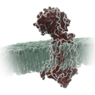
G Protein-Coupled Receptors (GPCRs) are a very large, diverse family of transmembrane receptors in eukaryotes. These receptors detect molecules outside the cell and activate internal signaling pathways by coupling with G proteins. Once a GPCR is activated, β-arrestins translocate to the cell membrane and bind to the occupied receptor, uncoupling it from G proteins and promoting its internalization.
Reporter tags are useful for studying the dynamics of GPCRs and associated proteins, but large tags can disrupt the receptors’ native functioning, and often overexpression of the tagged protein is required to obtain sufficient signal. Here is one example of how researchers have used the small, bright NanoLuc® luciferase to overcome these common challenges and answer questions about GPCRs.
It’s a question I’m asked probably once a week. “What wavelength do I select on my luminometer when performing a luciferase assay?” The question is a good and not altogether unexpected one, especially for those new to bioluminescent assays. The answer is that in most cases, you don’t and in fact shouldn’t select a wavelength (the exception to this rule is if you’re measuring light emitted in two simultaneous luciferase reactions). To understand why requires a bit of an explanation of absorbance, fluorescence, and luminescence assays, and the differences among them.
Absorbance, fluorescence, and luminescence assays are all means to quantify something of interest, be that a genetic reporter, cell viability, cytotoxicity, apoptosis, or other markers. In principle, they are all similar. For example, a genetic reporter assay is an indicator of gene expression. The promoter of a gene of interest can be cloned upstream of a reporter such as β-galactosidase, GFP, or firefly luciferase. The amount of each of these reporters that is transcribed into mRNA and translated into protein by the cell is indicative of the endogenous expression of the gene of interest.
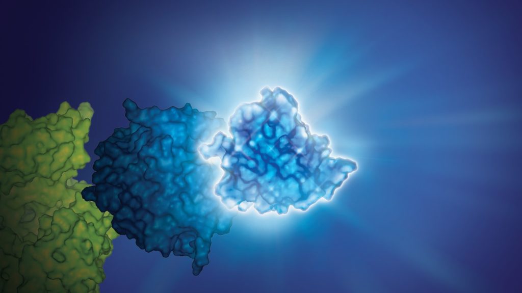
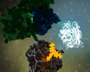
No protein is an island. Within a cell, protein-protein interactions (PPIs) are involved in highly regulated and specific pathways that control gene expression and cell signaling. The disruption of PPIs can lead to a variety of disease states, including cancer.
Two general approaches are commonly used to study PPIs. Real-time assays measure PPI activity in live cells using fluorescent or luminescent tags. A second approach includes methods that measure a specific PPI “after the fact”; popular examples include a reporter system, such as the classic yeast two-hybrid system.
XWe use cookies and similar technologies to make our website work, run analytics, improve our website, and show you personalized content and advertising. Some of these cookies are essential for our website to work. For others, we won’t set them unless you accept them. To learn more about our approach to Privacy we invite you to Read More
By clicking “Accept All”, you consent to the use of ALL the cookies. However you may visit Cookie Settings to provide a controlled consent.
We use cookies and similar technologies to make our website work, run analytics, improve our website, and show you personalized content and advertising. Some of these cookies are essential for our website to work. For others, we won’t set them unless you accept them. To find out more about cookies and how to manage cookies, read our Cookie Policy.
If you are located in the EEA, the United Kingdom, or Switzerland, you can change your settings at any time by clicking Manage Cookie Consent in the footer of our website.
Necessary cookies are absolutely essential for the website to function properly. These cookies ensure basic functionalities and security features of the website, anonymously.
Cookie
Duration
Description
cookielawinfo-checbox-analytics
11 months
This cookie is set by GDPR Cookie Consent plugin. The cookie is used to store the user consent for the cookies in the category "Analytics".
cookielawinfo-checbox-functional
11 months
The cookie is set by GDPR cookie consent to record the user consent for the cookies in the category "Functional".
cookielawinfo-checbox-others
11 months
This cookie is set by GDPR Cookie Consent plugin. The cookie is used to store the user consent for the cookies in the category "Other.
cookielawinfo-checkbox-advertisement
1 year
The cookie is set by GDPR cookie consent to record the user consent for the cookies in the category "Advertisement".
cookielawinfo-checkbox-necessary
11 months
This cookie is set by GDPR Cookie Consent plugin. The cookies is used to store the user consent for the cookies in the category "Necessary".
cookielawinfo-checkbox-performance
11 months
This cookie is set by GDPR Cookie Consent plugin. The cookie is used to store the user consent for the cookies in the category "Performance".
gdpr_status
6 months 2 days
This cookie is set by the provider Media.net. This cookie is used to check the status whether the user has accepted the cookie consent box. It also helps in not showing the cookie consent box upon re-entry to the website.
lang
This cookie is used to store the language preferences of a user to serve up content in that stored language the next time user visit the website.
viewed_cookie_policy
11 months
The cookie is set by the GDPR Cookie Consent plugin and is used to store whether or not user has consented to the use of cookies. It does not store any personal data.
Analytical cookies are used to understand how visitors interact with the website. These cookies help provide information on metrics the number of visitors, bounce rate, traffic source, etc.
Cookie
Duration
Description
SC_ANALYTICS_GLOBAL_COOKIE
10 years
This cookie is associated with Sitecore content and personalization. This cookie is used to identify the repeat visit from a single user. Sitecore will send a persistent session cookie to the web client.
vuid
2 years
This domain of this cookie is owned by Vimeo. This cookie is used by vimeo to collect tracking information. It sets a unique ID to embed videos to the website.
WMF-Last-Access
1 month 18 hours 24 minutes
This cookie is used to calculate unique devices accessing the website.
_ga
2 years
This cookie is installed by Google Analytics. The cookie is used to calculate visitor, session, campaign data and keep track of site usage for the site's analytics report. The cookies store information anonymously and assign a randomly generated number to identify unique visitors.
_gid
1 day
This cookie is installed by Google Analytics. The cookie is used to store information of how visitors use a website and helps in creating an analytics report of how the website is doing. The data collected including the number visitors, the source where they have come from, and the pages visted in an anonymous form.
Advertisement cookies are used to provide visitors with relevant ads and marketing campaigns. These cookies track visitors across websites and collect information to provide customized ads.
Cookie
Duration
Description
IDE
1 year 24 days
Used by Google DoubleClick and stores information about how the user uses the website and any other advertisement before visiting the website. This is used to present users with ads that are relevant to them according to the user profile.
test_cookie
15 minutes
This cookie is set by doubleclick.net. The purpose of the cookie is to determine if the user's browser supports cookies.
VISITOR_INFO1_LIVE
5 months 27 days
This cookie is set by Youtube. Used to track the information of the embedded YouTube videos on a website.
Performance cookies are used to understand and analyze the key performance indexes of the website which helps in delivering a better user experience for the visitors.
Cookie
Duration
Description
YSC
session
This cookies is set by Youtube and is used to track the views of embedded videos.
_gat_UA-62336821-1
1 minute
This is a pattern type cookie set by Google Analytics, where the pattern element on the name contains the unique identity number of the account or website it relates to. It appears to be a variation of the _gat cookie which is used to limit the amount of data recorded by Google on high traffic volume websites.