Improved Characterization and Quantification of Complex Cell Surface N-Glycans

MSextractcropped

N-Glycosylation is a common protein post-translational modification occurring on asparagine residues of the consensus sequence asparagine-X-serine/threonine, where X may be any amino acid except proline. Protein N-glycosylation takes place in the endoplasmic reticulum (ER) as well as in the Golgi apparatus.

Approximately half of all proteins typically expressed in a cell undergo this modification, which entails the covalent addition of sugar moieties to specific amino acids. There are many potential functions of glycosylation. For instance, physical properties include: folding, trafficking, packing, stabilization and protease protection. N-glycans present at the cell surface are directly involved in cell−cell or cell−protein interactions that trigger various biological responses.

The standard method used to profile the N-glycosylation pattern of cells is glycoprotein isolation followed by denaturation and/or tryptic digestion of the glycoproteins and an enzymatic release of the N-glycans using PNGase F followed by analysis mass spec. This method has been reported to yield high levels of high-mannose N-glycans that stem from both membrane proteins as well as proteins from the ER.(1,2)

Are you looking for proteases to use in your research?
Explore our portfolio of proteases today.

For those researchers interested in characterizing only cell surface glycans (i.e.,  complex N-glycans)  a recent reference has developed a model system using HEK-292 cells that demonstrates a reproducible, sensitive, and fast method to profile surface N-glycosylation from living cells (3). The method involves standard centrifugation followed by enzymatic release of cell surface N-glycans. When compared to the standard methods the detection and quantification of complex-type N-glycans by increased their relative amount from 14 to 85%.

  1. North, S. J. et al. (2012) Glycomic analysis of human mast cells, eosinophils and basophils. Glycobiology. 2012, 22, 12–22.
  2. Reinke, S. O. et al. (2011) Analysis of cell surface N-glycosylation of the human embryonic
    kidney 293T cell line. J. Carbohydr. Chem.  30, 218–232.
  3. Hamouda, H. et al. (2014) Rapid Analysis of Cell Surface N‑Glycosylation from Living Cells Using Mass Spectrometry. J of Proteome Res. 13, 6144–51.
Mass Spec-Compatible Proteome Reference Material

MSextractcropped

The complexity of biological samples places high demand on mass spec analytical capability. Adequate monitoring of instrument performance for proteomics studies requires equally complex reference material such as whole-cell extracts. However, whole-cell extracts available commercially are developed for general research (e.g., enzymatic or Western blot analysis) and contain detergents and salts that interfere with reverse phase liquid chromatography and mass spectrometry. Even after clean up, the extracts need to be digested, requiring time, labor and experience to generate samples for use in mass spectrometry. To address the need for complex protein material, we have developed whole-cell protein extracts from yeast and human cells. The yeast extract offers the convenience of a relatively small and well annotated proteome, whereas the human extract provides a complex proteome with large dynamic range. The human extract also serves as reference material for studies targeting the human proteome.

The extracts are free of compounds that interfere with reverse phase liquid chromatography-mass spectrometry (LC-MS), and have been reduced with DTT and alkylated with iodoacetamide then digested with Trypsin/Lys-C Mix and lyophilized. These digested extracts (tryptic peptides) can be readily reconstituted in trifluoroacetic acid (TFA) or formic acid and injected into an instrument. The same human and yeast whole-cell extracts also are provided in an intact (undigested) form for users who would like to develop an independent method for preparing protein mass spectrometry samples. For convenience, the intact extracts are provided as a frozen solution.

Consistent extract protein composition is ensured by tight control over cell culture conditions and manufacturing process. Lot-to-lot consistency of extracts is monitored by various protein and peptide qualitative and quantitation methods, including LC-MS. (Quality control results are provided upon request.) Our manufacturing process assures compatibility with reverse phase liquid chromatography and mass spectrometry, minimal nonspecific protein fragmentation, nonbiological post-translational modifi cations and,for digested extracts, minimal undigested peptides. The extracts are optimized for a high number of peptide and protein identifications in mass spectrometry analysis.

Novel Application for ProteaseMAX Surfactant: Cell Lysis

ProteaseMax Surfactant


The novel mass spectrometry compatible surfactant sulfonate-(sodium 3-((1-(furan-2-yl)undecyloxy) carbonylamino)-propane-1-sulfonate (i.e.ProteaseMAX) facilitates both in-gel and in-solution digestion applications by reducing the time required, enabling protein solubilization/denaturation and increasing peptide/protein identifications.

A new application was highlighted in a recent publication (1) which utilized ProteaseMAX to lyse cells prior to trypsin digestion and subsequent mass spec analysis. The composition of the buffer determines the overall efficiency of cell lysis, dissociation of protein complexes, protein solubility and ease of removal prior to LC/MS-MS analysis.

Are you looking for proteases to use in your research?
Explore our portfolio of proteases today.

When compared to lysis buffers containing either urea or SDC, ProteaseMAX provided the optimal number of identified peptides/proteins.
In addition it can be easily removed from the lysate by acidic precipitation.

Reference

  1. Pirmoradian, M. et al. (2013). Rapid and deep human proteome analysis by single-dimension shotgun proteomics. Mol. Cell. Prot. 12, 3330–8.

Testing for Neonatal Sepsis: The Next Generation of Biomarkers

newbornNeonatal sepsis is a systemic infection prevalent in preterm and very low birth weight infants and causes high morbidity. Most cases of neonatal sepsis are caused by pathogenic bacteria that invade the bloodstream, triggering an abrupt and overwhelming infection in the target organs accompanied by a systemic inflammatory response. Testing for neonatal sepsis is challenging because it does not affect a specific organ and presents multiple symptoms that are often confused with other related conditions (1). Current diagnostic tests for sepsis include those that identify markers of the host response to infection (e.g., procalcitonin, C reactive protein, cytokines, etc.) and those that detect bacterial infection in blood (bacteremia) (2). The lack of specific diagnostic biomarkers for early and accurate detection of neonatal sepsis has spurred the quest for next-generation biomarkers using powerful mass screening technologies such as proteomics. Continue reading “Testing for Neonatal Sepsis: The Next Generation of Biomarkers”
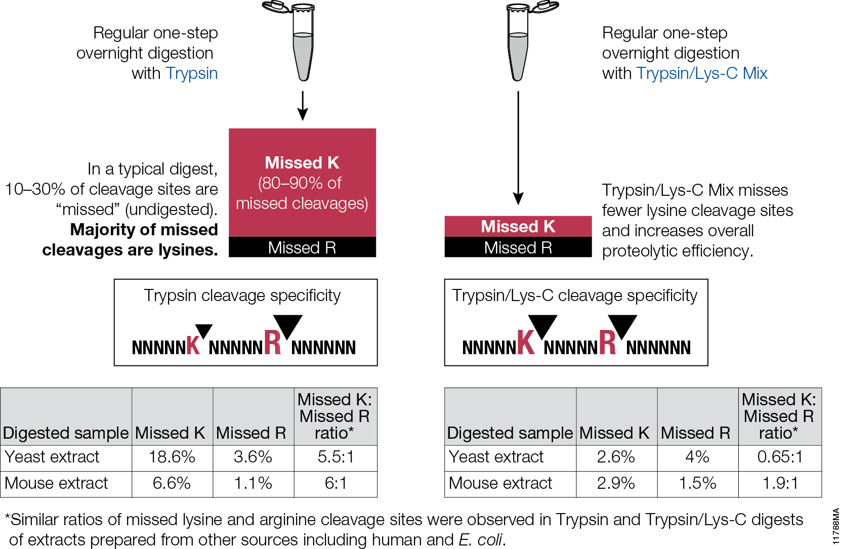
Trypsin/Lys-C Mix: Alternative for standard trypsin protein digestions

Trypsin/Lys-C Mix, Mass Spec Grade, is a mixture of Trypsin Gold, Mass Spectrometry Grade, and rLys-C, Mass Spec Grade. The Trypsin/Lys-C Mix is designed to improve digestion of proteins or protein mixtures in solution.It is a little known fact that trypsin cleaves at lysine residues with lesser efficiency than at arginine residues. Inefficient proteolysis at lysine residues is the major cause of missed (undigested) cleavages in trypsin digests.

11788MA


Supplementing trypsin with Lys-C enables cleavage at lysines with excepetional efficiency and specificity. Following the conventional trypsin digestion protocol (i.e., overnight incubation at nondenaturing conditions, reduction,alkylation, 25:1 protein:protease ratio [w/w], mix and incubate overnight at 37°C.) Replacing trypsin with Trypsin/Lys-C Mix in this conventional protocol leads to multiple benefits for protein analysis including more accurate mass spectrometry-based protein quantitation and improved protein mass spectrometry analytical reproducibility.

Are you looking for proteases to use in your research?
Explore our portfolio of proteases today.

Convenient, Non-Radioactive Detection of Isoaspartate

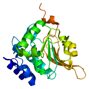
Structure of the PCMT1 protein. Based on PyMOL rendering of PDB 1i1n. Licensed under creative commons http://creativecommons.org/licenses/by-sa/3.0/deed.en

The ISOQUANT® Isoaspartate Detection Kit is intended for quantitative detection of isoaspartic acid residues in proteins and peptides, which can result from the gradual, nonenzymatic deamidation of asparagine or rearrangement of aspartic acid residues.

The ISOQUANT® Kit is designed to provide information regarding the global formation of isoaspartic acid residues at Asn and Asp sites, not at each site separately.

The deamidation of asparagine residues and rearrangement of aspartic acid residues is characterized by the formation of a succinimide intermediate that resolves to form a mixture of isoaspartic acid (typically 70–85%) and aspartic acid.
The rate and extent of isoaspartic acid formation can vary widely among proteins, depending on the amino acid sequence and size of the target protein. Deamidation of Asn residues has been observed most frequently at Asn-Gly and Asn-Ser sites within proteins.

The ISOQUANT® Isoaspartate Detection Kit uses the enzyme Protein Isoaspartyl ethyltransferase (PIMT) to specifically detect the presence of isoaspartic acid residues in a target protein. PIMT catalyzes the transfer of a methyl group from S-adenosyl-L-methionine (SAM) to isoaspartic acid. Spontaneous decomposition of this methylated intermediate results in the release of methanol and reformation of the succinimide.

References:

Wang, W. et al. (2012) Quantification and characterization of antibody deamidation by peptide mapping with mass spectrometry. Int. J. Mass. Spec. 312, 107–13.

Grappin, P. et al. (2011) New proteomic developments to analyze protein isomerization and their biological significance in plants. J. Proteomics, 74, 1475–82.

Yang, H. and Zubarev, R.A. (2010) Mass spectrometric analysis of asparagine deamidation and aspartate isomerization in polypeptides. Electrophoresis 31, 1764–71.

Sinha, S. et al. (2009) Effect of protein structure on deamidation rate in the Fc fragment of an IgG1 monoclonal antibody. Protein Sci. 18, 1573–84.

ProteaseMAX Surfactant: Enhanced In-solution Digestion Applications

ProteaseMax 11228MA

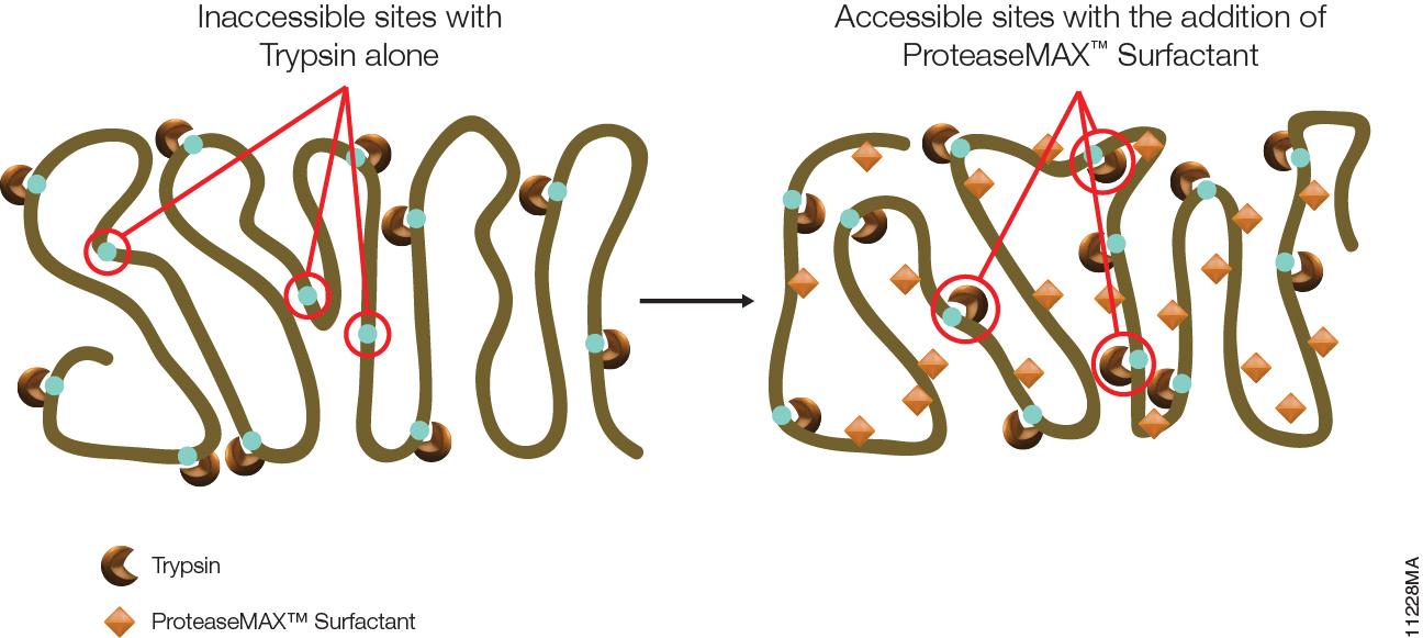
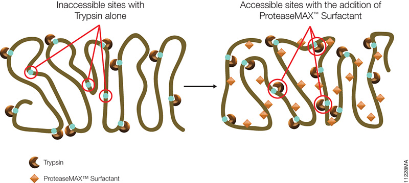
The primary advantage of ProteaseMAX™ Surfactant is that it improves identification of proteins in gel by enhanced protein digestion, increased peptide extraction, and minimized post digestion peptide loss. However, ProteaseMAX™ Surfactant can also facilitate in-solution digestion protocols.

ProteaseMAX™ Surfactant offers two major benefits for digesting proteins in solution.

Continue reading “ProteaseMAX Surfactant: Enhanced In-solution Digestion Applications”

PNGase F, a Novel Endoglycosidase

11123MA

PNGase F (Cat.# V4831) is a recombinant glycosidase cloned from Elizabethkingia meningoseptica and overexpressed in E. coli, with a molecular weight of 36kD.

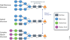
PNGase F catalyzes the cleavage of N-linked oligosaccharides between the innermost GlcNAc and asparagine residues of high mannose, hybrid, and
complex oligosaccharides from N-linked glycoproteins. PNGase F will not remove oligosaccharides containing alpha-(1,3)-linked core fucose,
commonly found on plant glycoproteins.

Applications
Determining whether a protein is in fact glycosylated is the initial step in glycoprotein analysis. Polyacrylamide gel electrophoresis in the
presence of sodium dodecyl sulfate (SDS-PAGE) has become the method of choice as the final step prior to mass spec analysis. Glycosylated proteins often migrate as diffused bands by SDS-PAGE. A marked decrease in band width and change in migration position after treatment with PNGase F is considered evidence of N-linked glycosylation.

Gel based data are often correlated with information obtained from mass spec analysis. Asn-linked type glycans can be cleaved enzymatically by PNGase F yielding intact oligosaccharides and a slightly modified protein in which Asn residues at the site of de-N-glycosylation are converted to Asp, by converting the previously carbohydrate-linked asparagine into an aspartic acid, a monoisotopic mass shift of 0.9840Da is observed. The deglycosylated peptides are then analyzed by tandem mass spectrometry (MS/MS), and software algorithms are used to correlate the experimental fragmentation spectra with theoretical tandem mass spectra generated from peptides in a protein database.

Enhancing Proteomics Data Using Arg-C Protease

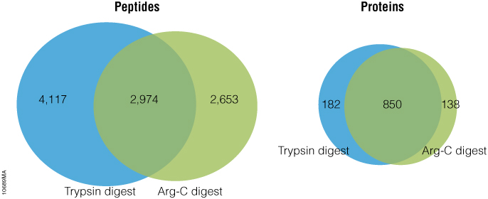
Arg-C (clostripain), Sequencing Grade (Cat.# V1881), is a specific endoproteinase isolated from the soil bacterium Clostridium histolyticum. It preferentially cleaves at the C-terminal side of arginine (R) residues. Unlike trypsin, Arg-C efficiently cleaves arginine sites followed by proline (P). This difference is important because every twentieth arginine is followed by proline. To illustrate this benefit, Arg-C was evaluated for protein analysis in two different experiments. In the first experiment, we studied the use of Arg-C for proteomic analysis. Yeast provides an excellent model proteome because its genome is well annotated. Yeast extract was digested in two parallel reactions, using trypsin in the first reaction and Arg-C in the second, using a conventional protocol consistent with LC-MS/MS analysis. As expected the trypsin digestion resulted in a high number of peptide and protein identifications (Figure 1). However, many peptides remained elusive. The parallel Arg-C digestion complemented the trypsin digestion by recovering an additional 2,653 peptides and providing a 37.4% increase in the number of identified peptides. Digesting with Arg-C also resulted in an increase in the number of identified proteins. In fact, 138 new proteins were identified in Arg-C digest compared to the parallel trypsin digest, offering a 13.4% increase in the overall number of identified proteins.

Figure 1. Side-by-side analysis of trypsin-digested and Arg-C digested yeast proteins.

Are you looking for proteases to use in your research?
Explore our portfolio of proteases today.

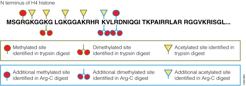
In a second experiment, the ability of Arg-C to analyze individual proteins was analyzed, selecting human histone H4 as a model protein. Like other histones, this protein is heavily modified post translational modifications (PTMs) that alter histone structure and regulate interaction with transcription factors. As a result, histone PTMs are implicated in gene regulation and associated with multiple disorders. Technical challenges, however, impede histone PTM analysis. Histone PTMs are complex and some, such as acetylation and methylation, prevent trypsin digestion, as shown by our data. In this experiment, trypsin digestion of histone H4 identified several PTMs (Figure 2). However, certain PTMs were missing. By digesting histone H4 with Arg-C, we were able to identify the missing PTMs including mono-, dimethylated and acetylated lysine and arginine residues. We speculate that the PTMs in human histone H4, which modified arginine and lysine residues, rendered trypsin unsuitable for preparing the corresponding histone regions for mass spectrometry. The problem was rectified by replacing trypsin with Arg-C.

Figure 2. Identification of histone h4 PTMs after Arg-C digestion.

Use of Nonspecific Proteases for Analysis of Proteins by Mass Spectrometry

mass spectrometry results

One of the approaches to identify proteins by mass spectrometry includes the separation of proteins by gel electrophoresis or liquid chromatography. Subsequently the proteins are cleaved with sequence-specific endoproteases. Following digestion the generated peptides are investigated by determination of molecular masses or specific sequence. For protein identification the experimentally obtained masses/sequences are compared with theoretical masses/sequences compiled in various databases.

Are you looking for proteases to use in your research?
Explore our portfolio of proteases today.

Nonspecific proteases such as pepsin, proteinase K, elastase and thermolysin can offer an alternative to traditional sequence-specific proteases for certain applications. The following references illustrate the use of nonspecific proteinases for the mass spec analysis of proteins:

Papasotiriou, D. et al. (2010) Peptide mass fingerprinting after less specific in-gel proteolysis using MALDI-LTQ-Orbitrap and 4-chloro-alpha-cyanocinnamic acid. J. Proteome. Res. 9, 2619–29. This reference demonstrates the use of either chymotrypsin, elastase, trypsin or proteinase K in combination with matrix CHCA for increase peptide identification and sequence coverage using MALDI.

Neue, K. et al. (2011) Elucidation of glycoprotein structures by unspecific proteolysis and direct nanoESI mass spectrometric analysis of ZIC-HILIC-enriched glycopeptides. J. Proteome. Res. 10, 2248–60. Notes use of thermolysin or elastase in combination with ZIC-HILIC enrichment as alternative method for the characterization of glycopeptides.

Baeumlisberger, D et al. (2011) Simple dual-spotting procedure enhances nLC-MALDI MS/MS analysis of digests with less specific enzymes. J. Proteome. Res. 10, 2889–94. Data noted that samples digested with elastase followed by nLC separation and subsequent alternative spotting on both MALDI-LTQ-Orbitrap and MALDL-TOF/TOF instruments resulted in 32% additional peptides.