T-Vector Cloning: Answers to Frequently Asked Questions

Blue/White colony screening helps you pick only the colonies that have your insert.

Q: Can PCR products generated with GoTaq® DNA Polymerase be used to for T- vector cloning?

A: Yes. GoTaq® DNA Polymerase is a robust formulation of unmodified Taq Polymerase. GoTaq®DNA Polymerase lacks 3’ →5’ exonuclease activity (proof reading) and also displays non-template–dependent terminal transferase activity that adds a 3′ deoxyadenosine (dA) to product ends. As a result, PCR products amplified using GoTaq® DNA Polymerase will contain A-overhangs which makes it suitable for T-vector cloning.

We have successfully cloned PCR products generated using GoTaq® and GoTaq® Flexi DNA Polymerases into the pGEM®-T (Cat.# A3600), pGEM®-T Easy (Cat.# A1360) and pTARGET™ (Cat.# A1410) Vectors.

Q: Can GoTaq® Long PCR Master Mix be used for T-Vector Cloning?

A: Yes it can. GoTaq® Long PCR Master Mix utilizes recombinant Taq DNA polymerase as well as a small amount of a recombinant proofreading DNA polymerase. This 3´→5´ exonuclease activity (proof reading) enables amplification of long targets. Despite the presence of a small amount of 3´→5´ exonuclease activity, the GoTaq® Long PCR Master Mix generates PCR products that can be successfully ligated into the pGEM®-T Easy Vector System.

We have demonstrated that GoTaq® Long PCR Master Mix successfully generated DNA fragments that could be ligated into pGEM®-T Easy Vector System without an A-tailing procedure, and with ligation efficiencies similar to those observed with the GoTaq® Green Master Mix.

For details refer to Truman, A., Hook, B. and Wieczorek, D. Using GoTaq® Long PCR Master Mix for T-Vector Cloning.

Tip: For cloning blunt-ended PCR fragments into T-vectors, use the A-tailing protocol discussed in the pGEM®-T and pGEM®-T Easy Technical Manual #TM042.

Q: How do I prepare PCR products for ligation? What products can be used to purify the DNA?

Continue reading “T-Vector Cloning: Answers to Frequently Asked Questions”

Beating the Odds of Cancer: Not Just a Tall Tale

elephants_webWhen it comes to combating cancer does size matter? If every cell in the body has the propensity to become cancerous, it should naturally follow that larger animals that pack greater number of body cells and that those whose cells undergo greater number of cell divisions are more likely to develop cancer. By the same logic, organisms with longer lifespans must also have a greater chance of accumulating mutations leading to cancer. Surprisingly, the risk of developing cancer is only 5% in elephants and 18% in whales whereas it is as high as 30% in humans and rodents.  The apparent lack of correlation between body mass, longevity and cancer- known as Peto’s paradox- has flummoxed scientists for several decades.1

A recent study published in Journal of the American Medical Association  by  Abegglen and colleagues has unlocked the secret weapon held by the pachyderms in fighting cancer2. While the weapon itself might not be new to cancer biologists, the stash carried by these marvelous animals is the highest recorded for any living species so far. To understand this weapon let’s revisit the coping mechanisms developed by cells to prevent cancer. When mammalian cells are exposed to cancer inducing treatments, such as UV radiation for example, a gene encoding TP53, kicks into gear making copies of the tumor suppressing protein of the same name. TP53 acts as a tumor suppressor, which means that it regulates cell division by keeping cells from growing and dividing too fast or in an uncontrolled way. It does so by either repairing any damage to the cells caused by the UV exposure or by killing off the cell by a self-destructing mechanism known as apoptosis which is akin to committing suicide.

Many mammals, including humans carry only two copies of this important gene; one copy or allele is inherited from each parent. If the TP53 gene is inactivated by mutations, the risk of developing cancer increases by several fold. A rare but lethal condition called Li-Fraumeni Syndrome marks patients who have only one working copy of TP53 with more than a 90 percent lifetime cancer risk from childhood into their adult years.  In a quest to investigate the unexplained resistance to cancer by elephants, the scientists combed through the elephant genome and stumbled upon 40 copies of genes that code for TP53. One pair was ancestral in origin, whereas the remainder appear to have diverged from the ancestral copy and were archived within the genome over the course of evolution as retrogenes. Continue reading “Beating the Odds of Cancer: Not Just a Tall Tale”

Removing Cancer’s Cloak of Invisibility

Copyright Promega.
Copyright Promega.
For decades scientists have been trying to harness the power of our immune system to fight cancer cells. It is not impossible to imagine that our immune system, which is sophisticated enough to fight against a multitude of invaders that threaten our health, should be able to tackle a deadly disease such as cancer. This formed the basis of testing a new type of cancer treatment known as immunotherapy. Immunotherapy for cancer means developing treatments to harness your immune system and using your own immune system to fight the cancerous cells.

But in reality it was hard to make this work. Because, as scientists discovered recently, cancer outsmarts the immune system by wearing a kind of “invisibility cloak”. Cancer is able to fool the immune system from recognizing that it is the enemy and in effect keeps the immune system from destroying it.

In a breakthrough discovery scientists have found a way around this treachery.

The breakthrough is in therapies called ‘checkpoint inhibitors’. Checkpoint inhibitors block the mechanisms that allow some tumor cells to evade the immune system. The drugs ensure that cancer cells are no longer be shielded from the immune system defenses, but are instead recognized as “foreign”. Continue reading “Removing Cancer’s Cloak of Invisibility”

MicroRNAs as Circulating Biomarkers

12097693_lMicroRNAs (miRNAs) are short strands of RNA averaging between 19-24 nucleotides in length that were first discovered in C.elegans and subsequently shown to exist in species ranging from algae to humans (1). Speculated to be merely “junk” more than a decade ago, miRNAs have emerged as powerful regulators of a wide array of cellular processes because of their influence on gene expression at the posttrancriptional level. Dysregulation of these miRNAs is also associated with life-threatening conditions such as cancer and cardiovascular disease, which points to a potential use of miRNAs in diagnosis and treatment. Recently, it has been demonstrated that miRNAs are present in circulating blood plasma, protected from degradation by inclusion in lipid or lipoprotein complexes. This opens up the possibility to exploit miRNA as a useful diagnostic tool in clinical samples. Continue reading “MicroRNAs as Circulating Biomarkers”

Angel’s Glow: Bioluminescence Uncovered on the Battlefield

1888 Chromolithograph of the Battle of Shiloh, American Civil War, produced by L. Prang & Co.

If battlegrounds could speak they would have many stories to tell.  In some cases the microbes found in those soils have lived on to separate fact from fiction. One such story has its origins in the Battle of Shiloh, which went down in history as one of the bloodiest battles fought during the American Civil War.  As the soldiers lay mortally wounded on the cold, hard grounds of Shiloh waiting for medical aid, they noticed a very strange phenomenon. Some of the wounds actually appeared to be glowing in the dark casting a faint light into the darkness of the battlefield. And the legend goes that soldiers with the glowing wounds had a better chance at survival and recovery from infections than their fellow brothers-in-arms whose wounds were not similarly luminescent. The seemingly protective effect of the mysterious light earned it the moniker “Angel’s Glow.”

Fast forward to the 21st century.

Continue reading “Angel’s Glow: Bioluminescence Uncovered on the Battlefield”

Review: Validation of a high throughput cell-based assay to screen compounds for mitochondrial toxicity

mitotox_covertip

Drug-induced mitochondrial toxicity is a concern for pharmaceuticals that was, until recently, limited by the availability of a cell-based assay that is amenable to rapid high-throughput screening. Incorporating high-throughput assay chemistry that can detect mitochondrial dysfunction early in drug discovery programs provides the opportunity to identify potential mitotoxicants before they reach clinical trials or the market population.

In the paper reviewed here (1) Swiss and colleagues have validated the use of a commercially available Mitochondrial ToxGlo™ Asssay (2) to replace traditional methods that not only require expensive reagents and/or equipment but also do not lend themselves easily to high-throughput screening formats.

Continue reading “Review: Validation of a high throughput cell-based assay to screen compounds for mitochondrial toxicity”

Testing for Neonatal Sepsis: The Next Generation of Biomarkers

newborn
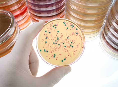
Neonatal sepsis is a systemic infection prevalent in preterm and very low birth weight infants and causes high morbidity. Most cases of neonatal sepsis are caused by pathogenic bacteria that invade the bloodstream, triggering an abrupt and overwhelming infection in the target organs accompanied by a systemic inflammatory response. Testing for neonatal sepsis is challenging because it does not affect a specific organ and presents multiple symptoms that are often confused with other related conditions (1). Current diagnostic tests for sepsis include those that identify markers of the host response to infection (e.g., procalcitonin, C reactive protein, cytokines, etc.) and those that detect bacterial infection in blood (bacteremia) (2). The lack of specific diagnostic biomarkers for early and accurate detection of neonatal sepsis has spurred the quest for next-generation biomarkers using powerful mass screening technologies such as proteomics. Continue reading “Testing for Neonatal Sepsis: The Next Generation of Biomarkers”