Science and the Wonderful Why

Curiosity.“Why? Why? Why?” Anyone who has been around small children has experienced the monotonous, often aggravating, seemingly endless barrage of the “W” word. Why does soap make bubbles? Why do feathers float and acorns fall to the ground? Why are baths important? Why are those flowers purple? Why can’t I be purple? Why do tigers have stripes and leopards have spots and lions don’t have anything (majestic manes not withstanding)? Why can rocks bounce (skip) off water? Why didn’t my rock bounce? Why does the plant in the window bend toward the light? Why are my eyes blue and my brother’s eyes brown?

It would seem that from a very young age people are hard wired to think like a scientist. It is not enough to simply know a feather will float slowly to the ground while the acorn will plummet, or that plants turn their leaves toward the sunlight. We want to know why.

I have watched nieces and nephews as well as my own children pass through the “Wonderful Why?” stage, and I have noticed that there is often a predictable progression to the questions: “Why do plants turn their leaves toward the light?” is quickly followed by: “How do they move their leaves to face the light?” and then “What if we took away the light?”

Ray Bradbury said,

Touch a scientist and you touch a child.

As children we are all scientists. It is just that some of us never grow up.

Rapid DNA Technology: Establishing your Identity in Less than Two Hours

DNA Sequence
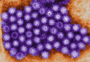
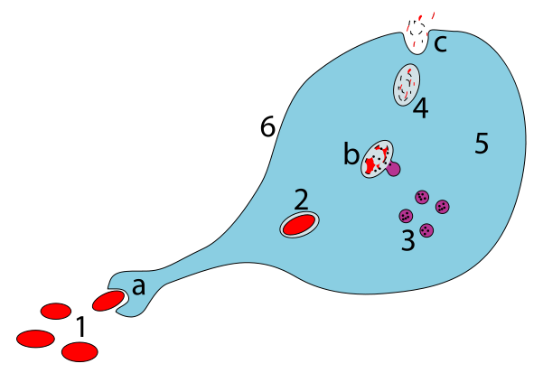
While the forensic and general communities continue to argue about the merits of the recent Supreme Court ruling on collection of samples from arrestees prior to conviction, I am fascinated by the technology that make this question relevant. The conventional way of generating a DNA profile from a sample by STR (short tandem repeat) analysis is a long process involving a series of steps that require sophisticated expensive equipment, trained personnel and, more importantly, time. The actual process of DNA analysis consists of a) sample collection, b) DNA extraction, c) PCR amplification using 16 or more unique fluorescently labeled primer sets d) capillary electrophoresis to size labeled DNA amplicons, e) software analysis to size DNA fragments and allele calls based on migration of allelic ladder fragments, and f) comparison to known profiles in the database. This entire workflow can typically take days or even weeks, and therefore it is not surprising that we see newspaper reports of backlogs of criminal and other property cases. With these time ranges, the sample collected at a site would be of no practical use to most ongoing investigations. Continue reading “Rapid DNA Technology: Establishing your Identity in Less than Two Hours”

Shining a Bright Light on Deep Questions in Biology with Bioluminescence

artists view inside a cell

Search the PubMed database for “dual-luciferase” and citations abound. The Dual-Luciferase® Reporter Assay is a powerful tool that allows researchers to ask a multitude of questions about gene control and expression in a system that itself could be normalized and internally controlled. For more than 15 years, firefly and Renilla luciferases  have formed the basis of a range of powerful assays and research tools for scientists who are asking questions about the deep and complex genetic and cellular story associated with cancer. Here we talk a bit of about bioluminescent chemistries, some of the newest bioluminescent tools available, and how some of these tools can be used to probe the deeper questions of cell biology, including cancer biology.

Continue reading “Shining a Bright Light on Deep Questions in Biology with Bioluminescence”

The Ocular Mood Ring

file9751262525646
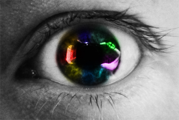
I asked my Facebook friends what my blog post should cover today. They gave me a list of very creative subjects that I will try to cover in the coming months. The winning suggestion for today is “why do my eyes change color depending on my mood?” In a simple Google search, it is apparent that many people have witnessed the phenomenon of their eyes changing color depending on their moods.There seem to be many explanations for this from light scattering to hormonal influences to psychic powers. What’s the real story? To get to the bottom of this, let’s take a closer look at how eye color is determined in the first place. Continue reading “The Ocular Mood Ring”

Rabbit Reticulocyte Lysate Translation Systems: Tools for the analysis of translational regulation

TEM of Norovirus particles. Photo Credit: Charles D. Humphrey, Centers for Disease Control and Prevention

Rabbit Reticulocyte Lysate Translation Systems are used in the identification of mRNA species, the characterization of their protein products and the investigation of transcriptional and translational control. Rabbit Reticulocyte Lysate is prepared from New Zealand white rabbits. After the reticulocytes are lysed, the extract is treated with micrococcal nuclease to destroy endogenous mRNA and thus reduce background translation to a minimum.

Untreated Lysate is prepared from New Zealand white rabbits in the same manner as treated lysates with the exception that it is not treated with micrococcal nuclease. Unlike a coupled system that initiates transcription/translation from DNA, the RNA-based rabbit reticulocyte can be used for the direct investigation of transcriptional/translational control and the replication of RNA-based viruses.


References

Characterization of translation regulation (i.e., UTRs, Capping, IRES)

  1. Nguyen, H-L .et al. (2013) Expression of a novel mRNA transcript for human microsomal epoxide hydrolase is regulated by short reading frames within it 5’ –untranslated region. RNA. 19, 752–66.
  2. Wei, J. et al. (2013) The stringency of start codon selection in the filamentous fungus Neurospora crass. J. Biol. Chem. 288, 9549–62.
  3. Paek Ki-Y. et al. (2012) Cap-Dependent translation without base-by-base scanning of an messenger ribonucleic acid. Nucl. Acid. Res. 40, 7541–51.
  4. Se, and NH. Su.W. et al. (2011) Translation, stability, and resistance to decapping of mRNA containing caps substituted in the triphosphate with BH3. RNA 17, 978–88.
  5. Anderson, D. et al. (2011) Nucleoside modifications in RNA limit activation of 2’-5’ oligoadenylate synthetase and increase resistance to cleavage by RNase L. Nucl. Acid. Res. 39, 9329-38.

RNA virus Characterization

  1. Vashist, S. et al. (2012) Identification of RNA-protein interaction networks involved in the Norovirus life cycle. J. Vir. 86, 11977–90.
  2. Soto-Rifo, R. et al. (2012) Different effects of the TAR structure on HIV-1 and HIV-2 genomics RNA translation. Nucl. Acids. Res. 40, 2653–67.
  3. Poyry, T. et al. (2011) Mechanisms governing the selection of translation initiation sites on Foot-and-Mouth Disease Virus RNA. J.Vir. 85, 10178–88.
  4. Cheng, E. et al. (2011) Characterization of the interaction between Hantavirus nucleopcapsid protein and ribosomal protein S19. J. Biol. Chem. 286, 11814–24.
  5. Vera-Otarola, J. et al. (2011) The Andes Hantavirus NSs Protein is expressed from the Viral mRMA by a leaky scanning mechanism. J. Vir. 86, 2176–87.

Priming an Effective T cell Response to Cancer

Cancer vaccines have been in progress for some time now. But a vaccine that is highly effective against cancer is not currently available.

However, an interesting report from Stanford University School of Medicine researchers, Dr. Irving Weissman, et al. shows some promise in a development of an altered means of stimulating the immune system, that could result in a stronger immune response and ultimately a better cancer vaccine. The paper by Weissman et al. was published electronically ahead of print in PNAS USA, May 20, 2013: “Anti-CD47 antibody-mediated phagocytosis of cancer by macrophages primes an effective antitumor T-cell response.”

Schematic of a macrophage engulfing, digesting and presenting parts of a pathogen or foreign cell to the cell surface.

Continue reading “Priming an Effective T cell Response to Cancer”

Peering into the History of Leprosy

Abbot Richard Wallingford
By user:Leinad-Z [Public domain], via Wikimedia Commons
What is it about the UK and graves and dead people to identify or bacteria to sequence? First, there are Black Death graves, then Richard III under a car park and now a leprosy grave site. While we do know leprosy has been a scourge of human beings for centuries, tracking the different strains and origins of the disease is relatively new. Taylor et al. were interested in this grave site near Winchester, located in southern Britain, north of Southampton and southwest of London, because little is known about the disease organism Mycobacterium leprae during this early medieval period (late 11th to 12th century). What could they learn about the strains that caused leprosy and the possible spread of the disease? Continue reading “Peering into the History of Leprosy”

About the Wild Life in Our Homes (at least the single-celled kind)

The initial paper from the Wild Life in Our Homes study by Dunn et al. found a correlation between the presence of dogs and specific bacterial communities on pillowcases and TV screens.

Back in the fall, I received a sampling kit, an Informed Consent form and instructions for collecting samples for the Wild Life In Our Homes citizen science project. I carefully swabbed the requested surfaces: exterior and interior door trim, kitchen counter tops, pillowcases, etc., and sent my samples in. I later received confirmation that my samples had been received and again later confirmation that they were being analyzed.

The first paper from this project has been published by Dunn et al. in PLOS ONE (Home Life: Factors Structuring the Bacterial Diversity Found within and between Homes). This initial report covers the first 40 homes sampled, all from the Raleigh-Durham, NC, USA area. Volunteers sampled their homes in the Fall of 2011, collecting specimens from nine areas: cutting boards, kitchen counters, refrigerator, toilet seat, pillowcase, door handle, TV screen, and interior and exterior door trim. The scientists used direct PCR and high-throughput sequencing to sequence the bacterial 16S rRNA gene from the submitted samples. By doing this they were able to estimate the diversity within each sample—they did not distinguish between live and dead organisms, and they did not sequence anything other than the bacterial 16SrRNA, so this study is limited to bacteria. Continue reading “About the Wild Life in Our Homes (at least the single-celled kind)”

OneZoom, The Fractal Phylogenic Tree Explorer

OneZoom_zommed_out

I am reminded daily that we live in an age of wonders. To find out where somebody lives, I plug in their address into any one of a number of mapping web applications, and instantly see their neighborhood, detailed satellite views, driving directions, even gas stations nearby should I need to stop by one. I can similarly figure out who people are and how I’m connected to them with a variety of social networks, and all these data are delivered painlessly: No flipping through gargantuan phonebooks, no need for obscure incantations to formulate database queries.

Scientific visualization has been catching up in fits and starts to this new world of ubiquitous and trivially accessible relationship data. This is partly due to the inherent complexity of scientific data, and partly due to the vastly smaller user base that would benefit from such an endeavor, and the limited resources available to researchers. There are certain scientific datasets, however, that are eminently suited to benefit from this new visualization paradigm.

Consider the phylogenetic tree of living creatures: representing how different species are related to each other. Long ago in school, for example, I was taught that tetrapods (vertebrates, except for the fishes) were grouped into amphibians, reptiles, birds and mammals, pretty much in that order and with very little sense of how little or much diversity each of those groups encompassed. Since then, genetic sampling has revolutionized our understanding of the tree of life. However I’m pretty confident that kids are still taught about amphibians, reptiles, birds, and mammals, in pretty much that order. Perhaps somebody mentions that the dividing lines aren’t quite as clear-cut anymore, but that probably just muddles things even more.

James Rosindell and Luke Harmon took on this problem of visualizing the modern, genetics-based understanding of phylogeny in a way that is accessible to the general public. Their approach was inspired by the navigation conventions of Google maps, and by the aesthetics of fractals, especially the tree-like L-systems. Continue reading “OneZoom, The Fractal Phylogenic Tree Explorer”

Peeking Inside the Chrysalis: Metamorphosis in 3-D

Emerging monarch

Metamorphosis. In the case of butterflies, it is nature’s version of the great makeover. A plump and slow caterpillar transforms itself into a chrysalis and, tucked snuggly away from the curious prying eyes of the world, metamorphoses into a colorful, graceful butterfly.

Growing up in Iowa, my sister and I had a summer tradition of stalking the milkweed plants in search of Monarch caterpillars. Once we captured our prey, we brought our new acquisitions home and placed them in lovingly crated Mason jar containers filled with milkweed leaves and sticks.  Over the next few days these lucky caterpillars lived in the lap of luxury with a constant supply of milkweed leaves. Once the time came for the caterpillar to transform into a chrysalis, we waited with baited breath for the butterfly to emerge.

As a child, those ten or so days I spent watching the unchanging chrysalis were filled with breathless speculation about what must be happening inside. Years later in biology class I learned all about the stages of butterfly development and what was really happening inside the shell of the chrysalis. This knowledge came with a little kernel of sadness though, because there was only one way for science to have figured out what was happening inside the chrysalis, and that way did not end well for the butterfly-to-be.

Wouldn’t it have been nice if we could take pictures of what was happening inside the chrysalis without disturbing nature’s makeover mid cycle?

Continue reading “Peeking Inside the Chrysalis: Metamorphosis in 3-D”