Fun Friday Science Videos

For your Friday entertainment, I am posting a couple of videos from my favorite chemists. These examples show slow motion views of some well-known chemical reactions.

The Iodine Clock

[youtube http://www.youtube.com/watch?v=KWJpKNQfXWo?feature=player_embedded&w=640&h=360]

Copper Sulfate in Slow Motion
[youtube http://www.youtube.com/watch?v=jxoHB_sTkI8?feature=player_embedded&w=640&h=360]

See more of these at the Periodic Table of Videos

Safety First (and Sensitivity too!): Diamond™ Nucleic Acid Dye

product photo for diamond™ nucleic acid dye

Gel electrophoresis and gel staining are common lab tasks that you may not think too much about.  It’s a fairly routine part of your day…purify DNA or RNA, check it on a gel.  As you probably know, interchelating agents like ethidium bromide can be used to visualize your nucleic acids on a gel for relatively low cost. The problem with ethidium bromide is that it’s highly mutagenic, making it less than ideal to work with and disposal of ethidium bromide can be quite costly. There are other commercially available alternatives to ethidium bromide that use fluorescent-based dyes to detect nucleic acids in gels. Some of these are touted to be safer than ethidium bromide; others are marketed as more sensitive.  If you are going to switch from an interchelating agent to something safer, you certainly don’t want to lose out on sensitivity.

To make your gel staining safer, more convenient, and more cost-effective, we’ve developed the Diamond™ Nucleic Acid Dye. This dye is not detectably genotoxic or cytotoxic at the 1:10,000 dilution recommended for gel staining, as determined by the Ames MPF™ Assay, is more sensitive than competing fluorescent-type “safe” dyes, and, in its concentrated form, is room-temperature stable for 90 days (1, 2).   If you are looking to switch to a safer, more sensitive way to stain your polyacrylamide or agarose gels to visualize your DNA or RNA, you may want to give the Diamond™ Nucleic Acid Dye a try.

  1.  Schagat, T. and Hendricksen, A. Diamond™ Nucleic Acid Dye is a Safe and Economical Alternative to Ethidium Bromide. [Internet] July 2013; tpub 125. [cited: 2013, July, 29].
  2. Truman, A., Hook, B. and Hendricksen, A. Diamond™ Nucleic Acid Dye: A Sensitive Alternative to SYBR® Dyes. [Internet] June 2013; tpub 121. [cited: 2013, July, 29].

Convenient, Non-Radioactive Detection of Isoaspartate

Structure of the PCMT1 protein. Based on PyMOL rendering of PDB 1i1n. Licensed under creative commons http://creativecommons.org/licenses/by-sa/3.0/deed.en

The ISOQUANT® Isoaspartate Detection Kit is intended for quantitative detection of isoaspartic acid residues in proteins and peptides, which can result from the gradual, nonenzymatic deamidation of asparagine or rearrangement of aspartic acid residues.

The ISOQUANT® Kit is designed to provide information regarding the global formation of isoaspartic acid residues at Asn and Asp sites, not at each site separately.

The deamidation of asparagine residues and rearrangement of aspartic acid residues is characterized by the formation of a succinimide intermediate that resolves to form a mixture of isoaspartic acid (typically 70–85%) and aspartic acid.
The rate and extent of isoaspartic acid formation can vary widely among proteins, depending on the amino acid sequence and size of the target protein. Deamidation of Asn residues has been observed most frequently at Asn-Gly and Asn-Ser sites within proteins.

The ISOQUANT® Isoaspartate Detection Kit uses the enzyme Protein Isoaspartyl ethyltransferase (PIMT) to specifically detect the presence of isoaspartic acid residues in a target protein. PIMT catalyzes the transfer of a methyl group from S-adenosyl-L-methionine (SAM) to isoaspartic acid. Spontaneous decomposition of this methylated intermediate results in the release of methanol and reformation of the succinimide.

References:

Wang, W. et al. (2012) Quantification and characterization of antibody deamidation by peptide mapping with mass spectrometry. Int. J. Mass. Spec. 312, 107–13.

Grappin, P. et al. (2011) New proteomic developments to analyze protein isomerization and their biological significance in plants. J. Proteomics, 74, 1475–82.

Yang, H. and Zubarev, R.A. (2010) Mass spectrometric analysis of asparagine deamidation and aspartate isomerization in polypeptides. Electrophoresis 31, 1764–71.

Sinha, S. et al. (2009) Effect of protein structure on deamidation rate in the Fc fragment of an IgG1 monoclonal antibody. Protein Sci. 18, 1573–84.

Back in My Day We Trudged Up Hill to the Lab…

Tube racks stacked in the refrigerator
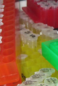
Remember the bubble getter? Siliconizing sequencing gel glass plates? Carrying out sequencing reactions in strip tubes? Diagramming, by hand, your cloning scheme and calculating the cut sizes with a hand-held calculator? Marking plates for plaque lifts with india ink?

This video is for all of you who were in the lab when life was “one gene, one graduate student”. What other oldie but goodies can you think of? Leave a comment or tweet @promega #backinmyday

[youtube=http://www.youtube.com/watch?v=voNepWllrMM]

The Power of One: Revealing Microbial Dark Matter Using Single-Cell Sequencing

abstract digital background
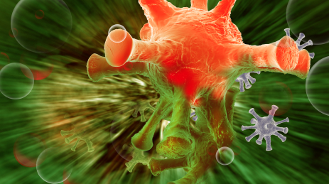
Microorganisms; they are the most abundant form of life. They are all around us, silent, unseen and undetected. The number of ‘species’ of archaea and bacteria climbs every year and is predicted to rise well past one million (1). Despite their abundance, we know very little about all but a small fraction of these diverse cellular life forms because we are unable to cultivate most in a laboratory setting. In fact, 88% of all our microbial isolates belong to just four bacterial phyla (Proteobacteria, Firmicutes, Actinobacteria and Bacterioidetes; 2). The remaining branches of the microbial phylogenetic tree range from underrepresented to virtually unknown and are collectively referred to as “microbial dark matter”.

If you want to target those shadowy, ill-defined branches where exotic and underrepresented organisms belong, you go to environments that might harbor them. Towards this end, Christian Rinke and a large coalition of co-authors collected samples from a wide and varied choice of habitats including the South Atlantic tropical gyre, the Homestake Mine in South Dakota, the Great Boiling Spring in Nevada, the sediment at the bottom of the Etoliko Lagoon in Greece and even a bioreactor. Continue reading “The Power of One: Revealing Microbial Dark Matter Using Single-Cell Sequencing”

Compensation 101: What You Need to Know Now

money in the handsCompensation is a bit of a mystery to most people outside of HR. We go to work to make money and receive benefits, but aren’t always sure how our salaries and benefits packages are decided. In order to understand if we are being paid fairly, negotiate an offer, or counsel a friend on a career change, we need to have some understanding of compensation. Interestingly, in most cases, the more people know about how they are being compensated, the better they feel about their pay and benefits.  I’m going to let you in on some secrets to help demystify compensation. Continue reading “Compensation 101: What You Need to Know Now”

Epigenetics and Exercise

Turning on some genes

If, like me, you sometimes need more motivation to exercise consistently—even though you know that it is good for you—you may be interested in the findings of a paper published recently in PLOS Genetics. The paper showed that consistent exercise over a 6-month period caused potentially beneficial changes in gene expression. In short, regular exercise caused expression of some “good” genes, and repression of “bad” ones, and these changes appeared to be controlled by epigenetic mechanisms.

Epigenetic changes are modifications to DNA that affect gene expression but don’t alter the underlying sequence. Perhaps the best understood example of an epigenetic change is DNA methylation—where methyl groups bind to the DNA at specific sites and alter expression, often by preventing transcription. Epigenetic changes have been shown to occur throughout all stages of development and in response to environmental factors such as diet, toxin exposure, or stress. The study of epigenetics is revealing more and more about how the information stored in our DNA is expressed in different tissues at different times and under different environmental circumstances. Continue reading “Epigenetics and Exercise”

ProteaseMAX Surfactant: Enhanced In-solution Digestion Applications

ProteaseMax 11228MA

The primary advantage of ProteaseMAX™ Surfactant is that it improves identification of proteins in gel by enhanced protein digestion, increased peptide extraction, and minimized post digestion peptide loss. However, ProteaseMAX™ Surfactant can also facilitate in-solution digestion protocols.

ProteaseMAX™ Surfactant offers two major benefits for digesting proteins in solution.

Continue reading “ProteaseMAX Surfactant: Enhanced In-solution Digestion Applications”

Ancient Samples Confirm the Cause of the 6th Century Plague Pandemic

When I started writing about research on Yersinia pestis and the Black Death, I was amazed at the ability to recover 14th century bacterial DNA from human remains, show Y. pestis was the caustive agent of the Black Death and then sequence the strain to compare to modern Y. pestis strains. The publications I read always mentioned the three waves of pandemics that devastated human populations in the introduction, and the Black Death was not the oldest one. The putative first pandemic was the Plague of Justinian in the 6th century, named after the Byzantine emperor. Like with the Black Death, there is debate about whether Y. pestis is the causative agent of the Plague of Justinian. The research published in PLOS Pathogens built on earlier work to isolate and genotype the suspected Y. pestis causative agent from human remains in 6th century graves, but this time with more stringent protocols enacted to answer critics who questioned the authenticity of earlier results.

Yersinia pestis by A.Myasnikov for Wiki (Self made work) [CC0], via Wikimedia Commons
Continue reading “Ancient Samples Confirm the Cause of the 6th Century Plague Pandemic”

Advice to Young Scientists: Obey the Passion

Do you remember what it was that first inspired in you your life’s passion for science? Was it collecting bugs, frogs or other creatures as a child? Or maybe that first chemistry set—the one that had your mother hovering behind you with a fire extinguisher. Perhaps it was a parent or teacher that first sparked something in you that never dimmed. Whatever, or whoever, it was that first kindled your interest in science, no doubt there have been times when you wished that someone would offer you some advice on how to navigate through the modern, rapidly changing world of science.

Written letter

On this past Saturday, I took a trip to my local library on a quest to get just exactly that. To be specific, I was going to check out a copy of Edward O. Wilson’s Letters to a Young Scientist (1). Although I haven’t had time to read more that the first chapter, the advice that he offers at the end of that chapter struck me as good advice to any young (or not so young) person:

It is quite simple: put passion ahead of training. Feel out in any way you can what you most want to do in science, or technology, or some other science-related profession. Obey that passion as long as it lasts.

Wilson is speaking specifically to young scientists, but it seems to me that this main point is more universal than that: It applies to the not-so-young scientist and the nonscientist as well.

I am really looking forward to reading the rest of the book, but for today I am challenging myself as well as all of you to “obey the passion”. Even if you can only do it for one day, put aside the demands of career or school, find that passion that started you on your journey in science and follow it!

Reference

  1. Wilson, E.O. (2013) Letters to a Young Scientist. Liveright Publishing, New York.