Because Timing Is Everything….

Camp Indian Trails Morning Flag Ceremony
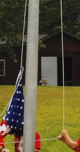
Timing is everything! I learned that the hard way just two weeks ago when I took my son to scout camp and thought I would try to capture the traditional American flag ceremony for posterity. I set up my camera for a panoramic shot and scanned the crowd.  Feeling very pleased with myself, I got home that evening ready to show my family the great camera skills I had honed over the Summer months. To my horror, I noticed that half of the scout troop was saluting the flag while the other half were standing to attention! I had got the timing horribly wrong (although the picture is still fun to look at in a strange sort of way).

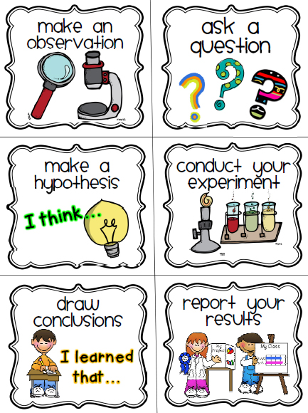
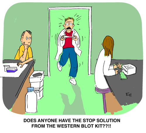
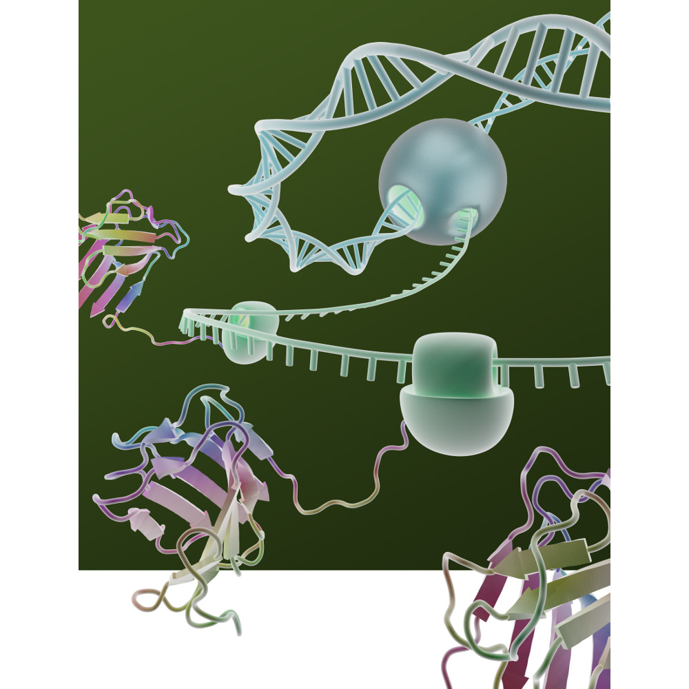
Timing is everything in science as well.  As a technical services scientist at Promega I have sung the ‘timing’ tune to many a biologist. No more so than in the study of apoptosis where Caspases activate each other in a choreographed cascade of molecular triggers that all have their place and time in a domino sequence of enzymatic cleavage events. I frequently talk to researchers about that ‘sweet spot’ of activity when any given Caspase is busily cleaving a peptide moiety off of the next Caspase in the sequence. Finding that sweet spot is anything but trivial and often requires a considerable amount of patience during the optimization phase of experimentation.

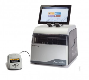
Promega has developed a comprehensive suite of systems (see here) designed to help get the timing right for the cell and compound combinations you might be working with.  The end result is that you have experiments that are timed so as to give you reliable information about what is really happening in your cells.

Novel Cell Surface Markers Identified that Differentiate White, Beige and Brown Adipocytes

2012 CDC-based data on U.S. obesity percentages by state.

Estimates of obesity in the U.S. range from 30% (Centers for Disease Control data) to 70% (persons selling online and television audience-focused weight-loss programs). We are a nation of fat or fat-obsessed persons, and rightfully so. CDC data shows that the cost of obesity, in 2008 dollars, was estimated at $147 billion. That amount of money would buy a lot of french fries or cheesecake or __ (name your poison).

We all help pay those high-dollar amounts in terms of rising healthcare costs, thus there is considerable interest in finding ways to not only avoid, but also to combat obesity.

In recent years researchers working to understand body fat biology have produced exciting information on differences in types of fat. For instance, we now understand that in addition to white adipose tissue, animals and humans also have brown and beige adipose tissue. White adipose tissue or WAT is commonly found in humans and mice subcutaneously and in visceral fat. Brown adipose tissue or BAT, and beige adipose, is less common, and in humans and mice, is found in deeper cervical, supraclavical and paraspinal areas.

Continue reading “Novel Cell Surface Markers Identified that Differentiate White, Beige and Brown Adipocytes”

Use of Mass Spectrometry to Quantitate Food Allergens


Food allergies are becoming increasingly prevalent among children. Credit: James Gathany, CDC

Food allergies are increasing worldwide and becoming a public health issue, especially among children are concerned. Children have a higher prevalence of food allergies, with about 4–8%, compared to adults (1–5%).  Currently antibody-based methods such ELISA (enzyme-linked immunosorbent assay) are the primary method for food allergen analysis. In most cases antibodies are only available for single well-known allergens. Often those that are commercially available are poorly characterized resulting cross-reactivity that leads to false-positive results in diagnostic tests.

A recent publication (1) presented a review of an alternative technology based on mass spec (i.e., multiple reaction monitoring, MRM) that circumvents the drawbacks of antibody based methods. MRM allows precise quantitative determination of target proteins in complex samples with broad dynamic range.  MRM also provides quantification of different isoforms. It is noted that tryptic digestion followed by mass spec analysis, has already identified several unique peptides for different allergens, including those found in crustaceans, eggs, fish, peanuts, soy and wheat. In summary the challenge is now to select the appropriate tryptic signature peptide(s) for the respective allergen and to develop well characterized standards (i.e., isotope labeled standards) to ensure accurate quanititation.

Citation:

Koeberl, M et al. (2014) Next Generation of Food Allergen Quantification using Mass Spectrometric Systems J. Proteome Research  13, 3499–509.

Freedom to Focus: Maxwell® Rapid Sample Concentrator

Wish I had one of these when I was at the lab bench...
Wish I had one of these when I was at the lab bench…

Back in the dark ages, when I was a graduate student, my idea of “automated” plasmid DNA extraction involved performing home-brew, “toothpick preps” in “strip tubes” or , if I was really feeling ambitious, a 96-well plate.

I would get just enough DNA to check for the presence of an insert, but the quality of the DNA was too low and the quantity too small to even consider using it for any other downstream experiments like amplification.

And increased throughput for other nucleic acid extraction needs? Nope. If I wanted genomic DNA, RNA or high-quality plasmid DNA, I spent time with columns and tubes, giving each sample my undivided individual attention.

Remember cesium chloride preps for RNA isolation? Even with the advent of column purification, which greatly simplified and standardized my protocols, nucleic acid purification was still a manual task that required a lot of time and effort to get the high-quality product I needed.

Doing the experiments that would answer the questions that I really wanted to ask (those “downstream experiments”), meant spending time at the bench performing careful (if tedious) work to isolate and clean up the highest quality nucleic acid possible. Even then inconsistency in sample prep could wreak havoc on downstream work.

Fortunately, for the modern scientist, personal, bench top automation, has progressed far beyond the toothpick and the strip tube to quality-tested, reliable nucleic acid extraction platforms like the Maxwell® Rapid Sample Concentrator (RSC).

The Maxwell® RSC improves sample preparation consistency, eliminating variability associated with manual handling, and your downstream results will reflect this consistency.  With the RSC you can extract DNA or RNA from up to 16 samples in approximately 1 hour and viral total nucleic acids in less than an hour.

The instrument is easy to use: simply load the sample, push a button and walk away. Cross contamination is minimized and the instrument is supported by the Promega technical support and service you have come to trust over the past 35 years. 


Want to know more about how the Maxwell® RSC can give you the freedom to focus on the work that interests you the most? To learn more, click here.


Broaden Your Horizons While Pursuing Your Doctorate—You Will Be Glad You Did

For this posting, I had promised to include some commentary on ACTION.

image credit: ComiCONNMitch via Wikimedia Commons

What can someone pursuing a doctorate in the biosciences DO during that time to widen the possibilities of employment in the future? In general, the process of obtaining the doctorate has been criticized for taking too long and not doing enough to prepare students for what they will do when they graduate. Considering these criticisms, it seems wrong to create additional check-boxes on the student to-do list leading up to graduation. Therefore, these things are not in addition to what is already expected, but are instead the same things that are already happening re-focused. Continue reading “Broaden Your Horizons While Pursuing Your Doctorate—You Will Be Glad You Did”

She’s Going Soft! – A commentary on “hard” and “soft” sciences

scientific-methodThis week I gave notice that I would be terminating my employment at Promega. This was a very difficult decision as I have really enjoyed the past six years here.  While I am leaving Biotech, I will not be leaving science all together.  Over the past few years, I have used my research, analytical, and organizational skills to assist various non-profit organizations in the community.  My primary focus will be on reform of the criminal justice system and racial disparities.  Spreading the word about this decision has resulted in a number of responses (overwhelmingly positive) including the comment that I am going soft! This got me thinking about where the terms hard and soft science came from. Continue reading “She’s Going Soft! – A commentary on “hard” and “soft” sciences”

Fun Friday: Western Blot Cartoon

As a fun followup to Wednesday’s blog, here is a cartoon for all you protein scientists out there.

Western Blot Cartoon by Ed Himelblau.

IF you would like to see more science cartoons by Ed Himelblau, you can find them in the Cartoon Lab section of the Promega PubHub.

Discovering the Complexity of the Human Proteome

Transcription TranslationI should preface this blog by stating that I am a nucleic acids gal. My years in the lab were spent with tubes of DNA and RNA. In fact my one and only tentative foray into the field of protein resulted in a Western Blot so ugly that those who witnessed it have been sworn to secrecy. Given all of this, the mapping of the human proteome might seem like an odd topic for me to write about. Except that it isn’t really, because the sequencing of the proteome offers answers to some of the questions that the sequencing of the genome didn’t.

First, let’s start with what a proteome is: A proteome is all the proteins expressed at a certain time point. It can be as limited as the proteome of a single cell or as all encompassing as the proteome of an entire genome. However, unlike the genome, which is genetic information encoded in an organism’s DNA or RNA, the makeup of a proteome can vary dramatically as a result of expression patterns, alternative splicing events and post-translational modifications.

The genome is a constant, what you see today is what will still be there tomorrow. The proteome, on the other hand, is a constantly changing landscape. Up regulation or down regulation of a gene can mean more or less protein is present. Alternative splicing and post-translational modifications can result in fundamental changes to the protein itself.

In other words, if the genome is a beautiful, pristine Ansel Adams print, then the proteome is that same scene as interpreted by Andy Warhol—in Technicolor and 3D. Continue reading “Discovering the Complexity of the Human Proteome”

PowerQuant System: Tool for informed casework sample processing decisions

CottonSwabsIdentification of a crime perpetrator on the basis of DNA fingerprinting is not as easy as some of the CSI shows on television make it out to be. A sample such as blood stain, touch sample or body fluid retrieved at a crime scene is often a challenge for DNA analysts. In many instances, the samples are limited in quantity, found in dirty conditions, exposed to harsh environmental factors and are mixtures of more than one DNA—human and/or non-human. One of the most important aspects of the workflow to successfully obtain a DNA fingerprint profile is accurate quantification of human amplifiable DNA. The more information gleaned from the sample, the better equipped the DNA analyst is to determine the best course of action for obtaining a usable short tandem repeat (STR) profile from challenging samples. Therefore, Promega has developed the PowerQuant™ System, a probe-based 4-target, 5-dye real-time PCR method to a) determine human and male DNA concentrations in a sample, b) detect possible PCR inhibitors c) identify possible mixtures and d) measure DNA integrity. Continue reading “PowerQuant System: Tool for informed casework sample processing decisions”

Angel’s Glow: Bioluminescence Uncovered on the Battlefield

New information has surfaced about this story, and we encourage you to read our updated blog from July 2024 (linked) for the latest on this story.

1888 Chromolithograph of the Battle of Shiloh, American Civil War, produced by L. Prang & Co.

If battlegrounds could speak they would have many stories to tell.  In some cases the microbes found in those soils have lived on to separate fact from fiction. One such story has its origins in the Battle of Shiloh, which went down in history as one of the bloodiest battles fought during the American Civil War.  As the soldiers lay mortally wounded on the cold, hard grounds of Shiloh waiting for medical aid, they noticed a very strange phenomenon. Some of the wounds actually appeared to be glowing in the dark casting a faint light into the darkness of the battlefield. And the legend goes that soldiers with the glowing wounds had a better chance at survival and recovery from infections than their fellow brothers-in-arms whose wounds were not similarly luminescent. The seemingly protective effect of the mysterious light earned it the moniker “Angel’s Glow.”

Fast forward to the 21st century.

Continue reading “Angel’s Glow: Bioluminescence Uncovered on the Battlefield”