Use of Cell-Free Technology to Evaluate Nuclease (TALEN) Activity on Target DNA

ImageSource=RCSB PDB; StructureID=1qpf; DOI=http://dx.doi.org/10.2210/pdb1qpf/pdb;

Transcriptional activator-like effector nucleases (TALENs) have rapidly become a technique of choice for precision genome engineering. TALENs are custom-designed nucleases that consist of a modular DNA-binding domain fused to a monomeric, C-terminal FokI nuclease domain (1). TALENs work in pairs and are designed to recognize and bind to tandem-oriented sequences in genomic DNA, separated by a short spacer (15–30 bp). TALEN binding causes dimerization and activation of the FokI nuclease domains, which results in cleavage of the DNA within the spacer region. Small insertions or deletions (indels) are frequently introduced at this site, as the result of errors made during DNA repair by nonhomologous end-joining (NHEJ). These indels can be up to several hundred base pairs in length and result in frameshift mutations that lead to the production of truncated or nonfunctional proteins.

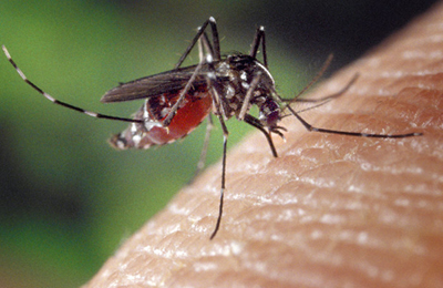
Successful use of TALENs for inducing targeted mutations has been reported in many conventional models, for example: mice, Xenopus and D. melanogaster. TALENs are also reported to be functional in a variety of other invertebrate arthropods, including mosquitos,silkworm and cricket. A recent publication (2) illustrates the use of TALEN technology for the genetic manipulation in P. dumerilii (marine ragworm).

Continue reading “Use of Cell-Free Technology to Evaluate Nuclease (TALEN) Activity on Target DNA”

Detecting Inhibition of Protein Interactions in vivo

Protein Interactions with NanoBRET

In a paper published in the September 2014 issue of ACS Medicinal Chemistry Letters, researchers from GlaxoSmithKline in the UK and Germany report on the discovery, binding mode and structure:activity relationship of a potent BRPF1 (bromodomain and PHD finger containing protein family) inhibitor. This paper came to our attention as it is one of the first publications to apply Promega NanoBRET technology in an vivo assay that reversibly measures the interaction of protein partners. The technology enabled the identification of a novel inhibitor compound that disrupts the chromatin binding of this relatively unstudied class of bromodomain proteins.

What exactly are bromodomains and why do they matter?
Bromodomains are regions (~100 amino acids) within chromatin regulator proteins that recognize and “read” acetylated lysine residues on histones. These acetylated lysines act as docking stations for regulatory protein complexes via binding of the bromodomain region. Because of their role in chromatin binding and gene regulation, bromodomains have attracted interest as potential targets for anti-cancer treatments. Although some bromodomain-containing proteins (e.g., those in the bromodomain and extraterminal domain (BET) subfamily) are well characterized and have been identified as potential therapeutic targets, others are less well understood.

Continue reading “Detecting Inhibition of Protein Interactions in vivo”

Riboprobes: RNA Probes Are Still Valuable Research Tools

9613ca[1]Riboprobes are RNA probes that can be produced by in vitro transcription of cloned DNA inserted in a suitable plasmid downstream of a viral promoter.
Viruses code for their own RNA polymerases, which are highly specific for the viral promoters. Using these enzymes, labeled NTPs, and inserts in both forward and reverse orientations, both sense and antisense riboprobes can be generated from a cloned gene.
Transcription of RNA is performed with the appropriate RNA polymerase (T3, T7 or SP6), depending on the RNA polymerase promoter sites present in the chosen vector. Because these polymerases are extremely promoter-specific (i.e., there is almost no transcriptional cross talk), virtually homogeneous RNA can be obtained using plasmid DNA as the template in a transcription reaction. When it is desirable to copy only insert DNA sequences, the plasmid is linearized at an appropriate restriction site before the transcription reaction and only discrete “run-off” transcripts are obtained, virtually free of vector sequences. RNA transcripts may be used to generate radioactive probes for hybridization to Northern and Southern blots, plaque and colony lifts as well as non-radioactive probes (i.e, labeled with digoxgenin)for in situ hybridization.

Recent references using riboprobes include: Continue reading “Riboprobes: RNA Probes Are Still Valuable Research Tools”

Improving Cancer Drug Screening with 3D Cell Culture

Differential contrast image of HCT116 colon cancer spheroid grown in a 96-well hanging-drop platform after seeding with 800 cells. Copyright Promega Corporation.
Tissue culture using primary or cultured cell lines has long been a mainstay of testing compounds for inhibiting cell growth or promoting apoptosis during screening for cancer drugs. However, the standard culture conditions result in monolayers of cells, dividing and growing across the bottom of a well, plate or flask in a single layer. The drawback of this technique is that organisms do not come in monolayers; a three-dimensional (3D) spheroid is closer to the in vivo state, especially if the spheroids are made up of more than one cell type like tumors in multicellular organisms. Even more beneficial would be using 3D cultured cells in high-throughput screening to facilitate compound profiling for target effectiveness and cytotoxicity. In a recent PLOS ONE article, researchers used normal and breast cancer cells both in monoculture and coculture to test a set of compounds and found results differed between 2D and 3D cultured cells. Continue reading “Improving Cancer Drug Screening with 3D Cell Culture”

To inject or not inject?

GloMax® Discover Multimode Reader with injectors.

Luciferase assays are useful tools for studying a wide range of biological questions. They can be performed easily by adding a reagent that provides components necessary to generate a luminescent signal directly to cells or a cell lysate. However, once this reagent has been added, how long you wait to measure the signal becomes a key consideration in generating consistent data. Dependent on which luciferase assay you use, you may need a luminometer that can use injectors to deliver the assay reagents. The reason for this is simple, but can be confusing to new users.

Let’s start by discussing two types of luciferase assays: “flash” vs. “glow”. Continue reading “To inject or not inject?”

DNA Reveals the Identity of Jack the Ripper?

A wanted poster for Jack the Ripper, who was also known as Leather Apron.  Image courtesy of the British Museum
A wanted poster for Jack the Ripper, who was also known as Leather Apron.
Image courtesy of the British Museum

In the late 1800s, Victorian England was mesmerized and horrified by a series of brutal killings in the crowded and impoverished Whitechapel district. The serial killer, who became known as “Jack the Ripper”, had murdered and mutilated at least five women, many of whom worked as prostitutes in the slums around London. None of these murders were ever solved, and Jack the Ripper was never identified, although investigators interviewed more than 2,000 people and named more than 100 suspects. Now, 126 years after the murders, a British author, who coincidentally has just published a book on the subject, is claiming that DNA analysis has revealed the identity of the notorious killer. DNA is often thought to be the “gold standard” of human identification techniques, so why is there so much skepticism surrounding this identification?
Continue reading “DNA Reveals the Identity of Jack the Ripper?”

Fold It Up and Discover a Whole New World

FIGURE 1: Foldscope design, components and usage. (A) CAD layout of Foldscope paper components on an A4 sheet. (B) Schematic of an assembled Foldscope illustrating panning, and (C) cross-sectional view illustrating flexure-based focusing. (D) Foldscope components and tools used in the assembly, including Foldscope paper components, ball lens, button-cell battery, surface-mounted LED, switch, copper tape and polymeric filters. (E) Different modalities assembled from colored paper stock. (F) Novice users demonstrating the technique for using the Foldscope. (G) Demonstration of the field-rugged design, such as stomping under foot.
FIGURE 1: Foldscope design, components and usage.
(A) CAD layout of Foldscope paper components on an A4 sheet. (B) Schematic of an assembled Foldscope illustrating panning, and (C) cross-sectional view illustrating flexure-based focusing. (D) Foldscope components and tools used in the assembly, including Foldscope paper components, ball lens, button-cell battery, surface-mounted LED, switch, copper tape and polymeric filters. (E) Different modalities assembled from colored paper stock. (F) Novice users demonstrating the technique for using the Foldscope. (G) Demonstration of the field-rugged design, such as stomping under foot.

Scientific inquiry —looking at the world and asking questions about what we observe—is a natural human behavior. Why is the sky blue? What would happen if I did this Mom? Ask any grade school teacher. Kids do science naturally. They are not afraid of questions. They are not afraid of nature. They are not afraid of experiments and data collection.

One other things kids do really well is: fold paper. I never cease to be amazed at the elaborate fortune tellers, hoppers, boats, hats and other creations that my daughter and her friends make at a moment’s notice out of virtually any scrap of paper they can find.

Recently members of the Prakash Lab at Standford University announced the Foldscope: an optical microscope that is printed and folded from a single flat sheet of paper. These microscopes, which can provide magnification of up to 2000X, can be produced for less than $1.00/each. Furthermore these scopes weigh less than 10g (a couple of coins), require no external power source, can be dropped from 3-stories without damage, and can even be stepped on.

These characteristics make the Foldscope ideal for field work, particularly in remote locations where access to power and other resources is difficult. Prakash and colleagues have published their work in a PLOS One paper and have demonstrated many uses for these Foldscopes including high-resolution brightfield microscopy, fluorescence microscopy, and darkfield microscopy. Continue reading “Fold It Up and Discover a Whole New World”

Friday Fun on the Blog

As we start a new year in our labs, we thought this cartoon reminding us of some lab etiquette might be useful. Enjoy!


eh73

Promising Treatment for Marburg Virus Hemorrhagic Fever

I admit to some trepidation about the diseases that may be harbored in my backyard. For example, do the mice in my yard and, despite my and my cats’ efforts, in my house carry deer ticks that harbor the bacterium Borrelia burgdorferi, which causes Lyme disease? Should I be keeping an eye on the vitality of the birds around my property and density of my local mosquito population for potential risk of West Nile Virus transmission? As troublesome as these infections can be, mortality is low for infected humans. Contrast that with the mortality rate of up to 90% for the filoviruses Ebola and Marburg. I find it easy to dismiss these viruses because the reservoir (asymptomatic host) is not in the Upper Midwest but rather Africa, but the tragedy of the Ebola outbreak in the West African countries of Liberia, Sierra Leone and Guinea demonstrates the number of lives lost in an epidemic. Currently, there is no therapy or vaccine to treat these deadly viruses other than transferring antibodies from survivors to those infected. Therefore, the article in Science Translational Medicine about an antiviral treatment that protected macaques injected with a lethal dose of Marburg virus was welcome news.

Continue reading “Promising Treatment for Marburg Virus Hemorrhagic Fever”

Chikungunya Virus and the Promise of a Virus-Like Particle Vaccine

My family and I just returned from a week-long camping trip along the North Shore of Lake Superior in Minnesota. It is beautiful country, filled with lakes, rivers, ponds—and mosquitoes, lots and lots of mosquitoes. We went prepared for the worse. We had a screen tent, head nets and tubes and tubes of insect repellent because in this area of the world, mosquitoes are a flying, buzzing, picnic-ruining, itch-inducing pest. In the US, though, a pest is really all they are. In other areas of the world they are a flying, buzzing, disease-carrying, deadly menace.

Image courtesy of James Gathany and the CDC

Mosquitos act as vectors for many diseases including malaria, Dengue fever, Yellow fever, encephalitis, West Nile Virus and chikungunya virus. Many of these diseases are deadly; in fact, mosquitoes are responsible for more human deaths than any other animal (~725,000 deaths annually). Although most of these diseases have a long and infamous history, two of them, West Nile virus (first identified in 1932) and chikungunya virus (first identified in 1950), are relative new comers on the world health stage. Continue reading “Chikungunya Virus and the Promise of a Virus-Like Particle Vaccine”