Using molecular biology and biochemistry to detect blood doping

Monitoring the use of performance-enhancing substances among athletes is complex and the requirements for tests and assays that detect use of such substances have changed significantly over the last few decades.
The haematological (blood) module of Athlete Biological Passport was adopted December 1, 2009 (ABP) by the World Anti-Doping Agency. The module sets out standard protocols to monitor doping of professional athletes by looking at changes in biological parameters, without relying on the detection of illegal compounds in body fluids. Such biological methods eliminate the need to develop and validate a test to detect every new compound that can be used for doping. The current version of the ABP, adopted in 2014, also adds monitoring of certain steroid use indicators from urine samples.

Blood doping which aims at increasing red blood cells so that more oxygen can be transported to muscles to increase stamina or performance is particularly difficult to detect. There are typically three ways that it is accomplished: use of erythropoietin (EPO) or synthetic oxygen carriers and blood transfusions. While transfusions of large volumes of blood or use of EPO can be detected, microdosing EPO or transfusing smaller volumes of packed red blood cells is much harder to detect.

Nicolas Leuenberger and colleagues at the Swiss Laboratory for Doping Analysis have developed a method to detect blood doping. In addition to addressing the detection of blood doping, his laboratory is also concerned about easing the transport and storage requirements for samples and ensuring that sample collection does not adversely affect athlete performance.

Improving Collection and Storage of Blood Samples

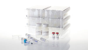
Because sample collection and storage are so critical to accurate test results, any new assays developed to detect blood doping benefit from ease of collection and storage. The Leuenberger laboratory investigated the use of the TAPโ„ข Push Button collection device, which is billed as a simple method for blood collection that is easy to use and eliminates the need for painful needle sticks or finger pricks that can affect athlete performance. After TAP collection, 20ยตl of blood from the device was placed on to filter paper and dried (dried blood samples; DBS), which are much easier to store and transport from collection site to laboratory.

An RNA Biomarker for Blood Doping

Blood withdrawal and autologous transfusion or recombinant human EPO injection stimulate erythropoiesis and immature red blood cells can be distinguished based on their gene expression profiles. One of the genes that is expressed by immature red blood cells is aminoleuvulinate synthase 2, a gene that encodes an enzyme ALAS2 involved in the synthesis of heme, a pathway active during RBC maturation. RNA transcripts are unstable and tend to degrade rapidly, so isolating linear RNA transcripts from a collected sample can be difficult. However circular RNAs (circRNAs) are a class of RNA molecule produced by the backsplicing of pre-mRNAs that are high in abundance, quite stable and maintain cell-type specific expression. The Leuenberger laboratory developed a method for measuring the linear and circular forms of ALAS2 RNA in DBS to monitor erythropoiesis.
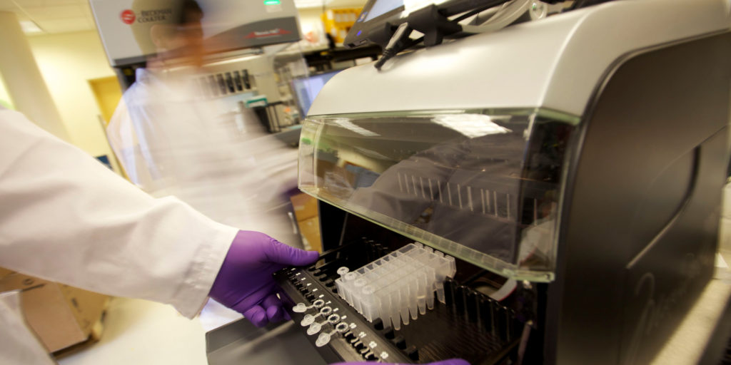
One of the greatest challenges in developing this protocol was achieving efficient RNA extraction from only 20ul of dried blood. Leuenberger and his colleagues adopted a two-step purification; beginning with a phenol:chloroform extraction on the DBS followed by a further purification on the Maxwellยฎ RSC automated instrument, using the Maxwell RSC miRNA Serum and Plasma kit. Switching from a manual to an automated method for the second step was crucial. It reduced chances of contamination as well reduced pipetting errors, without compromising good quality and yield of RNA therefore contributing to assay reproducibility. To normalize volumes within the blood spot, the protocol uses RNA produced by housekeeping genes. The work to automate the assay has been published in Bioanalysis.

Whatโ€™s Next

This protocol is being tested to see if microdosing of EPO or small transfusions can also be detected by monitoring ALAS2 RNA expression in DBS. The Swiss laboratory of Doping Analysis is also in the process of developing a method to detect gene doping by isolating plasmid DNA from whole blood samples, using the Maxwellยฎ RSC.

Additionally, the collection and storage methods used have implications for the clinic, especially for patients that need routine blood monitoring. The ability to isolate circular RNAs shows promise in forensic applications to identify body fluids. 


Want to know more about how the Maxwellยฎ RSC can give you the freedom to focus on the work that interests you the most? To learn more, click here.

Uncovering the Origins of the Commensal House Mouse

Figure of house mouse. Copyright George Shuklin.
📷: George Shuklin

When I encounter my cat fixated on specific locations in my kitchen, her behavior shows me that she has heard some mice in those areas. In fact, mice have been attributed as a reason that cats became companions to humans. Mice start gathering and reproducing so cats followed the food source and hunted the rodents, thus endearing themselves to humans, who were storing food for their own use. However, new evidence described in Scientific Reports has shown that mice have been associated with humans even before grain storage was widespread. In fact, by making our dwellings comfortable, we also created an inviting place for mice to live.

Continue reading “Uncovering the Origins of the Commensal House Mouse”

NanoBRET™ Assays to Analyze Virus:Host Protein:Protein Interactions in Detail

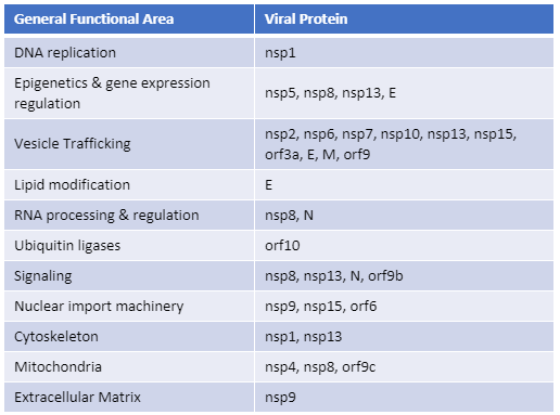
Recently, Gordon et al. published an atlas of protein:protein interactions of all proposed SARS-CoV-2 proteins expressed individually in HEK 293 cells (Table 1). The study tagged each of the viral proteins with an epitope tag and performed a pull-down of the expressed protein followed by trypsin digestion and mass spec analysis, a process referred to as affinity purificationโ€“mass spec analysis. The group identified 332 human proteins interacting with 27 SARS-CoV-2 proteins.

The interactions identified in the HEK 293 cells helped Appelberg et al. analyze interactions over time in SARS-CoV-2-infected Huh7 cells. Gordon et al. used the PPI data to identify FDA-approved drugs, drugs in clinical trials, and pre-clinical compounds that bound to the identified human proteins and labs in New York and Paris tested some of these drugs for antiviral effects.   

Table 1. The general functional area of human proteins identified to interact with individually expressed SARS-CoV-2 proteins as reported by Gordon, et al. (1). The SARS-CoV-2 proteins are classified as non-structural proteins (nsp#), structural proteins (E, M, and N) and accessory proteins (orf#).  
Continue reading “NanoBRET™ Assays to Analyze Virus:Host Protein:Protein Interactions in Detail”

Using Wastewater Surveillance to Track COVID-19 Outbreaks

Today’s blog is written by guest blogger, Sameer Moorji, Director, Applied Markets.

Even as countries are now gradually starting to reopen after lockdown, the COVID-19 pandemic is far from over. Researchers around the world continue to find new ways to monitor, prevent and treat the disease. One new way of monitoring COVID-19 outbreaks relies on a somewhat unexpected source: sewage water.

In March 2020, researchers at the KWR Water Research Institute found the presence of SARS CoV-2 RNA in wastewater samples collected near Schiphol airport in Amsterdam and several other sites in Netherlands. The result came within a week after the first case of COVID-19 in the country was confirmed. This study opened the door to the possibility of using wastewater-based epidemiology to determine population-wide infections of COVID-19.

What is Wastewater-based epidemiology?

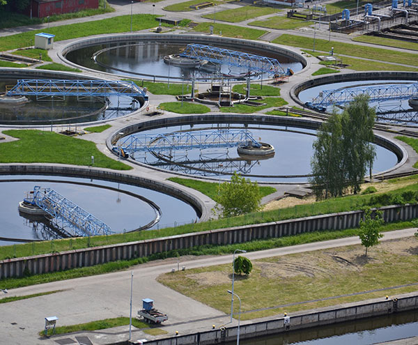
Wastewater based epidemiology (WBE), or sewershed surveillance, is an approach using analysis of wastewater to identify presence of biologicals or chemicals relevant for public health monitoring. WBE is not new, as wastewater has previously been used to detect the presence of pharmaceutical or industrial waste, drug entities (including opioid abuse), viruses and potential emergence of super bugs. In fact, several countries have been successful in containing Polio and Hepatitis A outbreaks within their geographic locations.

Continue reading “Using Wastewater Surveillance to Track COVID-19 Outbreaks”

Promega Leverages Long-Time Experience in MSI Detection with European Launch of CE-Marked IVD Assay for Microsatellite Instability

The genetic abnormality called microsatellite instability, or MSI, has been linked to cancer since its discovery in 1993 (1). MSI is the accumulation of insertion or deletion errors at microsatellite repeat sequences in cancer cells and results from a functional deficiency within one or more major DNA mismatch repair proteins (dMMR).  This deficiency, and the resulting genetic instability, is closely related to the carcinogenicity of tumors (2).

Historically MSI has been used to screen for Lynch Syndrome, a dominant hereditary cancer propensity. More recently, tumors with deficient MMR function have been identified as being more likely to respond to immune checkpoint inhibitor (ICI) therapies (3.). Because MSI can be the first evidence of an MMR deficiency, MSI-High status is predictive of a positive response to immunotherapies such as ICI therapies. (3).

Learn more about MSI in this short animation.
Continue reading “Promega Leverages Long-Time Experience in MSI Detection with European Launch of CE-Marked IVD Assay for Microsatellite Instability”

The Marine Biological Lab: Finding Answers Beneath the Ocean

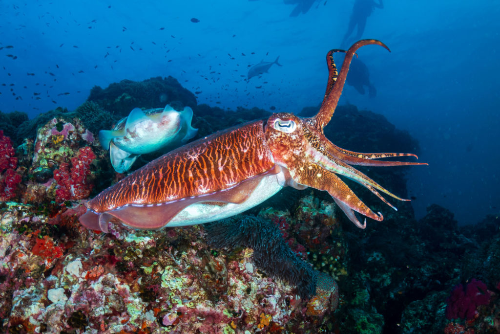
cuttlefish

Marine animals are fascinating. Not only are their appearances alien-like (think tentacles, suckers and bioluminescence). But many have also developed unique capabilities unlike anything you see on land.

In fact, most of the biodiversity of the world lies beneath the ocean. According to the World Register of Marine Species, there are more than 400,000 marine species, and it is estimated that 91% of marine species have yet to be identified. Studying marine animals may help us learn more about how we evolved and even lead to new ways to study and treat human diseases. At the forefront of marine biology research is the Marine Biological Lab (MBL), located in Woods Hole, Massachusetts.

Continue reading “The Marine Biological Lab: Finding Answers Beneath the Ocean”

Choices for Measuring Luciferase-Tagged Reporter Pseudotyped Viral Particles in Coronavirus Research

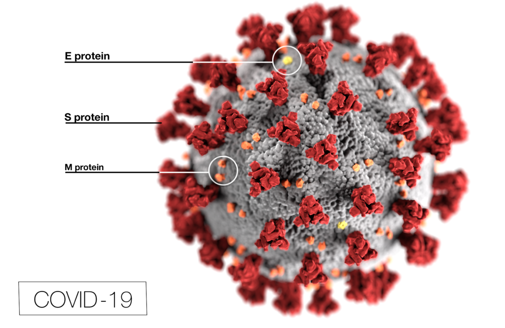
Coronavirus (CoV) researchers are working quickly to understand the entry of SARS-CoV-2 into cells. The Spike or S proteins on the surface of a CoV is trimer. The monomer is composed of an S1 and S2 domain. The division of S1 and S2 happens in the virus producing cell through a furin cleavage site between the two domains. The trimer binds to cell surface proteins. In the case of the SARS-CoV, the receptor is angiotensin converting enzyme 2. (ACE2). The MERS-CoV utilizes the cell-surface dipeptidyl peptidase IV protein. SARS-CoV-2 uses ACE2 as well. Internalized S protein goes though a second cleavage by a host cell protease, near the S1/S2 cleavage site called S2′, which leads to a drastic change in conformation thought to facilitate membrane fusion and entry of the virus into the cell (1).  

CDC / Alissa Eckert, MS; Dan Higgins, MAMS

Rather than work directly with the virus, researchers have chosen to make pseudotyped viral particles. Pseudotyped viral particles contain the envelope proteins of a well-known parent virus (e.g., vesicular stomatitis virus) with the native host cell binding protein (e.g., glycoprotein G) exchanged for the host cell binding protein (S protein) of the virus under investigation. The pseudotyped viral particle typically carries a reporter plasmid, most commonly firefly luciferase (FLuc), with the necessary genetic elements to be packaged in the particle. 

To create the pseudotyped viral particle, plasmids or RNA alone are transfected into cells and the pseudotyped viruses work their way through the endoplasmic reticulum and golgi to bud from the cells into the culture medium. The pseudoviruses are used to study the process of viral entry via the exchanged protein from the virus of interest. Entry is monitored through assay of the reporter. The reporter could be a luciferase or a fluorescent protein.   

Continue reading “Choices for Measuring Luciferase-Tagged Reporter Pseudotyped Viral Particles in Coronavirus Research”

The Surprising Landscape of CDK Inhibitor Selectivity in Live Cells

Cyclin-dependent kinases (CDKs) are promising therapeutic targets in cancer and are currently among the most intensely studied enzymes in drug discovery. The FDA has recently approved three drugs for breast cancer that target members of this kinase subfamily, fueling interest in the entire family. Although broad efforts in drug discovery have produced many CDK inhibitors (CDKIs), few have been characterized in living cells. So just how potent are these compounds in a cellular environment? Are these compounds selective for their intended CDK target, or do they bind many similar kinases in cells? To address these questions, teams at the Structural Genomics Consortium and Promega used the NanoBRET™ Target Engagement technology to uncover surprising patterns of selectivity for touted CDKIs and abandoned clinical leads (1). The results offer exciting opportunities for repurposing some inhibitors as selective chemical probes for lesser-studied CDK family members.

CDKs and CDKIs

nanobret technology for kinase target engagement

Cyclin-dependent kinases (CDKs) regulate a number of key global cellular processes, including cell cycle progression and gene transcription. As the name implies, CDK activity is tightly regulated by interactions with cyclin proteins. In humans, the CDK subfamily consists of 21 members and several are validated drivers of tumorigenesis. For example, CDKs 1, 2, 4 and 6 play a role in cell cycle progression and are validated therapeutic targets in oncology. However, the majority of the remaining CDK family is less studied. For example, some members of the CDK subfamily, such as CDKs 14โ€“18, lack functional annotation and have unclear roles in cell physiology. Others, such as the closely related CDK8/19, are members of multiprotein complexes involved broadly in gene transcription. How these kinases function as members of such large complexes in a cellular context remains unclear, but their activity has been associated with several pathologies, including colorectal cancer. Despite their enormous therapeutic potential, our knowledge of the CDK family members remains incomplete.

Continue reading “The Surprising Landscape of CDK Inhibitor Selectivity in Live Cells”

38 Years After First Release, RNasin Protects COVID-19 Tests

A protein first purified and sold by Promega almost four decades ago has emerged as a crucial tool in many COVID-19 testing workflows. RNasinยฎ Ribonuclease Inhibitor was first released in 1982, only four years after the company was started. At that time, the entire Promega catalog fit on a single sheet of 8.5 ร— 11โ€ paper, and RNasin was one of the first products to draw widespread attention to Promega. Today, the demand for this foundational product has skyrocketed as it supports labs responding to the COVID-19 pandemic.

What is RNasinยฎ Ribonuclease Inhibitor?

RNA is notoriously vulnerable to contamination by RNases. These enzymes degrade RNA by breaking the phosphodiester bonds forming the backbone of the molecule. To say that RNases are everywhere is barely an exaggeration โ€“ almost every known organism produces some form of RNase, and theyโ€™re commonly found in all kinds of biological samples. Theyโ€™re easily introduced into experimental systems, since even human skin secretes a form of RNase. Once theyโ€™re present, itโ€™s very hard to get rid of them. Even an autoclave can’t inactivate RNases; the enzymes will refold and retain much of their original activity.

RNasinยฎ Ribonuclease Inhibitor is a protein that has been shown to inhibit many common contaminating RNases, but without disrupting the activity of enzymes like reverse transcriptase that may be essential to an experiment. It works by binding to the RNase enzyme, prevent it from acting on RNA molecules. This is important for ensuring that RNA samples are intact before performing a complex assay.

Continue reading “38 Years After First Release, RNasin Protects COVID-19 Tests”

Understanding the Structure of SARS-CoV-2 Spike Protein

Glycosylation is the process by which aย carbohydrateย isย covalentlyย attached to targetย macromolecules, typicallyย proteins. This modification serves various functions including guiding protein folding (1,2), promoting protein stability (2), and participating signaling functions (3).

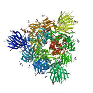
ribbon structure of SARS-CoV-2 protein
Ribbon Structure of SARS-CoV-2 Spike Protein

SARS-CoV-2 utilizes an extensively glycosylated spike (S) protein that protrudes from the viral surface to bind to angiotensin-converting enzyme 2 (ACE2) to mediate host-cell entry. Vaccine development has been focused on this protein, which is the focus of the humoral immune response. Understanding the glycan structure of the SARS-CoV-2 virus spike (S) protein will be critical in the development of glycoprotine-based vaccine candidates.

Continue reading “Understanding the Structure of SARS-CoV-2 Spike Protein”