Targeting MYC: The Need to Study Protein:Protein Interactions in Cells

Crystal Structure of MYC MAX Heterodimer bound to DNA ImageSource=RCSB PDB; StructureID=1nkp; DOI=http://dx.doi.org/10.2210/pdb1nkp/pdb;

In 1982, picked up because of its homology to chicken virus genes that could transform cells, MYC became one of the first human genes identified that could drive cellular transformation (1,2). Since that time countless laboratories have prodded and poked the human MYC gene, the MYC protein, their homologs in other animal models, and their transforming viral counterparts.

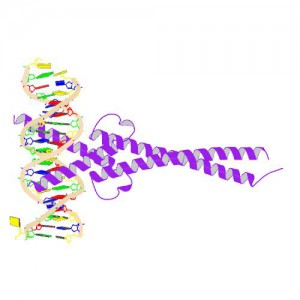
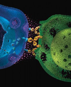
MYC is a transcription factor and forms heterodimers with a required protein partner, MAX, before binding to the E box sequences of DNA regulatory regions (3). MYC regulates gene expression of many targets through interactions with a host of proteins, often referred to as the MYC Interactome (2).  In fact, MYC is estimated to bind 10–15% of the genome, and it regulates the expression of genes that  are transcribed by by each of the three RNA polymerases (2).

MYC plays a central role in regulating cell growth, proliferation, apoptosis, differentiation and transformation, acting as a central integrator of cellular signals. MYC is tightly regulated at multiple levels from gene expression to protein stability. Dysregulation (usually upregulation) of the amount and stability of Myc protein is observed in many human cancers. Even in cancers in which MYC is not directly involved in transforming cells, its normal expression is often required to support the extracellular matrix and/or vascularization necessary for tumor growth and formation (4).

Because MYC is such a central player cancer pathology, it is an attractive target for cancer therapeutics  (2) .

Continue reading “Targeting MYC: The Need to Study Protein:Protein Interactions in Cells”

From Death Row to Exoneration Thanks to DNA Testing

Hands of man prisoner gripping rusty prison bars

Imagine being convicted of a crime for which you are not guilty—not some minor crime, but one of the most heinous crimes imaginable: the rape and murder of a young girl. Would you feel shock and anger at the injustice? Disappointment in the legal system that could make such a horrible error? Sadness and depression at the thought of spending time imprisoned for a crime that someone else committed? Probably all of those emotions and more. At your sentencing hearing, the situation gets worse; you are sentenced to death. Now, this horrible crime will prematurely claim the life of two innocents: the young girl and you.

This is the situation that Kirk Bloodsworth faced in 1985: a death sentence for the rape and murder of 9-year-old Dawn Hamilton. Although Bloodsworth didn’t know it at the time, DNA testing would eventually prove his innocence and save his life.

Continue reading “From Death Row to Exoneration Thanks to DNA Testing”

Choosing Primary and Control Reporters for Dual-Luciferase Assays

Dual-Reporter Assays give scientists the ability to simultaneously measure two reporter enzymes within a single sample. In dual assays, the activity of an experimental reporter is correlated with the effect of specific experimental conditions, while the activity of a control reporter relays the baseline response, providing an essential internal control that reduces variability caused by differences in cell viability or transfection efficiency. The Nano-Glo® Dual-Luciferase® Reporter (NanoDLR™) Assay provides a choice of two sensitive reporters (firefly and NanoLuc luciferases) for use in dual-assay format. Both reporters give state-of-the-art functionality, raising the question “Which luciferase should be the primary reporter and which should be the control?”

This infographic outlines the various NanoDLR dual-reporter assay choices and the situations where you would choose one format over another. Continue reading “Choosing Primary and Control Reporters for Dual-Luciferase Assays”

Thawing Out to Sing: The Story of the Wood Frog

Wood Frog_Northern Wisconsin
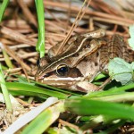
One of the hallmarks of the arrival of Spring in Wisconsin is the cacophony of evening croaks and calls from the Spring Peepers and Chorus frogs. Indeed frogs and toads are ubiquitous around the globe, and many of us who have become life scientists (even those of us who have relegated ourselves to the world of macromolecules, cell signaling networks, and nucleic acids) probably spent some time in our childhood chasing and catching frogs.

But what happens to those frogs and toads over the harsh winter months in places like Wisconsin? Well, their strategies are species-dependent, but at least some of them overwinter by freezing, and the story of one species, the Wood Frog, is quite amazing. Think about it. It freezes from the inside out. No heart beat, no circulation, completely dormant. Then in response to some unknown signal (day length? temperature? angle of the sun?), bodily functions slowly resume. What kind of cell signaling cascade controls that response?

Here is a video from NOVA about the Wood Frog and its amazing deicing event. The next time you are out on a Spring or Summer evening and you hear a chorus of frogs calling, you can think about the incredible molecular story behind the event and be even more impressed!

A NOVA Video about the Wood Frog:


Animal or Plant? FISH Labeling Reveals Horizontal Transfer of Algae Gene into Sea Slug Chromosome

There are times when I ask myself why I chose a career in science. This happens on what I call “grass is greener” days. On these days I dream of other careers—like National Geographic reporter or Caribbean tour guide–which all sound way more exciting than scientist. Admittedly these alternative careers are not ones that many people have the privilege of attaining, but sometimes reality gets to take a vacation. Fortunately, science is a fast-moving, always-changing field. As much as I might occasionally dream of exotic jobs in far away locations, science always pulls me back in with something new and unexpected. Because as much as we’d like to think we know, the truth is there is so much more that we don’t.

Image from: Pelletreau KN, Weber APM, Weber KL, Rumpho ME (2014) Lipid Accumulation during the Establishment of Kleptoplasty in Elysia chlorotica. PLoS ONE 9(5): e97477. doi:10.1371/journal.pone.0097477
The sea slug Elysia chlorotica. Image from: Pelletreau K.N., et al. (2014)  PLoS ONE 9: e97477.

A case in point—sea slugs. These unfortunately named, exotic looking creatures have some surprising secrets.

Continue reading “Animal or Plant? FISH Labeling Reveals Horizontal Transfer of Algae Gene into Sea Slug Chromosome”

Removing Cancer’s Cloak of Invisibility

Copyright Promega.
Copyright Promega.
For decades scientists have been trying to harness the power of our immune system to fight cancer cells. It is not impossible to imagine that our immune system, which is sophisticated enough to fight against a multitude of invaders that threaten our health, should be able to tackle a deadly disease such as cancer. This formed the basis of testing a new type of cancer treatment known as immunotherapy. Immunotherapy for cancer means developing treatments to harness your immune system and using your own immune system to fight the cancerous cells.

But in reality it was hard to make this work. Because, as scientists discovered recently, cancer outsmarts the immune system by wearing a kind of “invisibility cloak”. Cancer is able to fool the immune system from recognizing that it is the enemy and in effect keeps the immune system from destroying it.

In a breakthrough discovery scientists have found a way around this treachery.

The breakthrough is in therapies called ‘checkpoint inhibitors’. Checkpoint inhibitors block the mechanisms that allow some tumor cells to evade the immune system. The drugs ensure that cancer cells are no longer be shielded from the immune system defenses, but are instead recognized as “foreign”. Continue reading “Removing Cancer’s Cloak of Invisibility”

Cloning Tips for Restriction Enzyme-Digested Vectors and Inserts

Cartoon created and owned by Ed Himelblau
While T-vector cloning is commonly used for PCR-amplified inserts, restriction enzymes still have their uses. For example, you can ensure directional cloning if you digest a vector with the same two enzymes like BamHI and EcoRI that are used to digest your insert. This way, the insert can only be cloned in one direction. However, there are other cloning techniques that can be used to modify the end of vectors and inserts after restriction enzyme digestion and prior to ligation. Continue reading “Cloning Tips for Restriction Enzyme-Digested Vectors and Inserts”

HaloTag® Research Application: Detection of Cancer Biomarkers

10242TA
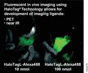
Antibodies labelled with radioisotopes or the sequential administrationof an antibody and a radioactive secondary agent facilitate the in vivo detection and/or characterisation of cancers by positron emission tomography (PET) or by single-photon emission computed tomography (SPECT) imaging.

There are drawbacks to both methods, including prolonged exposure to radiation and  ensuring that both the antibody and the radiolabelled secondary agent are suitably designed so that they bind rapidly upon contact at the tumor.

A recent publication (1) investigated a alternative method utilizing the HaloTag® dehalogenase enzyme HaloTag® is a dehalogenase enzyme (33 kDa) that contains an engineered cavity designed to accommodate the reactive chloroalkane group of a HaloTag® ligand (HTL). Upon entering the enzyme cavity, the terminal chlorine atom rapidly undergoes nucleophilic displacement, and a covalent adduct is formed, effectively anchoring the HaloTag® ligand in a precise location.

Three new HaloTag® ligands were synthesized and each labelled with the SPECT radionuclide indium-111  111In-HTL-1  and the dual-modality HaloTag® ligands,111In-HTL-2 and111;In-HTL-3 containing TMR which allows complementary imaging data).

For the validation of the pretargeting strategy based on these HaloTag® ligands, the target human epidermal growth factor receptor 2 (HER2)was selected. Trastuzumab (Herceptin®) was selected as the primary targeting agent and was modified with HaloTag® protein via the trans-cyclooctene/tetrazine ligation.

All three 111In-labelled HaloTa®g ligands exhibited significantly higher binding to the HER2 expressing when compared to negative controls.

Literature Cited

Knight, J. C et al.(2015) Development of an enzymatic pretargeting strategy for dual-modality imagingChem. Commun. 51, 4055–8.

Avoid False Hits During Compound Screening for Drug Discovery

One goal of drug discovery and research programs is to reduce false hits as early as possible in the process. Follow-up on false hits is costly in terms of time and resources, and the longer the false hits remain in the drug development pipeline, the more costly they are. So methods that can easily reduce the number of false hits during compound screening early in the discovery process are particularly sought after.

Reporter assays have proven to be invaluable tools for elucidating the mechanisms of action of small molecules or other agents on signaling pathways within cells, and the luciferase reporter assay has become a standard research tool in the biological research laboratory.

However, one caveat of using standard luciferase-based reporter assays for larger-scale compound screening efforts is the frequency of false hits that result from direct interaction of compounds with the luciferase reporter. This issue can be mitigated with a “coincidence reporter” system where two independent reporter proteins are produced from a single transcript. In this type of assay, a bicistronic transcript is stoichiometrically translated into two nonhomologous reporters by means of a 2A “ribosomal skipping” sequence. Since it is unlikely that compounds will interact with two distinct types of reporter, “coincident” responses will indicate on-target activity. Such a coincident reporter system provides an important control against costly false hits early in drug discovery research programs.

A paper published online in ACS Chem Biol in February describes the first successful application of the firefly/NanoLuc luciferase coincidence reporter system to identify new pathways that up-regulate PARK2 expression.

Continue reading “Avoid False Hits During Compound Screening for Drug Discovery”

Could This be the Next Generation Ebola Virus Vaccine?

Ebola virus has received a lot of press in the last year due to the extended epidemic outbreak in Africa. Ebola is part of the family of Filioviruses (filamentous virus) and causes hemorrhagic fever that leads to internal bleeding and loss of bodily fluids. As the epidemic in Africa has illustrated so starkly, once the virus infects a large enough population, the human suffering it causes is devastating to individuals and communities. Because no treatment other than palliative fluid support is available to those infected by Ebola virus, virologists have focused attention on potential therapeutics and vaccines. The vaccine strategies now in clinical trials are based on a single Ebola virus glycoprotein, GP, and involve a DNA-based vaccine or innoculation with an Ebola protein expressed from a viral vector. How effective and safe this approach may be for protection from Ebola virus infection is currently under investigation.

Based on the history of effective vaccines, Marzi et al. was interested in testing a whole-virus vaccine for Ebola (EBOV). A whole-virus-based vaccine like smallpox or measles uses an attenuated or inactivated virus. The advantage of this method is that all the proteins as well as the nucleic acid are available for immunological reaction, offering broader-based protection than a single protein. In the recently published Science report from Marzi et al., a replication-incompetent Ebola virus was used as the basis for a whole-virus vaccine that was tested for its efficacy in nonhuman primates.

Continue reading “Could This be the Next Generation Ebola Virus Vaccine?”