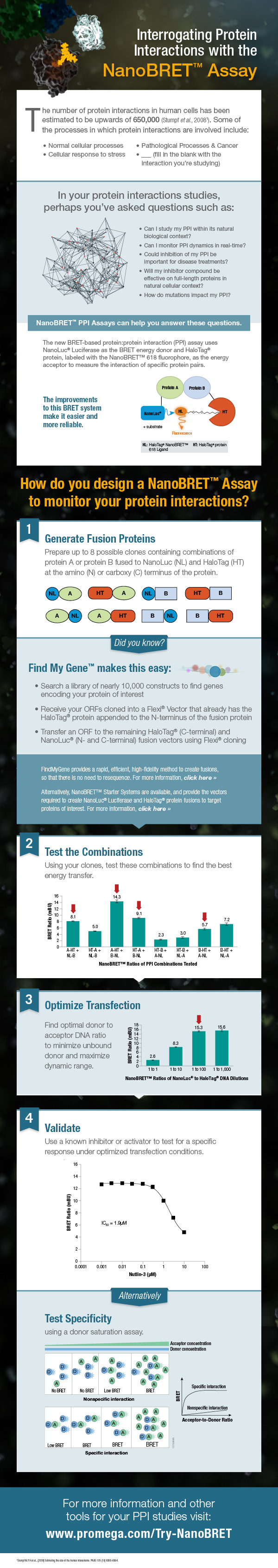
Increasing Drug Research and Development Efficiency Using a 4-point Screening Method to Determine Molecular Mechanism of Action

Fig 4. Four point MMOA screen for tideglusib and GW8510. Time dependent inhibition was evaluated by preincubation of TbGSK3β with 60 nM tideglusib and 6 nM GW-8510 with 10μM and 100μM ATP. (A). Tideglusib [60 nM] in 10μM ATP. (B). GW8510 [60 nM] in 10μM ATP. (C.) Tideglusib [60 nM] at 100μM ATP. (D.) GW8510 [60 nM] at 100μM ATP. All reactions preincubated or not preincubated with TbGSK3β for 30 min at room temperature. Experiments run with 10μM GSM peptide, 10μM ATP, and buffer. Minute preincubation (30 min) was preincubated with inhibitor, TbGSK3β, GSM peptide, and buffer. ATP was mixed to initiate reaction. No preincubation contained inhibitor, GSM peptide, ATP, and buffer. The reaction was initiated with TbGSK3β. Reactions were run at room temperature for 5 min and stopped at 80°C. ADP formed was measured by ADP-Glo kit. Values are mean +/- standard error. N = 3 for each experiment and experiments were run in duplicates. Control reactions contained DMSO and background was determined using a zero time incubation and subtracted from all reactions. Black = 30 min preincubation Grey = No preincubation.
Four point MMOA screen for tideglusib and GW8510.
Time dependent inhibition was evaluated by preincubation of TbGSK3β with 60 nM tideglusib and 6 nM GW-8510 with 10μM and 100μM ATP. (A). Tideglusib [60 nM] in 10μM ATP. (B). GW8510 [60 nM] in 10μM ATP. (C.) Tideglusib [60 nM] at 100μM ATP. (D.) GW8510 [60 nM] at 100μM ATP. All reactions preincubated or not preincubated with TbGSK3β for 30 min at room temperature.  Black = 30 min preincubation Grey = No preincubation.
The first small-molecule kinase inhibitor approved as a cancer therapeutic, imatinib mesylate (Gleevec® treatment), has been amazingly successful. However, a thorough understanding of its molecular mechanism of action (MMOA) was not truly obtained until more than ten years after the molecule had been identified.

Understanding the MMOA for a small-molecule inhibitor can play a major role in optimizing a drug’s development. The way a drug actually works–the kinetics of binding to the target molecule and how it competes with endogenous substrates of that target–ultimately determines whether or not a a candidate therapeutic can be useful in the clinic. Drugs that fail late in development are extremely costly.

Drug research and discovery for neglected tropical diseases suffer from a lack of a large commercial market to absorb the costs of late-stage drug development failures. It becomes very important to know as much as possible, simply and quickly, about MMOA for candidate molecules for these diseases that are devastating to large populations.

One such neglected topical disease is Human African trypanosomiasis (HAT, also known as sleeping sickness). Continue reading “Increasing Drug Research and Development Efficiency Using a 4-point Screening Method to Determine Molecular Mechanism of Action”

Inflammasome Research: A Tool to Aid Progress

Inflammasomes: A Few Basics

Inflammasomes are protein complexes composed of immune system receptors and sensor molecules. These complexes can respond to both infectious organisms and molecules derived from host proteins. When activated, a series of receptors and molecules signal via either pathogen-associate molecular patterns (PAMPs) induced by microbial pathogens, or danger-associated molecular patterns (DAMPs) induced as a result of endogenous stressors; the common next step in signaling is through pattern recognition receptors (PRR).

Inflammasome diagram.
Innate immune response in inflammation; a basic diagram.

Inflammasome activation is integral to the host immune response in mice and humans (1). The activation results in signaling that activates the caspase-1 scaffold, causing release of immune mediators such as interleukins IL-1β and IL-18. So, whether inflammation is host tissue- or pathogen-induced, inflammasome activation results in a cascade of receptor signaling and mediator release, of which caspase-1 is a critical component.

Continue reading “Inflammasome Research: A Tool to Aid Progress”

Optimizing Antibody Internalization Assays: pHAb Dyes

22255190_pHabAmine_3D_image_050615-final-large

Promega has recently developed a method that allows antibodies to be screened for their internalization properties in a simple, plate-based format. The method uses pH sensor dyes (pHAb dyes), which are not fluorescent at neutral pH but become highly fluorescent at acidic pH. When an antibody conjugated with pHAb dye binds to its antigen on the cancer cell membrane, the antibody-dye-antigen complex is not fluorescent, but upon internalization and trafficking into endosomal and lysosomal vesicles the pH drops, and the dye becomes fluorescent.

To demonstrate the broad utility of the pHAb dye for receptor mediated antibody internalization, two therapeutic antibodies, trastuzumab and cetuximab,which bind to HER2 and EGFR respectively, were selected for a case study (1). Both the antibodies, which are known to internalize were labeled with pHAb dyes using amine or thiol chemistry.

Parameters such as the impact of dye–to-antibody ratio on the antigen–antibody binding, change in fluorescence as a function of pH of free dye and labeled dye, and labeled antibody internalization as a function of pHAb conjugated antibody concentration were evaluated.

The results indicate that pHAb dyes are pH sensitive fluorescent dyes that enable the study of receptor-mediated antibody internalization.Internalization assays can be performed in a plate-based homogeneous format and allow endpoint assays as well as real-time monitoring of internalization. They further show that internalization can be monitored even at a very low amount of antibody which is very important during the early monoclonal antibody development phase when the amount of sample is limited and the antibody concentration in the samples is low. a complimentary approach, they  also showed that a secondary antibody labeled with pHAb dye can be used instead of labeling primary antibodies.

Literature cited

Nath, N. et al. (2016) Homogeneous plate based antibody internalization assay using pH sensor fluorescent dye J.  Immunol. Methods epub ahead of print

Making Drug Discovery More Efficient: Predicting Drug Side Effects in Early Screening Efforts

26911030-Laymans-KSPS-figure-WEB-R4Drug research and development is a complex and expensive process that begins with initial screening steps of candidate chemical compounds, and compounds that appear to have the desired potency against a specific cellular target or pathway are further evaluated. Candidate compounds that fail late in development or during clinical trials because of off-target effects are costly, and can be dangerous. Therefore drug developers not only need to ensure that a candidate compound is effective as a therapy, but also they need to predict any potential undesirable side effects due to off-target activities as early as possible in the drug discovery and development process. Continue reading “Making Drug Discovery More Efficient: Predicting Drug Side Effects in Early Screening Efforts”

Studying Mitochondrial Fission with NanoBiT Complementation Assay

3D depiction of NanoBiT Protein Complementation

Updated February 15, 2021

If you’re interrogating two proteins to understand the conditions under which they interact, a complementation system enables you to tag each protein. Interaction of the tagged proteins facilitates the complementation of the subunits, resulting in a signal. Here we discuss the NanoBiT complementation assay and describe its use to study mitochondrial fission.

Continue reading “Studying Mitochondrial Fission with NanoBiT Complementation Assay”

A NanoBRET™ Biosensor for GPCR:G protein Interaction with the Kinetics and Temporal Resolution of Patch Clamping

Electrophysiologists are talented scientists/artists who see into the events of the cell with amazing detail.
Electrophysiology experiments provide a view into the cell with amazing detail. The paper reviewed here describes a molecular reporter biosensor (NanoBRET) that can offer the same kind of temporal and spatial resolution traditionally reserved for extremely labor-intensive experiments like patch clamp analysis.

I confess that I struggled through biophysics, and my Bertil Hille textbook Ion Channels of Excitable Membranes lies neglected somewhere in a box in my basement (I have not tossed it into the recycle bin—I can’t bear too, I spent too much time bonding with that book in graduate school).

My struggles in that graduate class and my attendance at the seminars of my grad school colleagues who were conducting electrophysiological studies left me with a sincere awe and appreciation of both the genius and the artistry required to produce nice electrophysiology data. The people who are good at these experiments are artists—they have the golden touch when it comes to generating that megaohm seal between a piece of cell membrane and a finely pulled glass pipette. And, they are brilliant scientists, they really understand the physics, the chemistry and the biology of the cells they study from a perspective that very few scientists ever develop.

Electrophysiology data, which often demonstrate the gating of a single channel protein in response to a single stimulus in real time–ions crossing a membrane through a single protein–are amazing for their ability, unlike virtually any other experimental data for the story they can tell about what is going on in a cell in real time under physiological conditions.

So when I read the paper recently published by Mashuo et al. in Science SignalingDistinct profiles of functional discrimination among G proteins determine the action of G protein-coupled receptors”, this sentence really caught my attention:

When constructs were ectopically expressed in HEK 293T/17 cells, we obtained very similar kinetics for the GPCR-driven responses between NanoBRET™ biosensors and the patch clamp recordings.

They continue:

Indeed, the activation rates that we observed were very similar to those of GPCR-stimulated GIRKs [G protein-coupled, inwardly rectifying K+ channel] in native cells, suggesting that the conditions of this assay closely match the in vivo setting. This finding further demonstrates the ability of the system to resolve the fast, physiological relevant kinetics of GPCR signaling.

A reporter biosensor that can resolve events similarly to patch clamping?!  Amazing. Continue reading “A NanoBRET™ Biosensor for GPCR:G protein Interaction with the Kinetics and Temporal Resolution of Patch Clamping”

NanoBiT™ Assay: Transformational Technology for Studying Protein Interactions Named a Top 10 Innovation of 2015

21416943-wb-cr-uw-nanobit-pm-page-hero
For three out of the last four years, we have been honored to have one of our key technologies named a Top 10 Innovation by The Scientist. This year the innovative NanoBiT™ Assay (NanoLuc® Binary Technology) received the recognition. NanoBiT™ is a structural complementation reporter based on NanoLuc® Luciferase, a small, bright luciferase derived from the deep sea shrimp Oplophorus gracilirostris.

Using plasmids that encode the NanoBiT complementation reporter, you can make fusion proteins to “report” on protein interactions that you are studying. One of the target proteins is fused to the 18kDa subunit; the other to the 11 amino acid subunit. The NanoBiT™ subunits are stable, exhibiting low self-affinity, but produce an ultra-bright signal upon association. So, if your target proteins interact, the two subunits are brought close enough to each other to associate and produce a luminescent signal. The strong signal and low background associated with a luminescent system, and the small size of the complementation reporter, all help the NanoBiT™ assay overcome the limitations associated with traditional methods for studying protein interactions.

The small size reduces the chances of steric interference with protein interactions. The ultra bright signal, means that even interactions among proteins present in very low amounts can be detected and quantified–without over-expressing large quantities of non-native fusion proteins and potentially disrupting the normal cellular environment. And the NanoBiT™ assay can be performed in real time, in live cells.

The NanoBiT™ assay is already being deployed in laboratories to help advance understanding of fundamental cell biology. You can see how one researcher is already taking full advantage of this innovative technology in the video embedded below:

Visit the Promega web site to see more examples more examples how the NanoBiT™ assay can break through the traditional limitations for studying protein interactions in cells.

You can read the Top 10 article in The Scientist here.

A Better GTPase Assay for Drug Development

Cover of October Issue of Assay and Drug Development Technologies featuring GTPase-Glo™ Assay.The path to drug development is strewn with obstacles: Identifying targets; configuring assays to help identify targets or drugs; uncovering the right compound to affect the selected target without off-target effects and screening multiple compounds to eliminate or identify potential drugs. Without the right tools, compounds or target, identifying potential disease therapies becomes nearly impossible.

When it comes to a drug target for cancer, the Ras protein family is at the top of the list because the proteins are expressed ubiquitiously and found mutated in many types of cancer. Because Ras proteins are involved in transducing signals from the surface of cells, many of the resulting mutations produce an activated Ras, inducing uncontrolled expression of the genes that Ras controls. Ras proteins are small GTPases (20–25kDa) that comprise a larger superfamily of proteins divided into five subfamilies: Ras, Rho, Rab, Arf, and Ran. These proteins control diverse cellular activities, including cellular differentiation, proliferation, cell division, nuclear import and export, and vesicle transport. GTPases are guanosine-nucleotide-binding proteins with affinity for GDP or GTP and are able to hydrolyze GTP. When bound to GTP, GTPases are active (turned on) and interact with downstream proteins in the signaling cascade. When GTPases are bound to GDP, the proteins are inactivated (turned off) and no longer transduce signals. Continue reading “A Better GTPase Assay for Drug Development”

How Fruit Flies (and maybe Pigeons?) Navigate; A New Report

A rock dove, similar in plumage to a pigeon.

Several years ago an intriguing story of successful navigation in complex situation, by pigeons, the birds most often compared to rats, caught my eye.

Our backyard once had a coop full of pigeons, so I’m not a total stranger to their navigation abilities (nor am I a pigeon expert). My favorites were the tumbling pigeons.

But it didn’t take much time researching that article from 2012, to learn that one of the more hotly debated how-do-they-do-it topics is animal navigation, in particular, the ability of pigeons to navigate back to home/point A when released at point B.

So when it appeared online today, in Nature Materials, the story “A Magnetic Protein Biocompass” caught my eye.

Continue reading “How Fruit Flies (and maybe Pigeons?) Navigate; A New Report”

Interrogating Protein Interactions: An Infographic for NanoBRET™ Assay Design

Yesterday my fellow blogger, Kari, posted a review of the ACS Chemical Biology paper describing a new BRET platform for analyzing protein-protein interactions. If you are interested in studying induction and inhibition of protein interactions in real time, take a look at the infographic below to learn how to develop a NanoBRET™ Assay to monitor your protein of interest.

21421668-IG-CR-NanoBRET-Infographic-Final