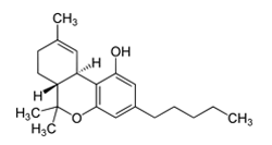
Synthetic cannabinoids (SCs) were originally created for the scientific investigation of two cannabinoid receptors, CB1 and CB2, but have made their way to the streets as “safe” and “legal” alternatives to marijuana.
The problem is that these SCs engage the cannabinoid receptors more completely and with higher affinity than anything derived from marijuana. As a result, SCs can produce serious side effects that often require medical attention. In fact, you are 30 times more likely to seek emergency medical attention following the use of an SC than with natural cannabinoid sources like marijuana.
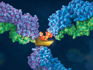
Therapeutic monoclonal antibodies (MAbs) are inherently heterogeneous due to a wide range of both enzymatic and chemical modifications, such as oxidation, deamidation and glycosylation which may occur during expression, purification or storage. For identification and functional evaluation of these modifications, stability studies are typically performed by employing stress conditions such as exposure to chemical oxidizers, elevated pH and temperature.
To characterize MAbs, a variety of analytical techniques are chosen, such as size exclusion chromatography and ion exchange chromatography. However, due to the large size of the intact MAbs, these methods lack structural resolution. Often, the chromatographic peaks resolved by SEC and IEC methods are collected and further analyzed by peptide mapping to obtain more detailed information. Peptide mapping, in which antibodies are cleaved into small peptides through protease digestion followed by LC–MS/MS analysis, is generally the method of choice for detection and quantitation of site-specific modifications. However sample preparation and lengthy chromatographic separation make peptide mapping impractical for the analysis of large numbers of samples. In contrast to peptide mapping analysis, the middle-down approach offers the advantage of high-throughput and specificity for antibody characterization.
Limited proteolysis of IgG molecules by the IdeS enzyme has been introduced for antibody characterization due to its high cleavage specificity and simple digestion procedure.
Not every lab has a tried and true transfection protocol that can be used by all lab members. Few researchers will use the same cell type and same construct to generate data. Many times, a scientist may need to transfect different constructs or even different molecules (e.g., short-interfering RNA [siRNA]) into the same cell line, or test a single construct in different cultured cell lines. One construct could be easily transfected into several different cell lines or a transfection protocol may work for several different constructs. However, some cells like primary cells can be difficult to transfect and some nucleic acids will need to be optimized for successful transfection. Here are some tips that may help you improve your transfection success.
Transfect healthy, actively dividing cells at a consistent cell density. Cells should be at a low passage number and 50–80% confluent when transfected. Using the same cell density reduces variability for replicates. Keep cells Mycoplasma-free to ensure optimal growth.
Transfect using high-quality DNA. Transfection-quality DNA is free from protein, RNA and chemical contamination with an A260/A280 ratio of 1.7–1.9. Prepare purified DNA in sterile water or TE buffer at a final concentration of 0.2–1mg/ml.
Antibiotic-resistant bacteria and their potential to cause epidemics with no viable treatment options have been in the news a lot. These “superbugs,” which have acquired genes giving them resistance to common and so-called “last resort” antibiotics, are a huge concern as effective treatment options dwindle. Less attention has been given to an infection that is not just impervious to antibiotics, but is actually enabled by them.
Clostridium difficile Infection (CDI) is one of the most common healthcare-associated infections and a significant global healthcare problem. Clostridium difficile (C. diff), a Gram-positive anaerobic bacterium, is the source of the infection. C. diff spores are very resilient to environmental stressors, such as pH, temperature and even antibiotics, and can be found pretty much everywhere around us, including on most of the food we eat. Ingesting the spores does not usually lead to infection inside the body without also being exposed to antibiotics.
Individuals taking antibiotics are 7-10 times more likely to acquire a CDI. Antibiotics disrupt the normal flora of the intestine, allowing C. diff to compete for resources and flourish. Once exposed to the anaerobic conditions of the human gut, these spores germinate into active cells that embed into the tissue lining the colon. The bacteria are then able to produce the toxins that can cause disease and result in severe damage, or even death.
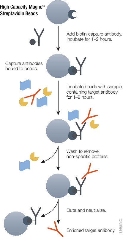
Schematic showing immuno-enrichment using High Capacity Magne® Streptavidin Beads.
During preclinical research and development of therapeutic antibodies, multiple variants of each antibody are assessed for pharmacokinetic (PK) characteristics across model systems such as rodents, beagles and primates. Ligand-binding assays (LBA) or liquid chromatography coupled to tandem mass spectrometry(LC–MS/MS)-based methods represent the two most common technologies used to perform the PK studies for mAb candidates(1,2).
Using either method it is essential to ensure accurate quantitative results that the initial enrichment of the target therapeutic antibody from serum or plasma be optimal. Biotinylated antibodies or antigens (against the therapeutic targets) immobilized onto high capacity streptavidin beads will enrich therapeutic antibody from serum or plasma samples. The affinity of biotin for streptavidin (Kd = 10–15) is one of the strongest and most stable interactions in biology therefore the biotin-streptavidin interaction cannot be reversed under non-denaturing conditions. Hence, it is possible to perform extensive washing to remove nonspecifically bound protein and elute therapeutic antibodies without also eluting the biotinylated component, thus improving the detection limit.
Magnetic based separation techniques have several advantages in comparison with standard separation procedures. This process is usually very simple, with only a few handling steps. All the steps of the purification procedure can take place in one single test tube. The magnetic separation techniques are also the basis of various automated procedures. Learn more about the High Capacity Magne™ Streptavidin Beads (Cat # V7820) .
RNA molecules have become a hot topic of research. While I was taught about messenger RNA (mRNA), ribosomal RNA (rRNA) and transfer RNA (tRNA), many more varieties have come into the nomenclature after I graduated with my science degrees. Even more interesting, these RNAs do not code for a protein, but instead have a role in regulating gene expression. From long non-coding RNA (lncRNA) to short interfering RNA (siRNA), microRNA (miRNA) and small nucleolar RNA (snoRNA), these classes of RNAs affect protein translation, whether by hindering ribosomal binding, targeting mRNA for degradation or even modifying DNA (e.g., methylation). This post will cover the topic of microRNAs, explaining what they are, how researchers understand their function and role in metabolism, cancer and cardiovascular disease, and some of the challenges in miRNA research.
What are microRNAs? MicroRNAs (miRNAs) are short noncoding RNAs 19–25 nucleotides long that play a role in protein expression by regulating translation initiation and degrading mRNA. miRNAs are coded as genes in DNA and transcribed by RNA polymerase as a primary transcript (pri-miRNA) that is hundreds or thousands of nucleotides long. After processing with a double-stranded RNA-specific nuclease, a 70–100 nucleotide hairpin RNA precursor (pre-miRNA) is generated and transported from the nucleus into the cytoplasm. Once in the cytoplasm, the pre-miRNA is cleaved into an 18- to 24-nucleotide duplex by ribonuclease III (Dicer). This cleaved duplex associates with the RNA-induced silencing complex (RISC), and one strand of the miRNA duplex remains with RISC to become the mature miRNA.
CRISPR is a hot topic right now, and rightly so—it is revolutionizing research that relies on editing genes. But what exactly is CRISPR? How does it work? Why is everyone so interested in using it? Today’s blog is a beginner’s guide on how CRISPR works with an overview of some new applications of this technology for those familiar with CRISPR.
Introduction to CRISPR/Cas9
Clustered Regularly Interspaced Short Palindromic Repeats (CRISPR) were discovered in 1987, but it took 30 years before scientists identified their function. CRISPRs are a special kind of repeating DNA sequence that bacteria have as part of their “immune” system against invading nucleic acids from viruses and other bacteria. Over time, the genetic material from these invaders can be incorporated into the bacterial genome as a CRISPR and used to target specific sequences found in foreign genomes.
CRISPRs are part of a system within a bacterium that requires a nuclease (e.g. Cas9), a single guide RNA (sgRNA) and a tracrRNA. The tracrRNA recruits Cas9, while sgRNA binds to Cas9 and guides it to the corresponding DNA sequence of the invading genome. Cas9 then cuts the DNA, creating a double-stranded break that disables its function. Bacteria use a Protospacer Adjacent Motif, or PAM, sequence near the target sequence to distinguish between self and non-self and protect their own DNA.
While this system is an effective method of protection for bacteria, CRISPR/Cas9 has been manipulated in order to perform gene editing in a lab (click here for a video about CRISPR). First, the tracrRNA and sgRNA are combined into a single molecule. Then the sequence of the guide portion of this RNA is changed to match the target sequence. Using this engineered sgRNA along with Cas9 will result in a double-stranded break (DSB) in the target DNA sequence, provided the target sequence is adjacent to a compatible PAM sequence.
In today’s post, guest blogger, Martha O’Brien, PhD, provides a preview of her upcoming AAI poster and block symposium talk on the inflammasome, caspase-1 activity and pyroptosis.
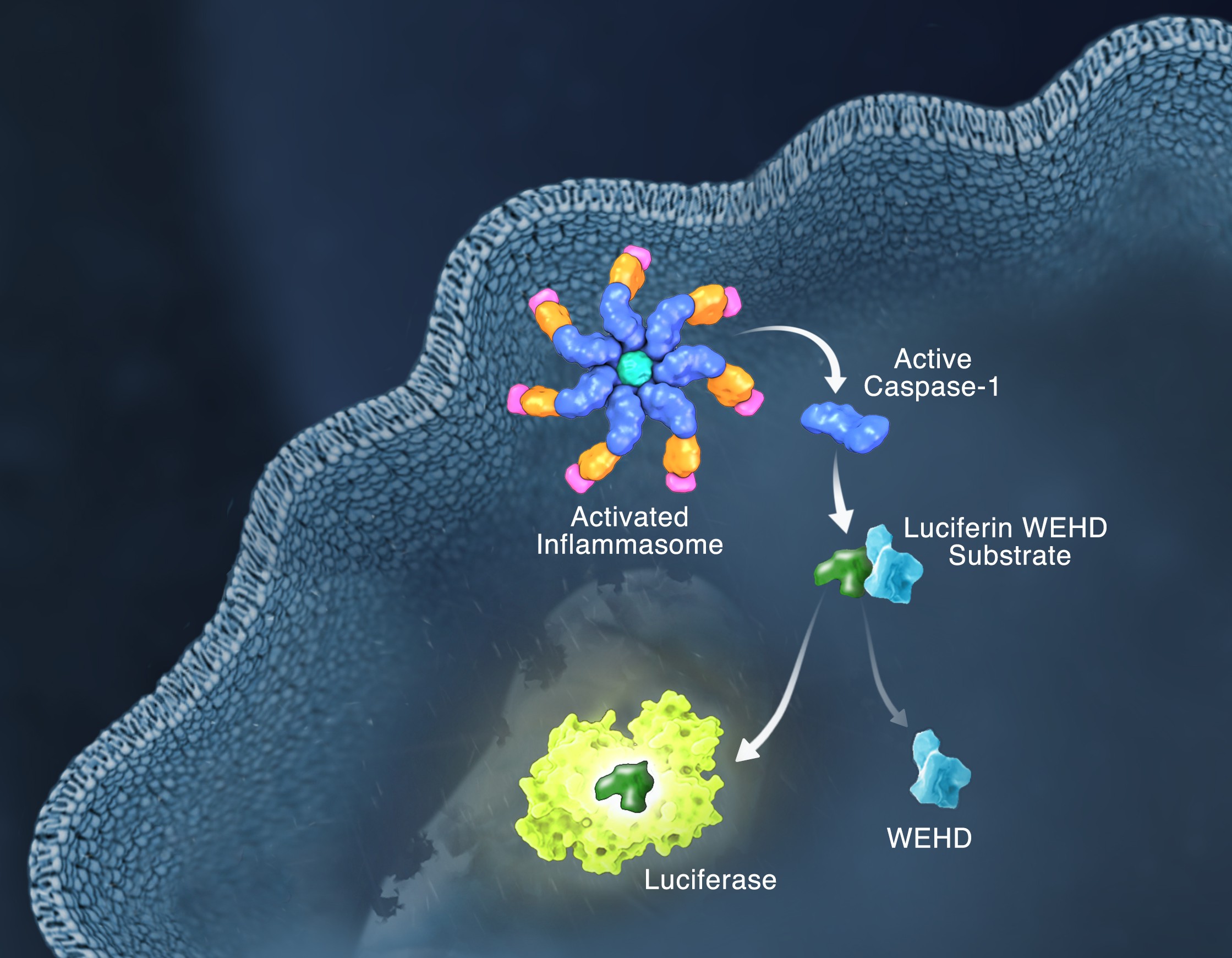
Schematic of the Caspase-Glo 1 Inflammasome Assay.
Responding rapidly to microbial pathogens and damage-associated molecular markers is critical to our innate immune system. Caspase-1 is pivotal in this process leading to processing and release of essential cytokines and an immunogenic form of cell death, termed pyroptosis. Upon sensing pathogen-associated and damage-associated molecular patterns (PAMPs and DAMPs), innate immune cells form inflammasome protein complexes that recruit and activate caspase-1 (canonical inflammasomes). In addition, other inflammatory caspases, 4 and 5 in humans and 11 in mice, directly bind bacterial lipopolysaccharides (LPS), triggering pyroptosis (non-canonical inflammasome). LPS-triggered non-canonical inflammasomes in mice and humans ultimately lead to canonical inflammasome engagement and caspase-1 activation (1–3). Caspase-1 was originally termed interleukin converting enzyme (ICE) for its well-established role in processing IL-1ß and IL-18, two important inflammation cytokines. How caspase-1 mediates pyroptosis is less well understood, but is beginning to be delineated. Recently, a substrate of the inflammatory caspases, gasdermin D, was identified and its processed fragment, gasdermin-N domain, was shown to be required for pyroptosis in non-canonical inflammasome circumstances (4, 5). The precise role of gasdermin D in canonical inflammasome-triggered pyroptosis is still under investigation. Linking inflammatory caspases directly to pyroptosis is a notable step in understanding the mechanism of this important form of cell death.
Pyroptosis is clearly one means of releasing processed IL-1ß and IL-18 from the cell. However depending on the cell type and stimulus, there is evidence for inflammasome engagement, caspase-1 activation, and release of IL-1ß in the absence of cell death (6, 7). On the flip-side there is also evidence for caspase-1 mediated pyroptosis that helps clear bacteria, independent of IL-1ß and IL-18 involvement (8). To enable further studies on the inflammasome and in particular, assessing the connections between caspase-1 activation, pyroptosis, and cytokine release, Promega developed a new tool to conveniently monitor caspase-1 activation, the Caspase-Glo® 1 Inflammasome Assay. This bioluminescent, plate-based assay is used to measure caspase-1 activity directly in cell cultures or to monitor released caspase-1 activity in culture medium from treated cells. This flexibility allows easy multiplexing to monitor all three outcomes of inflammasome stimulation; caspase-1 activity, pyroptosis, and release of IL-1ß and IL-18. Caspase-1 activation typically is monitored indirectly with western blots of processed caspase-1. Now the activity of the enzyme can be monitored directly, providing accurate information on temporal aspects of the inflammasome. The assay can be readily combined with real-time measures of cell death (e.g., CellTox™ Green Cytotoxicity Assay) and some of the culture medium can be removed for IL-1ß/IL-18 assessment, leaving the cells and remaining culture medium for caspase-1 activity measurements.
Macrophages. By NIAID (https://www.flickr.com/photos/niaid/17380707492/) [CC BY 2.0 , via Wikimedia CommonsMany think of glucose as something diabetics have to test each day using a blood monitor, or a quick energy boost for someone exercising intensely. However, the simple sugar glucose, a monosaccharide, fuels most of the cells in our bodies. Disaccharides that contain glucose (e.g., sucrose is comprised of glucose and fructose) and glucose polymers (e.g., starch and glycogen) are carbohydrates that are consumed by organisms from bacteria to humans to produce energy. These carbohydrates are broken down into component monosaccharides like glucose and lactose. The process of glycolysis generates the energy currency of cells, ATP, as well as precursor molecules for nucleotides, lipids and amino acids. Because glucose is the cell fuel source, the uptake of glucose and its subsequent metabolism is increased by cells that divide rapidly like cancer cells. The more energy and precursor molecules the cancer cell can create for itself, the more rapidly the tumor can grow.
Because glucose metabolism is central to cellular functioning, changes that decrease glucose uptake or increase glycolysis have a widespread effect on on both the cells and organism. How does a simple sugar molecule create such broad effects on health? For example, diabetes results from the inability to store glucose because of a lack of insulin, a hormone that draws glucose from the blood and stores it as glycogen in the liver, muscles and adipose tissue. High levels of sugar in the blood negatively affect the body over the long term, damaging blood vessels and eyesight, making the kidneys work harder to excrete the excess sugar and increasing the risk of stroke and coronary artery disease. Because cancer cells have such a high metabolic demand for glucose, many of the mutations in cancers affect pathways that regulate glucose uptake and glucose breakdown, allowing the cancer cells to survive and grow, crowding out nearby normal cells.
Glucose metabolism is altered by processes other than mutations or an reduced production of a hormone. Throughout its life cycle, a cell will vary its requirements for glucose. For example, the cells that comprise our innate immune response are typically in a quiescent or steady state. However, when these immune cells encounter an foreign invader, they become activated and increase their demand for glucose. To respond to a potential pathogen, the activated cells need glucose to fuel cell proliferation and the production of cytokines, chemicals that activate other immune cells and initiate an inflammatory response. The typical signs of inflammation are red inflamed area that may be painful to the touch, such as a cut that becomes infected. Most inflammation resolves when the infection is eliminated, leaving behind whole skin in the instance of a cut, and the activated immune cells become quiescent again.
An Interesting Observation about Glucose Metabolism in M2 Macrophages
Glucose uptake, immunity and metabolism are cellular pathways that are intertwined such that understanding how glucose is utilized in macrophages illuminates gene induction and regulation in activated macrophages. In a recently published eLife article, Covarrubias et al. studied how activation of murine bone marrow-derived macrophages (BMDMs) by interleukin-4 (IL-4), a signaling cytokine, altered glucose metabolism in the cells and regulated a subset of genes involved in macrophage activation. Continue reading “Finding a Connection Between Glucose Metabolism and Macrophage Activation”
Most, if not all, processes within a cell involve protein-protein interactions, and researchers are always looking for better tools to investigate and monitor these interactions. One such tool is the protein complementation assay (PCA). PCAs use a reporter, like a luciferase or fluorescent protein, separated into two parts (A and B) that form an active reporter (AB) when brought together. Each part of the split reporter is attached to one of a pair of proteins (X and Y) forming X-A and Y-B. If X and Y interact, A and B are brought together to form the active enzyme (AB), creating a luminescent or fluorescent signal that can be measured. The readout from the PCA assay can help identify conditions or factors that drive the interaction together or apart.
A key consideration when splitting a reporter is to find a site that will allow the two parts to reform into an active enzyme, but not be so strongly attracted to each other that they self-associate and cause a signal, even in the absence of interaction between the primary proteins X and Y. This blog will briefly describe how NanoLuc® Luciferase was separated into large and small fragments (LgBiT and SmBiT) that were individually optimized to create the NanoBiT® Assay and show how the design assists in monitoring protein-protein interactions.
XWe use cookies and similar technologies to make our website work, run analytics, improve our website, and show you personalized content and advertising. Some of these cookies are essential for our website to work. For others, we won’t set them unless you accept them. To learn more about our approach to Privacy we invite you to Read More
By clicking “Accept All”, you consent to the use of ALL the cookies. However you may visit Cookie Settings to provide a controlled consent.
We use cookies and similar technologies to make our website work, run analytics, improve our website, and show you personalized content and advertising. Some of these cookies are essential for our website to work. For others, we won’t set them unless you accept them. To find out more about cookies and how to manage cookies, read our Cookie Policy.
If you are located in the EEA, the United Kingdom, or Switzerland, you can change your settings at any time by clicking Manage Cookie Consent in the footer of our website.
Necessary cookies are absolutely essential for the website to function properly. These cookies ensure basic functionalities and security features of the website, anonymously.
Cookie
Duration
Description
cookielawinfo-checbox-analytics
11 months
This cookie is set by GDPR Cookie Consent plugin. The cookie is used to store the user consent for the cookies in the category "Analytics".
cookielawinfo-checbox-functional
11 months
The cookie is set by GDPR cookie consent to record the user consent for the cookies in the category "Functional".
cookielawinfo-checbox-others
11 months
This cookie is set by GDPR Cookie Consent plugin. The cookie is used to store the user consent for the cookies in the category "Other.
cookielawinfo-checkbox-advertisement
1 year
The cookie is set by GDPR cookie consent to record the user consent for the cookies in the category "Advertisement".
cookielawinfo-checkbox-necessary
11 months
This cookie is set by GDPR Cookie Consent plugin. The cookies is used to store the user consent for the cookies in the category "Necessary".
cookielawinfo-checkbox-performance
11 months
This cookie is set by GDPR Cookie Consent plugin. The cookie is used to store the user consent for the cookies in the category "Performance".
gdpr_status
6 months 2 days
This cookie is set by the provider Media.net. This cookie is used to check the status whether the user has accepted the cookie consent box. It also helps in not showing the cookie consent box upon re-entry to the website.
lang
This cookie is used to store the language preferences of a user to serve up content in that stored language the next time user visit the website.
viewed_cookie_policy
11 months
The cookie is set by the GDPR Cookie Consent plugin and is used to store whether or not user has consented to the use of cookies. It does not store any personal data.
Analytical cookies are used to understand how visitors interact with the website. These cookies help provide information on metrics the number of visitors, bounce rate, traffic source, etc.
Cookie
Duration
Description
SC_ANALYTICS_GLOBAL_COOKIE
10 years
This cookie is associated with Sitecore content and personalization. This cookie is used to identify the repeat visit from a single user. Sitecore will send a persistent session cookie to the web client.
vuid
2 years
This domain of this cookie is owned by Vimeo. This cookie is used by vimeo to collect tracking information. It sets a unique ID to embed videos to the website.
WMF-Last-Access
1 month 18 hours 24 minutes
This cookie is used to calculate unique devices accessing the website.
_ga
2 years
This cookie is installed by Google Analytics. The cookie is used to calculate visitor, session, campaign data and keep track of site usage for the site's analytics report. The cookies store information anonymously and assign a randomly generated number to identify unique visitors.
_gid
1 day
This cookie is installed by Google Analytics. The cookie is used to store information of how visitors use a website and helps in creating an analytics report of how the website is doing. The data collected including the number visitors, the source where they have come from, and the pages visted in an anonymous form.
Advertisement cookies are used to provide visitors with relevant ads and marketing campaigns. These cookies track visitors across websites and collect information to provide customized ads.
Cookie
Duration
Description
IDE
1 year 24 days
Used by Google DoubleClick and stores information about how the user uses the website and any other advertisement before visiting the website. This is used to present users with ads that are relevant to them according to the user profile.
test_cookie
15 minutes
This cookie is set by doubleclick.net. The purpose of the cookie is to determine if the user's browser supports cookies.
VISITOR_INFO1_LIVE
5 months 27 days
This cookie is set by Youtube. Used to track the information of the embedded YouTube videos on a website.
Performance cookies are used to understand and analyze the key performance indexes of the website which helps in delivering a better user experience for the visitors.
Cookie
Duration
Description
YSC
session
This cookies is set by Youtube and is used to track the views of embedded videos.
_gat_UA-62336821-1
1 minute
This is a pattern type cookie set by Google Analytics, where the pattern element on the name contains the unique identity number of the account or website it relates to. It appears to be a variation of the _gat cookie which is used to limit the amount of data recorded by Google on high traffic volume websites.