Optimizing Antibody Internalization Assays: pHAb Dyes

22255190_pHabAmine_3D_image_050615-final-large

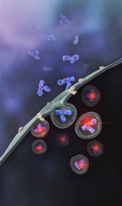
Promega has recently developed a method that allows antibodies to be screened for their internalization properties in a simple, plate-based format. The method uses pH sensor dyes (pHAb dyes), which are not fluorescent at neutral pH but become highly fluorescent at acidic pH. When an antibody conjugated with pHAb dye binds to its antigen on the cancer cell membrane, the antibody-dye-antigen complex is not fluorescent, but upon internalization and trafficking into endosomal and lysosomal vesicles the pH drops, and the dye becomes fluorescent.

To demonstrate the broad utility of the pHAb dye for receptor mediated antibody internalization, two therapeutic antibodies, trastuzumab and cetuximab,which bind to HER2 and EGFR respectively, were selected for a case study (1). Both the antibodies, which are known to internalize were labeled with pHAb dyes using amine or thiol chemistry.

Parameters such as the impact of dye–to-antibody ratio on the antigen–antibody binding, change in fluorescence as a function of pH of free dye and labeled dye, and labeled antibody internalization as a function of pHAb conjugated antibody concentration were evaluated.

The results indicate that pHAb dyes are pH sensitive fluorescent dyes that enable the study of receptor-mediated antibody internalization.Internalization assays can be performed in a plate-based homogeneous format and allow endpoint assays as well as real-time monitoring of internalization. They further show that internalization can be monitored even at a very low amount of antibody which is very important during the early monoclonal antibody development phase when the amount of sample is limited and the antibody concentration in the samples is low. a complimentary approach, they  also showed that a secondary antibody labeled with pHAb dye can be used instead of labeling primary antibodies.

Literature cited

Nath, N. et al. (2016) Homogeneous plate based antibody internalization assay using pH sensor fluorescent dye J.  Immunol. Methods epub ahead of print

Helix® Personified

Have you ever thought about life from the point of view of a Helix® On-Site Smart Inventory Unit? We did, and this is what we imagined…

helixtouchsceenisolatedWow! There were a lot of people in and out of here today.

The post doc who never goes anywhere without her earbuds in. I wonder what her research playlist is, she’s always dancing her way in and out of the hall. The new grad student in the virology lab next door. He’s looking a little lost right now.

And of course there’s the PI with the big boots. Never looks where he’s going and constantly almost plowing people down. It’s kind of funny actually, the near misses I’ve seen between him and grad students running out of here with that key reagent they need to finish those last few reaction tubes.

There was a big-name speaker today for the lunch time journal club, so there was a lot of activity on the hall this afternoon. Lots of talking about the presentation. Everyone sounded pretty excited about what they heard.

But now it’s quiet. Most people have gone home for the evening.

Except for the security lighting, the hall is dark.

Oh wait, I hear footsteps heading my way. Apparently, not everyone has stopped work for the day, apparently.

Oh, it’s you. One of my favorite after-hour scientists.

You open the door to the common room, and check the product list on your phone: 100bp DNA ladder … there’s one left. Drawer D.

You pull the pass out of your pocket. I scan it. Click. The door unlocks. You grab your package and let the door close.

I’ll take it from here so that you can get back to work.

Your pass tells me that you’re a post doc in a lab upstairs. After you close the door, I check to see what you took. My scan reads the tag on each reagent. G2101: 100bp DNA ladder. Drawer D.

That was the last one. I update the inventory list. More will arrive in the next resupply shipment.

I send you an email with the details.

Time to check my systems.

Power? Check.
Connection to Promega? Check.
Door closed and locked? Check.
Storage temperature holding steady? Check.
All reagents present? Check.

The lights go dark as I transmit the report back home.

There and Back Again: Part IV (South Island, New Zealand)

In 2014, Promega created a special incentive to reward field science consultants who help the scientific community take advantage of our on-site stocking program. The winners had to meet ambitious criteria to receive 2 round-trip tickets to anywhere in the world, a week of paid vacation and spending money. Our four winners from 2014 will share photos and stories about their journeys in a semi-regular Friday feature on the Promega Connections Blog.

Today’s travelogue is Part IV and the final installment of the adventures of Mica Zaragoza, a senior client rep in Chicago, IL, who used his award to travel to Australia and New Zealand.

26061846-Helix-Travel---Image-1-WEBQueenstown, New Zealand 

Walking out of our room into the open air, Queenstown instantly provided a lasting impression.  Nestled along Lake Wakatipu, watched over by the Remarkable Mountains, the city feels like Aspen on steroids—it’s no wonder there’s such an international pull for young travelers and skiers.

We jumped right into exploring with a gondola ride. This photo is my best attempt at capturing Queenstown from high above. Continue reading “There and Back Again: Part IV (South Island, New Zealand)”

Dino Protein: New Methods for Old (Very) Samples

Hadrosaurus skeleton vintage engraving.

Brachylophosaurus was a mid-sized member of the hadrosaurid family of dinosaurs living about 78 million years ago, and is known from several skeletons and bonebed material from the Judith River Formation of Montana and the Oldman Formation of Alberta. Recent fossil evidence indicates structures similar to blood vessels in location and morphology, have been recovered after demineralization of multiple dinosaur cortical bone fragments from multiple specimens, some of which are as old as 80 Ma. These structures were hypothesized to be either endogenous to the bone (i.e., of vascular origin) or the result of biofilm colonizing the empty  network after degradation of original organic components (i.e., bacterial, slime mold or fungal in origin).  Cleland et al. (1) tested the hypothesis that these structures are endogenous and thus retain proteins in common with extant archosaur blood vessels that can be detected with high-resolution mass spectrometry and confirmed by immunofluorescence.

Continue reading “Dino Protein: New Methods for Old (Very) Samples”

There and Back Again 3: Christchurch, New Zealand (South Island) Travelogue

In 2014, Promega created a special incentive to reward field science consultants who help the scientific community take advantage of the our on-site stocking program. The winners had to meet ambitious criteria to receive 2 round-trip tickets to anywhere in the world, a week of paid vacation and spending money. Our four winners from 2014 will share photos and stories about their journeys in a semi-regular Friday feature on the Promega Connections Blog.

Today’s travelogue is Part III of the adventures of Mica Zaragoza, a senior client rep, who used his award to travel to Australia and New Zealand.

Helix-Travel-Blog---1-WEBAfter a day of travel, including a quick stop-off in Sydney, we arrived late in New Zealand the evening.  New Zealand is an island nation with strict customs requirements, having been stung in the past by a decision  to import the opossum.

Jumping full into the experience, we rented a car (5-speed) and headed into the City to scope out our hotel ( more on the car later).

During our time in Australia, we had spoken with several travelers who had visited Christchurch for their recommendations.  Without fail, their response was negative, using words like “destroyed” and “rubble” to describe the city.  They referred to a  series of earthquakes in 2011 that crippled the city of Christchurch; the residents were not aware of the risk and structures in the city had not been constructed to withstand such devastation. Continue reading “There and Back Again 3: Christchurch, New Zealand (South Island) Travelogue”

There and Back Again: Part II (Cairns, Australia)

In 2014, Promega created a special incentive to reward field science consultants who help the scientific community take advantage of the our on-site stocking program. The winners had to meet ambitious criteria to receive 2 round-trip tickets to anywhere in the world, a week of paid vacation and spending money. Our four winners from 2014 will share photos and stories about their journeys in a semi-regular Friday feature on the Promega Connections Blog.

Today’s travelogue is Part II of the adventures of Mica Zaragoza, a senior client rep, who used his award to travel to Australia and New Zealand.

Cairns, Australia

24254847-Aussie-Kiwi-1Jumping from down South to way up North, the city of Cairns seems like a touristy beach town, sporting a laid-back feel with lots of cafes, pubs, and restaurants.  The locals drop the hard ‘R’s in most words, making the name of the city hit the ear more like “cans”.  Popping into town, we stopped by the city center and soaking pools along the Esplanade to relax.
With the Esplanade pools right next to the shoreline, I asked one of the locals why no one went swimming in the ocean. The answer was simple: underwater mangroves, mudflats, and saltwater crocs… more on that later. Continue reading “There and Back Again: Part II (Cairns, Australia)”

ProteaseMAX: A Surfactant for the Most Complex Mixtures

Alternate Proteases Cover

Here we provide two examples of “atypical” experiments that take advantage of the properties of the ProteaseMAX™ Surfactant to improve studies involving digestion of complex protein mixtures.

Example 1
Clostridium difficile spores are considered the morphotype of infection, transmission and persistence of C. difficile infections. A recent publication (1) illustrated a novel strategy using three different approaches  to identify proteins of the exosporium layer of C. difficile spores and complements previous proteomic studies on the entire C. difficile spores.

Continue reading “ProteaseMAX: A Surfactant for the Most Complex Mixtures”

Real-Time (quantitative) qPCR for Quantitating Library Prep before NGS

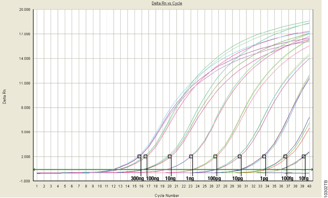
Real-Time (or quantitative, qPCR) monitors PCR amplification as it happens and allows you to measure starting material in your reaction.

This the last in a series of four blogs about Quantitation for NGS is written by guest blogger Adam Blatter, Product Specialist in Integrated Solutions at Promega.

When it comes to nucleic acid quantitation, real-time or quantitative (qPCR) is considered the gold standard because of its unmatched performance in senstivity, specificity and accuracy. qPCR relies on thermal cycling, consisting of repeated cycles of heating an cooling for DNA melting and enzyamtic replication. Detection instrumentation is capable of measuring the accumulation of DNA product after each round of amplification in real time.

Because PCR amplifies specific regions of DNA, the method is highly sensitive, specific to DNA, and it can determine whether a sample is truly able to be amplified. Degraded DNA or free nucleotides, which might otherwise skew your quantiation, will not contribute to the signal, and your measurement will be more accurate.

However, while qPCR does provide technical advantages, the method requires special instrumentation, specialized reagents and is a more time-consuming process. In addition, you will probably need to optimize your qPCR assay for each of your targets to achieve your desired results.

Because of the added complexity and cost, qPCR is a technique suited for post-library quantitation when you need to know the exact amount of amplifiable, adapter-ligated DNA.  PCR is the only method capable of specifically targeting these library constructs over other DNA that may be present. This specificity is important because accurate normalization is especially critical for producing even coverage in multiplex experiments where equimolar amounts of several libraries are added to a pooled sample. This normalization process is essential  if your are screening for rare variants that might be lost in background and go undetected if underrepresented in a mixed pool.


Read Part 1: When Every Step Counts: Quantitation for NGS

Read Part 2: Nucleic Acid Quantitation by UV Absorbance: Not for NGS

Read Part 3: Fluorescence Dye-Based Quantitation: Sensitive and Specific for NGS Applications

ISO 18385: The Creation of a “Forensic Grade” Standard

Today’s blog is from guest blogger Charles Stollberg. Charlie is a Promega production scientist in the Genetic Identity group and is focused on manufacturing inventory material and production process improvements. He’s been with the company for about 4 years. He graduated from UW-Whitewater in 2007 with a bachelor’s degree in cell biology. Prior to Promega, he worked in a small genetics lab studying lethal recessive traits in cattle.

ForensicGradeLogoForensic DNA laboratories rely on reagent and plastics manufacturers to supply high-quality products with minimal interference from contaminating DNA. With the increasing sensitivity of short tandem repeat (STR) amplification systems, levels of DNA that were previously undetected may now generate partial profiles. To address the concern of laboratories worldwide regarding the potential of low-level DNA contamination in consumables, ISO 18385 was developed to provide requirements for minimizing the risk of human DNA contamination events during the manufacturing process. Many of you may not have heard of ISO 18385, so I’d like to give you an introduction to how the standard came to be.

Continue reading “ISO 18385: The Creation of a “Forensic Grade” Standard”

Fluorescence Dye-Based Quantitation: Sensitive and Specific for NGS Applications

This is the third post in a series of blogs on quantitation for NGS applications written by guest blogger Adam Blatter, Product Specialist in Integrated Solutions at Promega.

Fluorescent dye-based quantitation uses specially designed DNA binding compounds that intercalate only with double stranded DNA molecules. When excited by a specific wavelength of light, only dye in the DNA-bound state will fluoresce. These aspects of the technique contribute to low background signal, and therefore the ability to accurately and specifically detect very low quantities of DNA in solution, even the nanogram quantities used in NGS applications.

For commercial NGS systems, such as the Nextera Rapid Capture Enrichment Protocol by Illumina, this specificity and sensitivity of quantitation are critical. The Nextera protocol is optimized for 50ng of total genomic DNA. A higher mass input of genomic DNA can result in incomplete tagmentation, and larger insert sizes, which can adversely affect enrichment. A lower mass input of genomic DNA or low-quality DNA can generate smaller than expected inserts, which can be lost during subsequent cleanup steps, giving lower diversity of inserts.

Continue reading “Fluorescence Dye-Based Quantitation: Sensitive and Specific for NGS Applications”