High-throughput screening (HTS) has been a central approach in small-molecule drug discovery since the late 1980s. It gives researchers a systematic way to find hits: compounds that act on a target and can be refined into drug candidates. The assays used in those screens matter, and cell-based assays are especially valuable because they bridge biochemical assays and in vivo models. Biochemical assays give direct readouts but miss the cellular context, while in vivo models are informative, but costly and slow. Cell-based assays enable scientists to study signaling pathways, target modulation and cytotoxicity in a more native environment, at a practical scale.
Many cell-based screens depend on engineered reporter systems: cells genetically modified so a pathway of interest produces a measurable signal, such as light or fluorescence. These cell lines take significant time and effort to build and maintain, and because they’re modified, they may not reflect disease-relevant cell states. As drug discovery moves toward primary cells and patient-derived lines, assays that detect endogenous proteins without genetic manipulation become more appealing. Those assays work across more cell types and are faster to set up.
Lumit® Immunoassay Cellular Systems (Lumit® ICS) offer an alternative to engineered reporters. These assays measure the cell’s own signaling proteins in unmodified cells, using a simple add-mix-read workflow. Here, we describe a collaboration between Promega and Novartis Biomedical Research to test whether one assay from the system, Lumit® p-ERK, could move from 96-well to 1536-well plates and still provide reliable, high-quality data in an automated screen of about 7,000 compounds.
Choosing The Test Case: The Lumit® p-ERK Assay
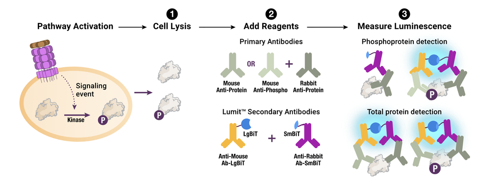
Lumit® ICS is a group of assays built on the same chemistry, each designed to detect a different signaling protein. The assay principle (Figure 1) is based on proximity: light is produced only when two antibodies bind the same target protein, close enough together for the luciferase fragments attached to them to combine. The same chemistry can detect either a phosphorylated protein or the total amount of that protein, depending on which primary antibodies are used.
To see how the chemistry performs at screening density, the team chose one assay, Lumit® p-ERK, as a test case. It measures phosphorylated ERK, the form that signals the pathway is active. ERK is a central signaling node in the MAPK pathway, sitting downstream of receptor tyrosine kinases, RAS, and RAF to regulate cell proliferation, differentiation and survival.
The MAPK pathway is frequently hyperactivated in human cancers. Because of this, it has been pursued extensively as a drug target, leading to several FDA-approved MEK inhibitors. Because the Lumit® p-ERK assay reports on overall pathway activity rather than a single target, it fits two roles: primary screens to find compounds that affect ERK signaling, and mechanistic studies to determine how those compounds work.
Miniaturizing the Assay: Re-Optimizing for 384-Well Plates
Fitting an assay into 1536-well plates means shrinking the reaction volumes while preserving assay performance. The team did this in two steps: first from 96- to 384-well plates, then to 1536-well plates. Moving to 384-well plates wasn’t a matter of shrinking every volume in proportion. The team had to re-optimize the lysis buffer concentration (lysis buffer breaks open the cells so the antibodies can reach their target protein), lowering it from 1X to 0.8X. They also reduced the antibody amount from 0.3 µL to 0.06 µL per well. Neither change followed a simple, linear pattern.
“Every assay scales a little differently. Some transfer almost proportionally; others need real optimization; you just have to test and see. What impressed me here is that the underlying chemistry held up no matter how we pushed it.”
Hicham Zegzouti, Ph.D
Senior Research Scientist
Assay Design Group (Promega)
Before moving to 1536-well plates, the team tested the optimized 384-well assay in a small, manual screen of 190 FDA-approved drugs. The assay had an average Z′ factor of 0.69, a measure of how cleanly the positive and negative controls separate (above 0.5 is considered robust), and a signal window of about 32-fold, meaning stimulated cells produced roughly 32 times more bioluminescence than cells treated with a MEK inhibitor control. The screen also picked out known kinase inhibitors that act on the MAPK pathway, a sign the assay was detecting real pathway biology.
Changing Hands: Screening 7,000 Compounds with Novartis
Internal testing shows an assay is sound in the developer’s hands. The next question was whether the results held outside of Promega’s R&D lab. At Novartis, researchers screened about 7,000 compounds from the company’s Mechanism of Action (MoA) library, a collection of well-characterized chemical probes with known targets, in 1536-well plates. Despite a fourfold increase in well density over the 384-well format, the assay held a Z′ of 0.60 and a signal window of about 18-fold. The screen produced hit rates of 3.4% for inhibitors and 1.3% for activators, and 95% of inhibitor hits confirmed in concentration-response testing. Of the 67 confirmed inhibitors profiled in an orthogonal TR-FRET p-ERK assay, 65 were active there too.
The hits also made biological sense. The screen was enriched for compounds annotated as MEK, ERK and PKC inhibitors, and the RAF/MEK inhibitor avutometinib was potent in the Lumit assay (IC50 of 0.4 nM) while a structurally related analog was more than 3,000-fold weaker (1,300 nM).
“A Promega-only validation proves the assay works,” says Hicham. “Having Novartis run it on their own automation and library, and get concordant biology out the other end, proves it’s trustworthy in someone else’s hands.”
Read the full study in SLAS Technology.
Learn more about the Lumit® Immunoassay Cellular Systems.

Anna Bennett
Latest posts by Anna Bennett (see all)
- Adapting a Cell-Based Immunoassay for HTS: From the Bench to a 7,000-Compound Screen - October 19, 2026
- The Tilcayo: A Tiny, Scrunch-Faced Cat With a Complicated Family Tree - September 24, 2026
- Midges on the Move: Tracking The Oropouche Virus Into New Territory - August 20, 2026
