Ten Validation Tips You Need to Know

Forensic lab validations can be intimidating, so Promega Technical Services Support and Validation teams shared these tips for making the process go more smoothly.

  1.  Prepare Your Lab. Make sure all of your all of your instrumentation (CEs, thermal cyclers, 7500s, centrifuges) and tools (pipettes, heat blocks) requiring calibration or maintenance are up to date.
  2.  Start with Fresh Reagents. Ensure you have all required reagents and that they are fresh before beginning your validation. This not only includes the chemistry being validated, but any preprocessing reagents or secondary reagents like, polymer, buffers, TE-4 or H2O.
  3. Develop a Plan. Before beginning a validation, take the time to create plate maps, calculate required reagent volumes, etc. This up-front planning may take some time initially, but will greatly improve your efficiency during testing.
  4. Create an Agenda. After a plan is developed, work through that plan and determine how and when samples will be created and run. Creating an agenda will hold you to a schedule for getting the testing done.
  5. Determine the Number of Samples Needed to Complete Your Validation. Look at your plan and see where samples can be used more than once.  The more a sample can be used, the less manipulation done to the sample and the more efficient you become.
  6. Select the Proper Samples for Your Validation. Samples should include those you know you’ll obtain results with be similar to the ones you’ll most likely be using, and your test samples should contain plenty of heterozygotes. When you are establishing important analysis parameters, like thresholds, poor sample choice may cause more problems and require troubleshooting after the chemistry is brought on-line.
  7. Perform a Fresh Quantitation of Your Samples. This will ensure the correct dilutions are prepared. Extracts that have been sitting for a long time may have evaporated or contain condensation, resulting in a different concentration than when first quantitated.
  8. Stay Organized. Keep the data generated in well-organized folders. Validations can contain a lot of samples, and keeping those data organized will help during the interpretation and report writing phase.
  9. Determine the Questions to Be Answered. While writing the report, determine the questions each study requires to be answered. Determining what specifically is required for each study will prevent you from calculating unnecessary data.  Do you need to calculate allele sizes of your reproducibility study samples when you showed precision with your ladder samples?
  10. Have fun! Remember, validations are not scary when approached in a methodical and logical fashion. You have been chosen to thoroughly test something that everyone in your laboratory will soon be using. Take pride in that responsibility and enjoy it.

Need more information about validation of DNA-typing products in the forensic laboratory? Check out the validation resources on the Promega web site for more information for the steps required to adopt a new product in your laboratory and the recommended steps that can help make your validation efforts less burdensome.
What I Learned at My First Branch Meeting: Gratitude, Service and Collaboration

Today’s blog post is written by guest blogger Sarah Kolb, Marketing Coordinator for our North America Branch, and new employee at Promega.

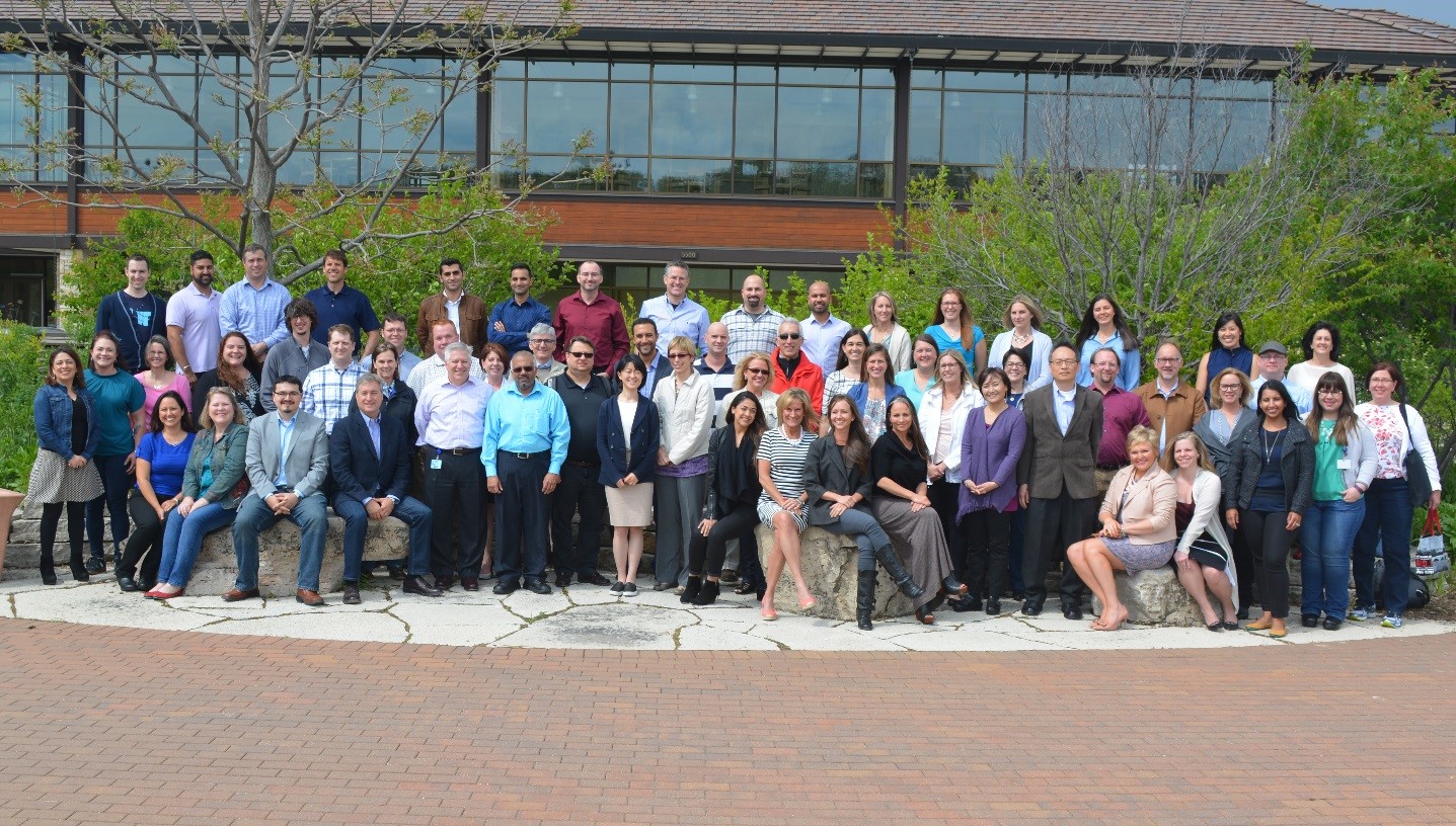
As a new member to the North America Marketing team, I was unsure of what to expect going into my first national sales meeting with Promega, but what I took away from this meeting was incredibly eye opening. The North America Branch Sales meeting is an opportunity to get all of the members of the North American branch together to learn about new products, connect with the different strategic business units about product application and network with each other to learn how to better the lives of our customers. The year’s meeting occurred in May in the Ideation room at Promega Headquarters in Madison, Wisconsin.

NA branch meeting

The room itself is not your typical conference space. An antique car resides in the space, and you can find art-work from all over world nestled in corners, and on the walls and shelves. All around the room, collections of unique furniture are arranged to stimulate conversation. Ideation created an atmosphere of creativity, community and collaboration, which contributed to the overall success of the meeting.

I felt excitement Monday morning as the 62 attendees gathered in the space. Everyone greeted each other with big smiles and hugs, asking about families, travels and already discussing business. Continue reading “What I Learned at My First Branch Meeting: Gratitude, Service and Collaboration”

Optimizing Antibody Enrichment for Pharmacokinetic Assays

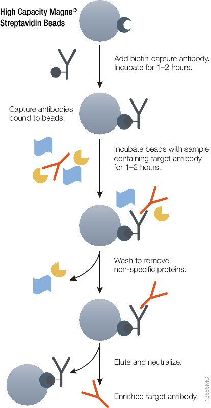
Schematic showing immuno-enrichment using High Capacity Magne® Streptavidin Beads.

During preclinical research and development of therapeutic antibodies, multiple variants of each antibody are assessed for pharmacokinetic (PK) characteristics across model systems such as rodents, beagles and primates. Ligand-binding assays (LBA) or liquid chromatography coupled to tandem mass spectrometry(LC–MS/MS)-based methods represent the two most common technologies used to perform the PK studies for mAb candidates(1,2).

Using either method it is essential to ensure accurate quantitative results that the initial enrichment of the target therapeutic antibody from serum or plasma be optimal. Biotinylated antibodies or antigens (against the therapeutic targets) immobilized onto high capacity streptavidin beads will enrich therapeutic antibody from serum or plasma samples. The affinity of biotin for streptavidin (Kd = 10–15) is one of the strongest and most stable interactions in biology therefore the biotin-streptavidin interaction cannot be reversed under non-denaturing conditions. Hence, it is possible to perform extensive washing to remove nonspecifically bound protein and elute therapeutic antibodies without also eluting the biotinylated component, thus improving the detection limit.

Magnetic based separation techniques have several advantages in comparison with standard separation procedures. This process is usually very simple, with only a few handling steps. All the steps of the purification procedure can take place in one single test tube. The magnetic separation techniques are also the basis of various automated procedures. Learn more about  the High Capacity Magne™ Streptavidin Beads (Cat # V7820) .

References

Discovering the Truth About the Dozier School for Boys

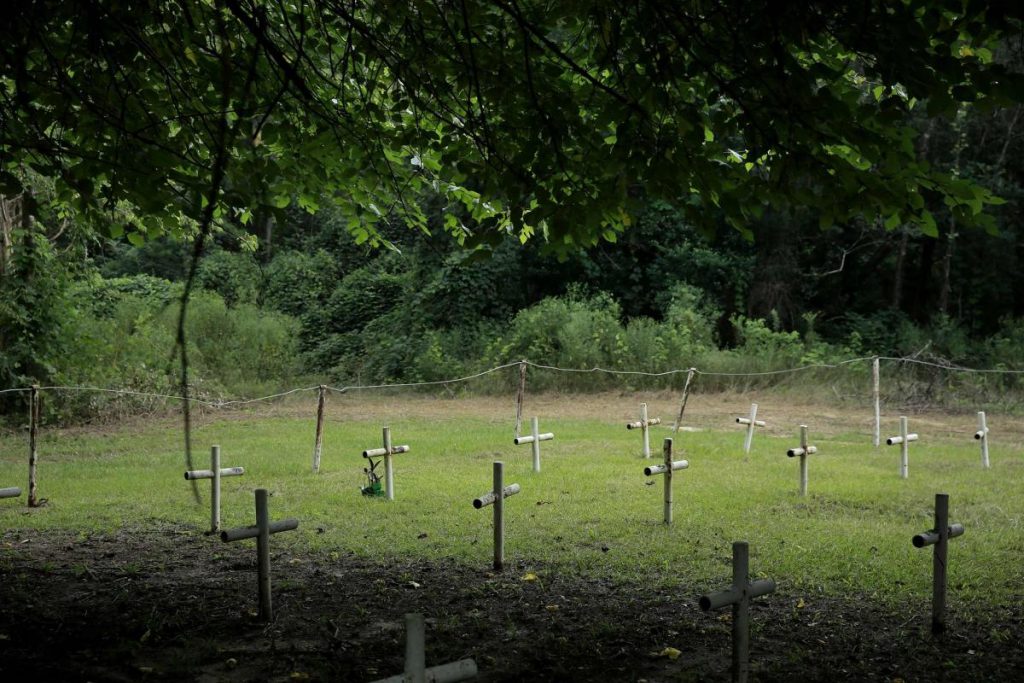
Dozier School for Boys gravesite
Photograph from The Tampa Bay Times

The Dozier School for Boys had cemeteries instead of playgrounds.

The stories of abuses that took place at the reformative school in Marianna, Florida are nothing short of a plot for the TV series American Horror Story. The beatings and other punishments administered to students throughout the school’s 111-year history contributed to the deaths for some of the nearly 100 deceased.

A 2010 investigation by the Florida Department of Law Enforcement did not lead to criminal charges against the school because there was “no tangible physical evidence for allegations of physical and sexual abuse.” The full report is available on The White House Boys Survivors Organization’s website, a name derived from the shed where the boys were beaten with wooden panels and leather straps. At the time, only 32 unmarked graves were known in the school’s cemetery.

Continue reading “Discovering the Truth About the Dozier School for Boys”

Awakened Consciousness in Review: Revisiting What We Learned

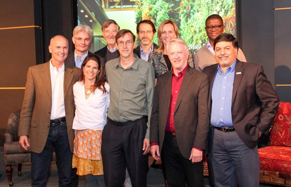
FORUM PRESENTERS Front row l-to-r: Chip Conley, Malynn Utzinger, John Roulac, Bill Linton, Raj Sisodia; Back row l-to-r: Steve Paulson, Mike Mears, Tim Weitzel, Betsy Myers, Martin Kalungu-Banda
FORUM PRESENTERS
Front row l-to-r: Chip Conley, Malynn Utzinger, John Roulac, Bill Linton, Raj Sisodia; Back row l-to-r: Steve Paulson, Mike Mears, Tim Weitzel, Betsy Myers, Martin Kalungu-Banda

Co-coordinated and co-hosted by the BioPharmaceutical Technology Center Institute (BTC Institute) and Promega Corporation, the International Forum on Consciousness – Awakened Consciousness and the Evolution of Business, was held on May 5–6, 2016.

The Forum is designed to bring together people from diverse perspectives and professions to facilitate public dialogue regarding complex and challenging issues.  This year, our intent was to respond to voices of wisdom and action that call for a shift in the consciousness of organizations that affect the lives of so many and planet Earth.  Key among the questions we asked: How does the self-actualized business become a model and advocate for change?

Our sense is that those who joined us left both more knowledgeable and inspired.  Comments from participants illustrate the importance of offering this opportunity, as well as deep appreciation for our presenters’ experience and insights:

audience

Diverse perspective relatable to many – excellent… overall, engaging, fascinating, action oriented…

Thank you so much for the wonderful program.  The diversity of speakers, viewpoints and topic was really enjoyable and provided great room for thought.  It was truly impressive.  Wonderful & kudos to the organizers and all the work and thought to create this program.

panel

An amazing experience, potentially life changing (we’ll see…).  Really top notch, felt like a series of TED talks, but with the added benefit of in-person interaction and a cohesiveness of topics.

Sound interesting?  There are several ways you can learn more about the Forum and get a meaningful taste of what these attendees are describing:

If you’re not on our mailing list and would like to be, just let me know (karin.borgh@btci.org) – hopefully, we’ll be offering a program of interest to you in the days ahead!

Exploring the Land of the Silver Fern: South Island of New Zealand—Part I

In 2014, Promega created a special incentive to reward field science consultants who help the scientific community take advantage of our on-site stocking program. The winners had to meet ambitious criteria to receive 2 round-trip tickets to anywhere in the world, a week of paid vacation and spending money. Our four winners from 2014 will share photos and stories about their journeys in a semi-regular Friday feature on the Promega Connections Blog.

Today’s travelogue is Part I of the adventures of Sarah Theos, a client support consultant, who used her award to travel to New Zealand.

Introduction

We went to New Zealand the first two weeks in December and were surprised that there was hardly anyone there. We had long stretches of the road entirely to ourselves and felt like we were the only ones around on many of the hikes. It was a magical feeling to take in the gorgeous scenery without another soul for miles. We, of course, purchased a book that told us about all of the “off the beaten path” hikes and sights that we must do and tried to check them all off the list! The locals were also extremely friendly and eager to chat. If we did come across any locals on the trails, they always stopped to talk and were eager to hear about where we were from and how we like the island so far. It was the beginning of summer so the days were long with the sun rising before 6am and setting around 10pm. We had plenty of daylight hours to explore.

Day 1: Christchurch

Starting in Washington, DC, we traveled over 29 hours door to door to get to Christchurch, NZ. Since we arrived around 7am, we decided to fight off the jetlag and go out to explore the city. I wasn’t quite sure what to expect in Christchurch, knowing that it suffered two devastating back to back earthquakes in 2010 and 2011. My first impression was that there were a lot of gravel parking lots everywhere and boarded up high rises. It looked like a ghost town. We checked into our hotel (built after the earthquakes) and took a walk down to the City Centre. The once beautiful cathedral sat in ruins, surrounded by large buildings in various states of decay. It was a sad sight to see. We then walked further down the street to the botanical gardens. The gardens are beautiful and we spent quite a while admiring the stunning Rose Garden. After exploring the gardens, we decided to drive up to the gondola to explore the city from a different angle.

The gondola takes you up to the top of Port Hills, about 500 meters above sea level. At the top you can see a 360 degree view from the Pacific Ocean and Christchurch to the Southern Alps and the surrounding Canterbury plains. We hiked to the top of Cavendish Bluff to take in the beauty. After the gondola ride, we decided to drive a little ways up the Banks Peninsula to Governor’s Bay where we found a cute little pub. Exhaustion set in around 5pm so we decided to head to a grocery store to stock up on snacks and water in preparation for our drive to the wild, west coast the next day. We passed out around 8pm. Continue reading “Exploring the Land of the Silver Fern: South Island of New Zealand—Part I”

High-Throughput Screening for Potential Biomarkers Using Cerebrospinal Fluid (CSF)

3240CA02_1A_rename_3Cerebrospinal fluid (CSF) is a bodily fluid present around the brain and in the spinal cord. It acts as a protective cushion against shocks and participates in the immune response in the brain. Analysis of total CSF protein can be used for diagnostic purposes, as, for instance, a sign of a tumor, bleeding, inflammation, or injury. Considering the high value of CSF as a source of potential biomarkers for brain-associated damages and pathologies, the development of robust automated platform for CSF proteomics is of great value.
The scalable automated proteomic pipeline (ASAP2)  was initially developed with the purpose of (i) discovering protein biomarkers in plasma (1). A summary of the ASAP2 process is as follows:As a first step, abundant-protein immuno-affinity depletion is performed with antibody-based columns and LC systems equipped with a refrigerated autosampler and fraction collector. This block is linked to and followed by buffer exchange performed in a 96-well plate format by manual operations that require <1 h to be completed. The rest of the process is fully automated and includes (i) reduction, alkylation, enzymatic digestion.; (ii) tandem mass tag (TMT) labeling and pooling (processing time of ); (iii) RP solid-phase extraction (SPE) purification ; and (iv) strong cation-exchange (SCX) SPE purification.

A recent reference (2) validated the use of ASAP2 for sample preparation and proteomic analysis of human CSF samples was performed. CSF samples were first depleted from abundant proteins by multiplexed immuno-affinity. Subsequently, reduction, alkylation, protein digestion (using Trypsin/Lys-C), TMT 6-plex labeling, pooling, and sample cleanup were performed in a 96-well-plate format using a liquid-handling robotic platform. Ninety-six  identical CSF samples were prepared using the highly automated ASAP2 procedure. Proteome coverage consistency, quantitative precision, and individual protein variability, were determined. Results indicated that, ASAP2 is efficient in analyzing large numbers of human CSF samples and would be a valuable tool for biomarker discovery.

References

  1. Dayon, L et al. (2014) Comprehensive and Scalable Highly Automated MS-Based Proteomic Workflow for Clinical Biomarker Discovery in Human Plasma. J of Proteome Res. 13, 3837–45
  2. Galindo, M-N. et al. (2015) Proteomics of Cerebrospinal Fluid: Throughput and Robustness Using a Scalable Automated Analysis Pipeline for Biomarker Discovery. Anan. Chem. 87, 10755–61

Thank a Tech or Assistant

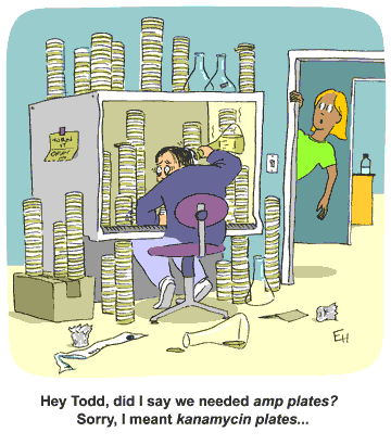
Today’s #FridayFeeling is one of gratitude for all of those people who do the things that make our lives easier: lab techs, work-study students, undergraduate assistants. They put up with our requests and changes of mind and help keep our laboratory glassware clean, solutions sterile and experiments running. Do you have someone who helps you keep your experiments up and running?eh26

Inflammasomes and Pyroptosis

In today’s post, guest blogger,  Martha O’Brien, PhD, provides a preview of her upcoming AAI poster and block symposium talk on the inflammasome, caspase-1 activity and pyroptosis.

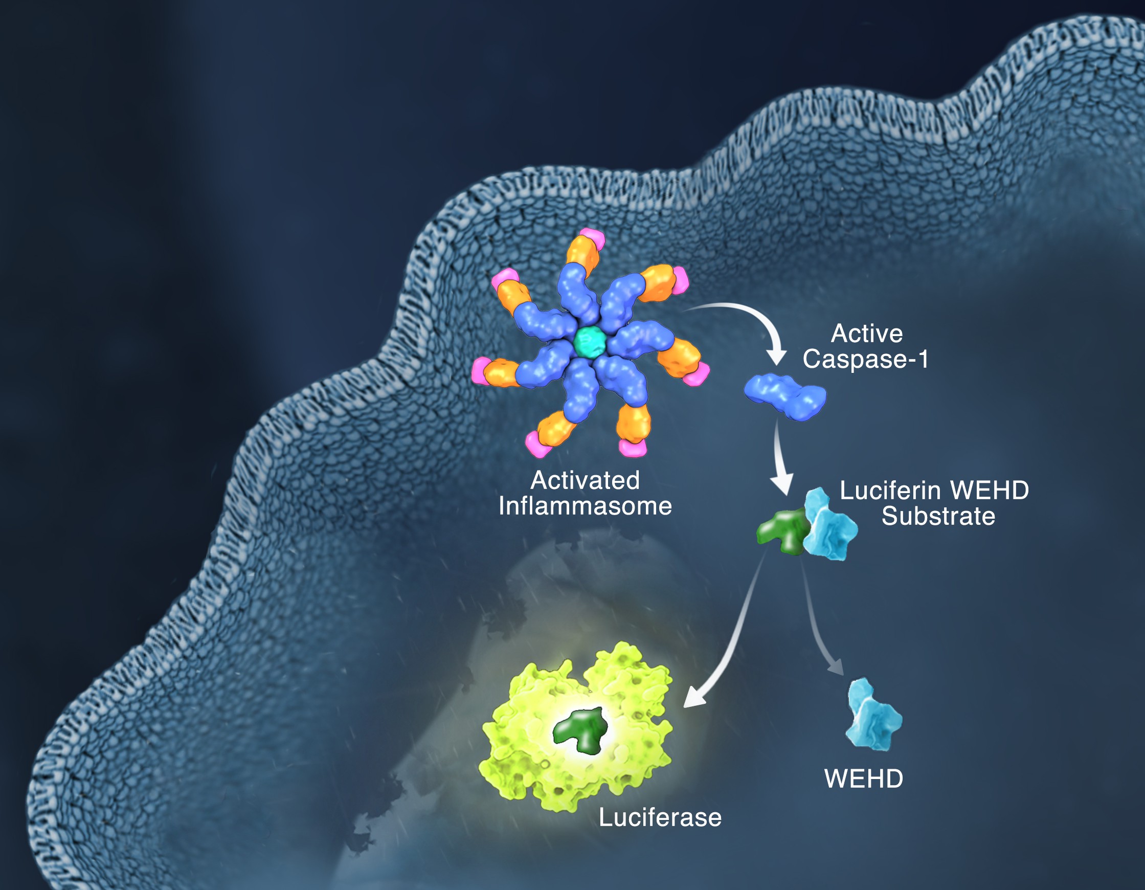
Schematic of the Caspase-Glo 1 Inflammasome Assay.

Responding rapidly to microbial pathogens and damage-associated molecular markers is critical to our innate immune system. Caspase-1 is pivotal in this process leading to processing and release of essential cytokines and an immunogenic form of cell death, termed pyroptosis. Upon sensing pathogen-associated and damage-associated molecular patterns (PAMPs and DAMPs), innate immune cells form inflammasome protein complexes that recruit and activate caspase-1 (canonical inflammasomes). In addition, other inflammatory caspases, 4 and 5 in humans and 11 in mice, directly bind bacterial lipopolysaccharides (LPS), triggering pyroptosis (non-canonical inflammasome). LPS-triggered non-canonical inflammasomes in mice and humans ultimately lead to canonical inflammasome engagement and caspase-1 activation (1–3).  Caspase-1 was originally termed interleukin converting enzyme (ICE) for its well-established role in processing IL-1ß and IL-18, two important inflammation cytokines. How caspase-1 mediates pyroptosis is less well understood, but is beginning to be delineated. Recently, a substrate of the inflammatory caspases, gasdermin D, was identified and its processed fragment, gasdermin-N domain, was shown to be required for pyroptosis in non-canonical inflammasome circumstances (4, 5). The precise role of gasdermin D in canonical inflammasome-triggered pyroptosis is still under investigation. Linking inflammatory caspases directly to pyroptosis is a notable step in understanding the mechanism of this important form of cell death.

Pyroptosis is clearly one means of releasing processed IL-1ß and IL-18 from the cell. However depending on the cell type and stimulus, there is evidence for inflammasome engagement, caspase-1 activation, and release of IL-1ß in the absence of cell death (6, 7). On the flip-side there is also evidence for caspase-1 mediated pyroptosis that helps clear bacteria, independent of IL-1ß and IL-18 involvement (8). To enable further studies on the inflammasome and in particular, assessing the connections between caspase-1 activation, pyroptosis, and cytokine release, Promega developed a new tool to conveniently monitor caspase-1 activation, the Caspase-Glo® 1 Inflammasome Assay.  This bioluminescent, plate-based assay is used to measure caspase-1 activity directly in cell cultures or to monitor released caspase-1 activity in culture medium from treated cells. This flexibility allows easy multiplexing to monitor all three outcomes of inflammasome stimulation; caspase-1 activity, pyroptosis, and release of IL-1ß and IL-18. Caspase-1 activation typically is monitored indirectly with western blots of processed caspase-1. Now the activity of the enzyme can be monitored directly, providing accurate information on temporal aspects of the inflammasome. The assay can be readily combined with real-time measures of cell death (e.g., CellTox™ Green Cytotoxicity Assay) and some of the culture medium can be removed for IL-1ß/IL-18 assessment, leaving the cells and remaining culture medium for caspase-1 activity measurements.

References

  1. Schmid-Burgk et al. (2015) Caspase-4 mediates non-canonical activation of the NLRP3 inflammasome in human myeloid cells. J. Immunol. 45, 2911–7.
  2. Baker et al. (2015) NLRP3 inflammasome activation downstream of cytoplasmic LPS recognition by both caspase-4 and caspase-5. J. Immunol. 45, 2918–26.
  3. Ruhl, S. and P. Broz (2015) Caspase-11 activates a canonical NLRP3 inflammasome by promoting K+ Eur. J. Immunol. 45, 2927–36.
  4. Shi et al. (2015) Cleavage of GSDMD by inflammatory caspases determines pyroptotic cell death. Nature 526, 660–5.
  5. Kayagaki et al. (2015) Caspase-11 cleaves gasdermin D for non-canonical inflammasome signaling. Nature 526, 666–71.
  6. Gaidt et al. (2016) Human monocytes engage an alternative inflammasome pathway. Immunity 44, 833–46.
  7. Chen et al. (2014) The neutrophil NLRC4 inflammasome selectively promotes IL-1ß maturation without pyroptosis during acute. Salmonella Cell Reports 8, 570–82.
  8. Miao et al. (2010) Caspase-1-induced pyroptosis is an innate immune effector mechanism against intracellular bacteria. Nature Immunology 11, 1136–42.

Characterizing Unique Protein: DNA Interactions Using Cell-Free Protein Expression

Molecular model of human telomere DNA

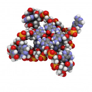
The POT1 protein plays a critical role in telomere protection and telomerase regulation. POT1 binds single-stranded 5′-TTAGGGTTAG-3′ and forms a dimer with the TPP1 protein. Human POT1 contains two Oligonucleotide/Oligosaccharide Binding (OB) fold domains, OB1 and OB2, which make physical contact with the DNA. OB1 recognizes 5′-TTAGGG whereas OB2 binds to the downstream TTAG-3′ (1,2). Several recent studies from other species have shown that some of these proteins are able to recognize a broader variety of DNA ligands than expected (3). A recent reference reexamined the sequence-specificity of the Human POT1 protein (4).
SELEX (Systematic Evolution of Ligands through Exponential Enrichment) was used  to re-examine the DNA-binding specificity of human POT1 (5).

Continue reading “Characterizing Unique Protein: DNA Interactions Using Cell-Free Protein Expression”