Insects and Science: Optimizing Work with Sf9 Insect Cells

Insects are a keystone species in the animal kingdom, often providing invaluable benefits to terrestrial ecosystems and useful services to mankind. While many of them are seen as pests (think mosquitos), others are important for pollination, waste management, and even scientific research.

Insect biotechnology, or the use of insect-derived molecules and cells to develop products, is applied in a diverse set of scientific fields including agricultural, industrial, and medical biotechnology. Insect cells have been central to many scientific advances, being utilized in recombinant protein, baculovirus, and vaccine and viral pesticide production, among other applications (5).

Therefore, as the use of insect cells becomes more widespread, understanding how they are produced, their research applications, and the scientific products that can be used with them is crucial to fostering further scientific advancements.

Primary Cell Cultures and Cell Lines

Cell culture - Cell lines - Insect Cells

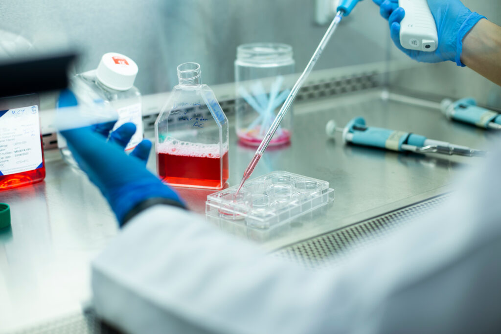
In general, experimentation with individual cells, rather than full animal models, is advantageous due to improved reproducibility, decreased space requirements, less ethical concerns, and a reduction in expense. This makes primary cell cultures and cell lines essential contributors to basic scientific research.

Continue reading “Insects and Science: Optimizing Work with Sf9 Insect Cells”

Firefly Luciferase Sheds Light on Development of New Malaria Treatments

field of fireflies at night; researchers are using firefly luciferase as a tool to power screening assays for new malaria treatments

Despite significant advancements in antimalarial drugs and widespread efforts to prevent transmission over the past decade, deaths from malaria remain high, particularly in younger children. New drugs with novel modes of action are urgently needed to continue reducing mortality and address drug resistance in the malaria parasite, Plasmodium falciparum. While tens of thousands of compounds have been identified as potential candidates through massive screening efforts, scalable methods for identifying the most effective compounds are needed.

The goal is to find a drug that is potent during all stages in the life cycle of P. falciparum and kills the parasite quickly. Focusing on assessing whether a compound can rapidly eliminate initial parasite burden, Paul Horrocks, PhD, and his colleagues developed a validated bioluminescence-based assay that rapidly determines the initial rate of kill for discovery antimalarials. One key to developing their assay was figuring out how to monitor when the parasite dies after introducing the drug. While measuring DNA content can be used to monitor parasite burden, it is too stable to use for a relevant time course assay.

See how Dr. Paul Horrocks uses a firefly luciferase-based system to understand the dynamics of drug action in the development of new malaria treatments.

Enter firefly luciferase, a dynamic reporter tool to investigate drug action. By creating transgenic P. falciparum that express the luc reporter gene, the researchers could monitor drug action over time. When the parasite is killed, it stops making the luciferase reporter. Since there is no new production of luciferase, levels fall quickly after the parasite dies, and a luciferase assay can determine how fast each drug killed the parasite.

Continue reading “Firefly Luciferase Sheds Light on Development of New Malaria Treatments”

Bioluminescence and Biotechnology: Shining Nature’s Cool Light on Biology

Imagine you’re taking a refreshing night swim in the warm blue waters of Vieques in Puerto Rico. You splash into the surf and head out to some of the deeper waters of the bay, when what to your wondering eyes should appear, but blue streaks of light in water that once was clear. Do you need to get your eyes checked? Are you hallucinating? No! You’ve just happened upon a cluster of dinoflagellates, harmless bioluminescent microorganisms called plankton, that emit their glow when disturbed by movement. These dinoflagellates are known to inhabit waters throughout the world but are generally not present in large enough numbers to be noticed. There are only five ecosystems in the world where these special bioluminescent bays can be seen, and three of them are in Puerto Rico. 

Bioluminescent plankton exhibit a blue glow when disturbed.
Bioluminescent plankton in the ocean

But you don’t have to travel to Puerto Rico or swim with plankton to see bioluminescence. There are bioluminescent organisms all over the world in many unexpected places. There are bioluminescent mushrooms, bioluminescent sea creatures—both large and small (squid, jellyfish, and shrimp, in addition to the dinoflagellates)—and bioluminescent insects, to name a few. Bioluminescence is simply the ability of living things to produce light.

Continue reading “Bioluminescence and Biotechnology: Shining Nature’s Cool Light on Biology”

From Viral Outbreak to Vaccine Development: Our Top 10 Most Viewed Blog Posts of 2020

This illustration, created at the Centers for Disease Control and Prevention (CDC), reveals ultrastructural morphology exhibited by coronaviruses. Photo Credit:  Alissa Eckert, MS; Dan Higgins, MAM CDC It is one is used in several of our top 10 most viewed blogs of 2020
Illustration from CDC; Photo Credit: Alissa Eckert, MS; Dan Higgins, MAM

When you look at our top 10 most viewed blog posts of 2020, there’s no surprise that all relate to COVID-19. We have come a long way since the beginning of the year, thanks to tireless scientists and researchers around the globe. They have led the way in COVID-19 research, treatment, and testing. Let’s take a closer look at this top 10 list:

10. Tips to Maintain Physical Distance in the Lab 

The spread of COVID-19 forced us to adapt and adjust to new ways in life, in work, and for this blog post, in the lab. In response to the pandemic, some labs shut down completely. Others have stayed open, especially those involving coronavirus research. This post provides 10 helpful distancing tips for researchers to stay safe and productive while working in the lab.  

9. Investigation of Remdesivir as a Possible Treatment for SARS-2-CoV (2019 nCoV) 

Scientists have worked hard to determine possible treatment for COVID-19. This blog post focuses on Remdesivir (RDV or GS-5734), an encouraging treatment used for the first case in the United States. It provides an in-depth look at numerous studies and clinical trials on Remdesivir as treatment for COVID-19. One key finding is that RDV needed to be administered either before or shortly after infection to limit lung damage. 

Continue reading “From Viral Outbreak to Vaccine Development: Our Top 10 Most Viewed Blog Posts of 2020”

Just What Is an RLU (Relative Light Unit)?

This post was contributed by guest blogger, Scott Messenger, Technical Support Scientist 2 at Promega Corporation.

It’s always an exciting time in the lab when you find a new assay to answer an important research question. Once you get your hands on the assay, it is always good to confirm it will work for your experimental setup. Repeating the control experiment shown in the technical manual is a great way to test the assay in your hands.

After running that first experiment of your assay, it looks pretty good. The trends of control and treatment are consistent. Time to get on with the experiments…but wait—the RLUs (Relative Light Units) are two orders of magnitude lower than the example data! I can’t show this data to my colleagues; it doesn’t match. What did I do wrong?

Relative Light Units, Measuring, Luminescence

This is a concern that we in Technical Services hear frequently. The concern is real, and I had this same thought when doing some of my first experiments using luminescence. When a question like this comes in, a Technical Service Scientist will make sure the experiment was performed as we described, and in most cases it is. We then start talking about RLUs (Relative Light Units).

Continue reading “Just What Is an RLU (Relative Light Unit)?”

The Path Brightens for Vaccine Researchers: Luminescent Reporter Viruses Detect Neutralizing Antibodies

Developing a vaccine that is safe, effective, easily manufactured and distributed is a daunting task. Yet, that is exactly what is needed in response to the COVID-19 pandemic.

Computer generated 3D image of coronavirus

Vaccine development, safety and efficacy testing take time. The mumps vaccine is thought to be the quickest infectious disease vaccine ever produced, and its development required four years from sample collection to licensing (2). However, there are many reasons to anticipate quicker development for a COVID-19 vaccine: Researchers are collaborating in unprecedented ways, and most COVID-19 scientific publications are free for all to access and often available as preprints. As of August 11, 2020, researchers around the globe have more than 165 vaccine candidates in development, 30 of which are in some phase of human clinical trials (1). The range of vaccine formulations available to scientists has expanded to include RNA and DNA vaccines, replication-defective adenovirus vaccines, inactivated or killed vaccines and subunit protein vaccines. Equally important is that vaccine developers and researchers have greater access to powerful molecular biology tools like bioluminescent reporters that enable quicker testing and development.

Continue reading “The Path Brightens for Vaccine Researchers: Luminescent Reporter Viruses Detect Neutralizing Antibodies”

NanoLuc: Tiny Tag with a Big Impact

Synthetic biology—genetically engineering an organism to do or make something useful—is the central goal of the iGEM competition each year. After teams conquer the challenge of cloning their gene, the next hurdle is demonstrating that the engineered gene is expressing the desired protein (and possibly quantifying the level of expression), which they may do using a reporter gene.

Reporters can also play a more significant role in iGEM projects when teams design their organism with reporter genes to detect and signal the presence of specific molecules, like environmental toxins or biomarkers. Three of the iGEM teams Promega sponsored this year opted to incorporate some version of NanoLuc® Luciferase into their projects.

NanoLuc® luciferase is a small monomeric enzyme (19.1kDa, 171 amino acids) based on the luciferase from the deep sea shrimp Oplophorus gracilirostris. This engineered enzyme uses a novel substrate, furimazine, to produce high-intensity, glow-type luminescence in an ATP-independent reaction. Unlike other molecules for tagging and detecting proteins, NanoLuc® luciferase is less likely to interfere with enzyme activity and affect protein production due to its small size.

NanoLuc® Luciferase has also been engineered into a structural complementation reporter system, NanoBiT® Luciferase, that contains a Large subunit (LgBiT) and two small subunit options: low affinity SmBiT and high affinity HiBiT. Together, these NanoLuc® technologies provide a bioluminescent toolbox that was used by the iGEM teams to address a diverse set of biological challenges.

Here is an overview of each team’s project and how they incorporated NanoLuc® technology.

Continue reading “NanoLuc: Tiny Tag with a Big Impact”

Why You Don’t Need to Select a Wavelength for a Luciferase Assay

Promega kit depicted; test involves wavelength for a luciferase assay.

It’s a question I’m asked probably once a week. “What wavelength do I select on my luminometer when performing a luciferase assay?” The question is a good and not altogether unexpected one, especially for those new to bioluminescent assays. The answer is that in most cases, you don’t and in fact shouldn’t select a wavelength (the exception to this rule is if you’re measuring light emitted in two simultaneous luciferase reactions). To understand why requires a bit of an explanation of absorbance, fluorescence, and luminescence assays, and the differences among them.

Absorbance, fluorescence, and luminescence assays are all means to quantify something of interest, be that a genetic reporter, cell viability, cytotoxicity, apoptosis, or other markers. In principle, they are all similar. For example, a genetic reporter assay is an indicator of gene expression. The promoter of a gene of interest can be cloned upstream of a reporter such as β-galactosidase, GFP, or firefly luciferase. The amount of each of these reporters that is transcribed into mRNA and translated into protein by the cell is indicative of the endogenous expression of the gene of interest.

Continue reading “Why You Don’t Need to Select a Wavelength for a Luciferase Assay”

When Proteins Get Together: Shedding (Blue) Light on Cellular LOV

NanoBRET
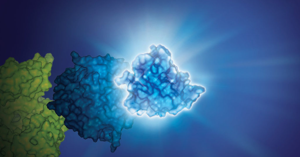
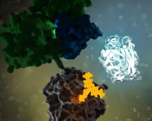
No protein is an island. Within a cell, protein-protein interactions (PPIs) are involved in highly regulated and specific pathways that control gene expression and cell signaling. The disruption of PPIs can lead to a variety of disease states, including cancer.

Two general approaches are commonly used to study PPIs. Real-time assays measure PPI activity in live cells using fluorescent or luminescent tags. A second approach includes methods that measure a specific PPI “after the fact”; popular examples include a reporter system, such as the classic yeast two-hybrid system.

Continue reading “When Proteins Get Together: Shedding (Blue) Light on Cellular LOV”

Eight Considerations for Getting the Best Data from Your Luminescent Assays

The stage is set. You’ve spent days setting up this experiment. Your bench is spotless. All the materials you need to finally collect data are laid neatly before you. You fetch your cells from the incubator, add your detection reagents, and carefully slide the assay plate into the luminometer. It whirs and buzzes, and data begin to appear on the computer screen. But wait!

Bad data
These data are garbage!

Don’t let this dramatic person be you. Here are 8 tips from us on things to watch out for before you start your next luminescent assay. Make sure you’ll be getting good data before wasting precious sample!

Continue reading “Eight Considerations for Getting the Best Data from Your Luminescent Assays”