Shocking Revelation about Starfish Anatomy: Just a Head

Two starfish on the beach
Recent research reveals that starfish anatomy is even stranger than previously thought

Most animals in the world are what biologists refer to as “bilateral”—their left and right sides mirror one another. It is also typically easy to tell which part of most animals is the top and which is the bottom. The anatomical arrangements of certain other animals, however, are slightly more confounding, for instance in the case of echinoderms, which include sea urchins, sand dollars and starfish. These animals are “pentaradial”, with five identical sections of the body radiating from a central axis. The question of how these creatures evolved into such a state has been a puzzle pondered by many a biologist, with little progress made until recently. In a new study published in Nature, scientists closely examining the genetic composition of starfish point to some key evidence that suggests a starfish is mostly just a head.

Starfish are a deuterostome, belonging to the superphylum Deuterostomia. Most deuterostomes are bilateral, leading scientists to believe that, despite their peculiar body plan, starfish evolved from a bilateral ancestor. This is supported by the fact that starfish larvae actually start out bilateral, and eventually transform into the characteristic star shape. But where the head of the starfish is, or whether it even has one, has proved difficult for scientists to parse out, especially since their outward structure offers no real clues.

There have been a number of theories posited, such as the duplication hypothesis—where each of the five sections of a starfish could be considered “bilateral”, placing the head at the center—and the stacking hypothesis, which asserts that the body is stacked atop the head. In a bilateral body plan, anterior genes broadly code for the front, or the head-region, and posterior genes code for the trunk, or the “torso”, and tail. Researchers in this new study looked at the expression of these genes throughout the body plan as a possible source of clarity as to which part of the starfish is its head and which parts comprise the body.

To this end, researchers used advanced molecular and genetic sequencing techniques including RNA tomography and in situ hybridization. RNA tomography allowed them to create a three-dimensional map of gene expression throughout the limbs of the sea star Patiria miniate. In situ hybridization is a fluorescent staining technique that offered them a means by which to examine where exactly anterior or posterior genes are expressed in the sea star’s tissue, providing a clearer picture of genetic body patterning.

Remarkably, scientists found that anterior or head-coding genes were expressed in the starfish’s skin, including head-like regions appearing in the center, or midline, of each arm, while tail-coding genes were only seen at the outer edges of the arms. Perhaps even more remarkable was the lack of genetic patterning accounting for a trunk or torso, leading scientists to the conclusion that starfish are, for the most part, just heads.

Whether this holds true for other echinoderms remains to be proven, and further investigations into starfish anatomy may seek to pinpoint where in the timeline the trunk was lost. Overall, research like this helps scientists understand how life came to look the way it does. Oddly shaped creatures like the humble starfish can offer insight into the strange evolutionary processes that result in such rich biodiversity across the animal kingdom.


Works cited:

  1. ‘A disembodied head walking about the sea floor on its lips’: Scientists finally work out what a starfish is | Live Science
  2. Molecular evidence of anteroposterior patterning in adult echinoderms | Nature
  3. Starfish Are Heads–Just Heads – Scientific American
  4. Study reveals location of starfish’s head | Stanford News
The Human Cell Atlas: Mapping a Cellular Landscape

From macrophages that seek out and destroy infectious agents to fibroblasts that hold tissues and organs together, cells give form and function to our bodies. However, despite their foundational roles in our biology, there is still much we don’t know about cells—like where different cell types are localized, what states a given cell type may take on, how the molecular characteristics of cells change over a person’s lifetime and more. Addressing these questions will provide a deeper understanding about the cellular and genetic basis of human health and disease.

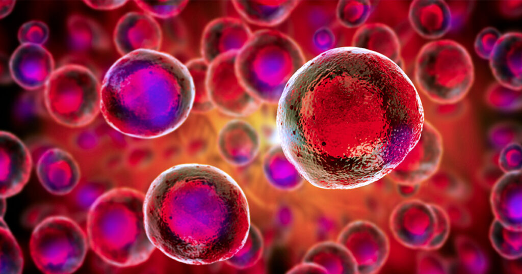
Image contains several cells with a hazy outline of a DNA molecule in the background and one cell is highlighted
Continue reading “The Human Cell Atlas: Mapping a Cellular Landscape”

RNA-Protein Interactions: A New Frontier for Drug Discovery

Almost 90% of the human genome is transcribed into RNA, but only 3% is ultimately translated into a protein. Some non-translated RNA is thought to be useless, while some play a significant yet often mysterious role in cancer and other diseases. Despite its abundance and biological significance, RNA is rarely the target of therapeutics.

“We say it’s undruggable, but I would say that ‘not-yet-drugged’ is a better way to put it,” says Amanda Garner, Associate Professor of Medicinal Chemistry at the University of Michigan. “We know that RNA biology is important, but we don’t yet know how to target it.”

Amanda’s lab develops systems to study RNA biology. She employs a variety of approaches to analyze the functions of different RNAs and study their interactions with proteins. Her lab recently published a paper describing a novel method for studying RNA-protein interactions (RPI) in live cells. Amanda says that with the right tools, RPI could become a critical target for drug discovery.

“It’s amazing that current drugs ever work, because they’re all based on really old approaches,” Amanda says. “This isn’t going to be like developing a small molecule kinase inhibitor. It’s a whole new world.”

Continue reading “RNA-Protein Interactions: A New Frontier for Drug Discovery”

Buckling Down to Scale Up: Providing Support Through the Pandemic

The past year has been a challenge. Amidst the pandemic, we’re thankful for the tireless work of our dedicated employees. With their support, we have continuously stayed engaged and prepared during all stages of the COVID-19 pandemic so that we can serve our customers at the highest levels.

How We Got Here

The persistent work by our teams has made a great impact on the support we can provide for scientists and our community during the pandemic. From scaling up manufacturing to investing in new automation, every effort has helped.

Promega has a long history of manufacturing reagents, assays, and benchtop instruments for both researching and testing viruses. When the pandemic began in 2020, we responded quickly and efficiently to unprecedented demands. In the past year, we experienced an approximately 10-fold increase in demand for finished catalog and custom products for COVID-19 testing. In response to these demands, we increased production lines. One year ago, we ran one shift five days per week. Currently, we run three shifts seven days per week. This change has allowed 50 different Promega products to support SARS-CoV-2 testing globally in hospitals, clinical diagnostic laboratories, and molecular diagnostic manufacturers. Additionally, our clinical diagnostics materials make up about 2/3 of COVID-19 PCR tests on the global market today. Since January 2020, Promega has supplied enough reagents to enable testing an estimated 700 million samples for SARS-CoV-2 worldwide.

Developments and Advances

Promega products are used in viral and vaccine research. This year, our technologies have been leveraged for virtually every step of pandemic response from understanding SARS-CoV-2 to testing to research studies looking at vaccine response.

Promega product: The Lumit™ Dx SARS-CoV-2 Immunoassay

Who Got Us Here

We are extremely grateful for our employees. In the past year, we hired over 100 people and still have positions open today. While welcoming newcomers, this challenging year also reinforced the importance of our collaborative culture. Relationships at Promega have been built over multiple years. The long history of our teams allows us to stay coordinated while prioritizing product distribution to customers across the globe. It also leads to effective communication with colleagues and vendors. Those leading our manufacturing operations team, for example, have an average tenure of 15 years. Their history in collaborating through challenging situations helps them quickly focus where needed most.

Our 600 on-site employees support product manufacturing, quality, and R&D. They do it all while remaining COVID-conscious by social distancing, wearing masks, working split shifts, and restricting movement between buildings. While we continue to practice physical safety precautions, we also prioritize our employees’ mental health and wellness. Promega provides a variety of wellness resources including phone and video mental health sessions, virtual fitness and nutrition classes, and stress and anxiety tools.

What’s to Come

While we acknowledge that the COVID-19 is not over, we are proud of the support we have been able to provide to customers working both on pandemic research and critical research not related to COVID-19. Our policies of long-term planning and investing in the future has allowed us to respond quickly and creatively and learn from the experience.


Related Posts

In Vitro Transcription and the Use of Modified Nucleotides

In vitro transcription
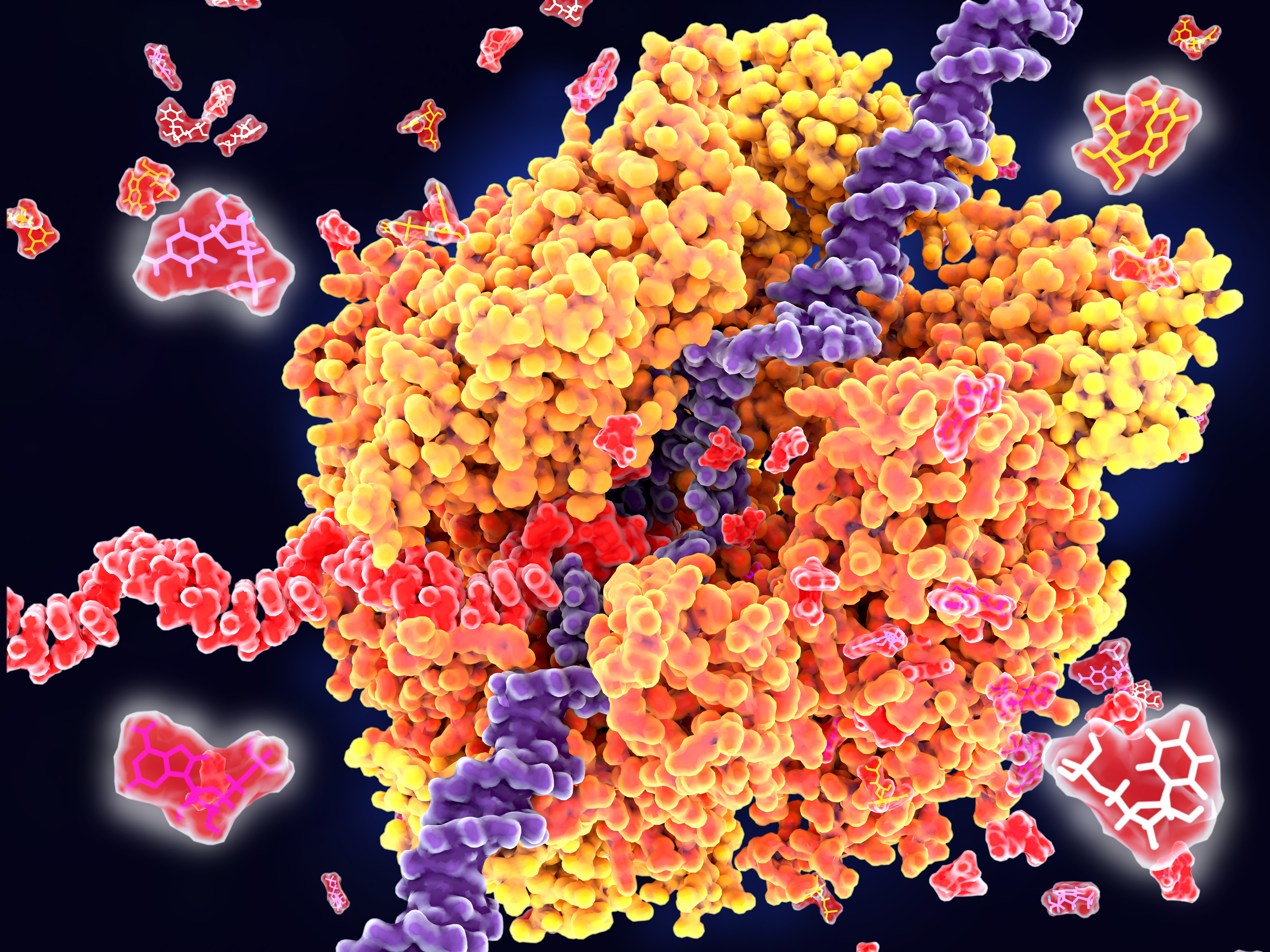
RNA polymerase unwinds DNA strands for transcription.

Transcription is the production of RNA from a DNA sequence. It’s a necessary life process in most cells. Transcription performed in vitro is also a valuable technique for research applications—from gene expression studies to the development of RNA virus vaccines.

During transcription, the DNA sequence is read by RNA polymerase to produce a complimentary, antiparallel RNA strand. This RNA strand is called a primary transcript, often referred to as an RNA transcript. In vitro transcription is a convenient method for generating RNA in a controlled environment outside of a cell.

In vitro transcription offers flexibility when choosing a DNA template, with a few requirements. The template must be purified, linear, and include a double stranded promoter region. Acceptable template types are plasmids or cloning vectors, PCR products, synthetic oligos (oligonucleotides), and cDNA (complimentary DNA). 

In vitro transcription is used for production of large amounts of RNA transcripts for use in many applications including gene expression studies, RNA interference studies (RNAi), generation of guide RNA (gRNA) for use in CRISPR, creation of RNA standards for quantification of results in reverse-transcription quantitative PCR (RT-qPCR), studies of RNA structure and function, labeling of RNA probes for blotting and hybridization or for RNA:protein interaction studies, and preparation of specific cDNA libraries, just to name a few!

In vitro transcription can also be applied in general virology to study the effects of an RNA virus on a cell or an organism, and in development and production of RNA therapeutics and RNA virus vaccines. The large quantity of viral RNA produced through in vitro transcription can be used as inoculation material for viral infection studies. Viral mRNA transcripts, typically coding for a disease-specific antigen, can be quickly created through in vitro transcription, and used in the production of vaccines and therapeutics.

Continue reading “In Vitro Transcription and the Use of Modified Nucleotides”

High-Throughput Purification with Experts Included

Implementing automated nucleic acid purification or making changes to your high-throughput (HT) workflow can be complicated and time-consuming. There are also many barriers to success such as challenging samples types and maintaining desirable downstream results that can add to the stress, not to mention actually getting the robotic instrumentation to do what you want it to. All of this makes it easy to understand why many labs avoid automating or own expensive instrumentation that goes unused. Continue reading “High-Throughput Purification with Experts Included”

A Crash Course in CRISPR

CRISPR is a hot topic right now, and rightly so—it is revolutionizing research that relies on editing genes. But what exactly is CRISPR? How does it work? Why is everyone so interested in using it? Today’s blog is a beginner’s guide on how CRISPR works with an overview of some new applications of this technology for those familiar with CRISPR.

Introduction to CRISPR/Cas9

27850283-June-13-CRISPR-image-WEB

Clustered Regularly Interspaced Short Palindromic Repeats (CRISPR) were discovered in 1987, but it took 30 years before scientists identified their function. CRISPRs are a special kind of repeating DNA sequence that bacteria have as part of their “immune” system against invading nucleic acids from viruses and other bacteria. Over time, the genetic material from these invaders can be incorporated into the bacterial genome as a CRISPR and used to target specific sequences found in foreign genomes.

CRISPRs are part of a system within a bacterium that requires a nuclease (e.g. Cas9), a single guide RNA (sgRNA) and a tracrRNA. The tracrRNA recruits Cas9, while sgRNA binds to Cas9 and guides it to the corresponding DNA sequence of the invading genome. Cas9 then cuts the DNA, creating a double-stranded break that disables its function. Bacteria use a Protospacer Adjacent Motif, or PAM, sequence near the target sequence to distinguish between self and non-self and protect their own DNA.

While this system is an effective method of protection for bacteria, CRISPR/Cas9 has been manipulated in order to perform gene editing in a lab (click here for a video about CRISPR). First, the tracrRNA and sgRNA are combined into a single molecule. Then the sequence of the guide portion of this RNA is changed to match the target sequence. Using this engineered sgRNA along with Cas9 will result in a double-stranded break (DSB) in the target DNA sequence, provided the target sequence is adjacent to a compatible PAM sequence.

Continue reading “A Crash Course in CRISPR”

How to Isolate RNA like a Pro

Ribbon diagram of RNA’s biggest threat: a ribonuclease

Back in graduate school, I purified a lot of RNA, and after a while, I became fairly successful at it. My yields were good, and the RNA was intact. However, many of my early attempts at RNA isolation yielded degraded RNA that did not work well in many downstream applications. In my case, successfully isolating high-quality RNA required practice. During my trials and tribulations, I learned a lot of tricks and tips about how to obtain high-quality RNA. Here I share some of these tricks to help you speed through that “practice makes perfect” phase so that you can isolate RNA like a pro.

Continue reading “How to Isolate RNA like a Pro”

Methods for Quantitating Your Nucleic Acid Sample

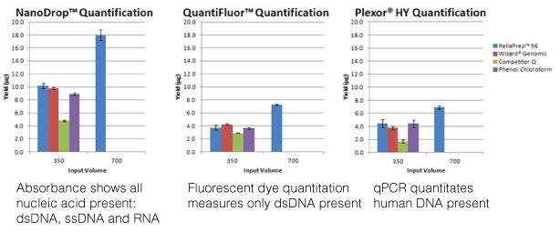
Nucleic acid quanitation webinarFor most molecular biology applications, knowing the amount of nucleic acid present in your purified sample is important. However, one quantitation method might serve better than another, depending on your situation, or you may need to weigh the benefits of a second method to assess the information from the first. Our webinar “To NanoDrop® or Not to NanoDrop®: Choosing the Most Appropriate Method for Nucleic Acid Quantitation” given by Doug Wieczorek, one of our Applications Scientists, discussed three methods for quantitating nucleic acid and outlined their strengths and weaknesses. Continue reading “Methods for Quantitating Your Nucleic Acid Sample”

Don’t Let Ribonucleases Ruin Your Week(end): Establish a Ribonuclease-free Environment

buffers_image1_274x218

My very first job in science was in a lab that worked exclusively with RNA, and it was only after I moved on to a different job that I learned just how much different the world of DNA research is from that of RNA. When working with DNA, for example, you rarely if ever have the sample you have labored over reduced to a fuzzy blur at the bottom of a gel because it has been degraded beyond rescue. With RNA, unfortunately, this happens all too frequently. In fact, a labmate of mine once put up a poll on the door to our lab asking if it was better to discover that your RNA sample was degraded on a Monday or a Friday.

The culprits in this scenario are Ribonucleases (RNases). They are everywhere. They are incredibly stable and difficult to inactivate. And, if you work with RNA, they are your enemy. Take heart though, they can be defeated if you follow some pretty simple steps.

Continue reading “Don’t Let Ribonucleases Ruin Your Week(end): Establish a Ribonuclease-free Environment”